. 2025 Jul 18;17(1):40.
doi: 10.1186/s11689-025-09621-9.

Brain functional connectivity correlates of autism diagnosis and familial liability in 24-month-olds

Collaborators, Affiliations

Brain functional connectivity correlates of autism diagnosis and familial liability in 24-month-olds

John R Pruett Jr et al. J Neurodev Disord. .

Abstract

Background: fcMRI correlates of autism spectrum disorder (ASD) diagnosis and familial liability were studied in 24-month-olds at high (older affected sibling) and low familial likelihood for ASD.

Methods: fcMRI comparisons of high-familial-likelihood (HL) ASD-positive (HLP, N = 23) and ASD-negative (HLN, N = 91), and low-likelihood ASD-negative (LLN, N = 27) 24-month-olds from the Infant Brain Imaging Study (IBIS) Network were conducted, employing object oriented data analysis (OODA), support vector machine (SVM) classification, and network-level fcMRI enrichment analyses.

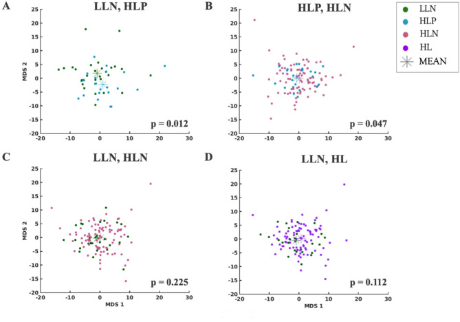
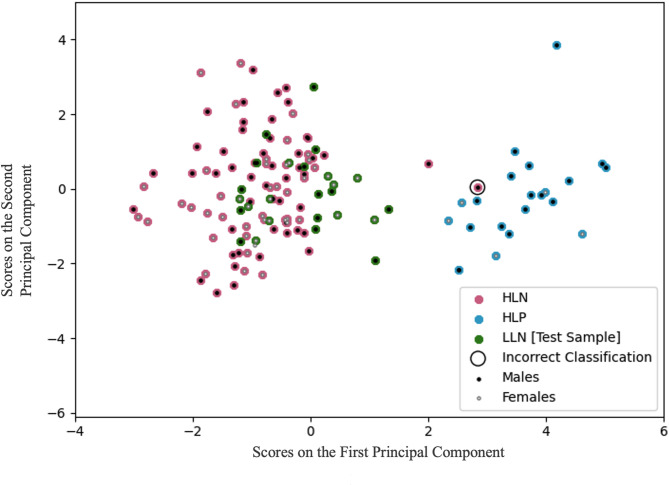
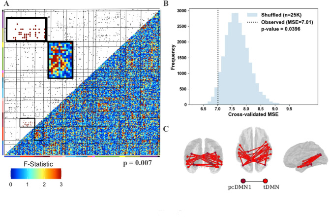
Results: OODA (alpha = 0.0167, 3 comparisons) revealed differences in HLP and LLN fcMRI matrices (p = 0.012), but none for HLP versus HLN (p = 0.047) nor HLN versus LLN (p = 0.225). SVM distinguished HLP from HLN (accuracy = 99%, PPV = 96%, NPV = 100%), based on connectivity involving many networks. SVM accurately classified (non-training) LLN subjects with 100% accuracy. Enrichment analyses identified a cross-group fcMRI difference in the posterior cingulate default mode network 1 (pcDMN1)- temporal default mode network (tDMN) pair (p = 0.0070). Functional connectivity for implicated connections in these networks was consistently lower in HLP and HLN than in LLN (p = 0.0461 and 0.0004). HLP did not differ from HLN (p = 0.2254). Secondary testing showed HL children with low ASD behaviors still differed from LLN (p = 0.0036).

Conclusions: 24-month-old high-familial-likelihood infants show reduced intra-DMN connectivity, a potential neural finding related to familial liability, while widely distributed functional connections correlate with ASD diagnosis.

Keywords: Default mode network; Familial; Functional connectivity; Infant; MRI.


Conflict of interest statement

Declarations. Ethics approval and consent to participate: This study protocol was reviewed and approved by the internal review boards of Washington University School of Medicine, IRB IDs 201103140 and 201301110, the University of Washington, IRB IDs 12317 and STUDY00012991, The Children’s Hospital of Philadelphia, IRB ID 07-005689, and the University of North Carolina at Chapel Hill, IRB ID 05-2293. Informed consent was signed by all study participants. Competing interests: Dr. Robert McKinstry serves on the advisory board of Nous Imaging, Inc. and receives funding for meals and travel from Siemens Healthineers and Philips Healthcare. Abraham Z. Snyder is a consultant for Sora Neuroscience, LLC. All other authors report no financial relationships with commercial interests.

Figures

Fig. 1
Fig. 1
Functional network architecture in 24-month-old infants. A Average correlations for 230 spherical regions of interest (ROIs). The ROIs are sorted by network. B ROIs brain map (dorsal, lateral, and medial views). Colors correspond to network assignments. C Cerebellar ROIs on a flattened cerebellar surface [62]. aDMN1: anterior default mode network 1; aDMN2: anterior default mode network 2; aFP: anterior frontoparietal network; Aud: auditory network; CO-SubC: cingulo-opercular-subcortical network; DAN: dorsal attention network; MotM: motor-mouth network; pcDMN1: posterior cingulate default mode network 1; pcDMN2: posterior cingulate default mode network 2; pFP: posterior frontoparietal network; Sal: salience network; SM1: somatomotor network 1; SM2: somatomotor network 2; SM3: somatomotor network 3; tDMN: temporal default mode network; US: unspecified (not assigned); Vis1 and Vis2: visual 1 and 2
Fig. 2
Fig. 2
Multi-dimensional scaling (Euclidean distance) indicates (A) some separation (p = 0.012) between fcMRI correlation matrix cluster means (colored asterisks) for high-likelihood positive (HLP) and low-likelihood negative (LLN); but (B) not for HLP versus high-likelihood negative subjects (HLN; p = 0.047); nor for (C) HLN versus LLN subjects (p = 0.225). D Overall, the HL and LL groups did not differ significantly from one another (p = 0.112). Group label colors in figure inset
Fig. 3
Fig. 3
Scores on the first two principal components, computed on the set of features retained by the crystallized support vector machine classifier. The classifier, trained on high-likelihood positive and high-likelihood negative subjects only, was highly accurate (overall = 99.1%, males = 98.5%, females = 100%). LLN subjects were excluded from the training and were all correctly classified as unaffected. There is clear separation of HLP and HLN/LLN subjects, with only one incorrect positive classification (circled). Negative predictive values: overall = 100%, male = 100%, female = 100%. Positive predictive values: overall = 95.6%, male = 94.4%, female = 100%
Fig. 4
Fig. 4
Connections important for accurate support vector machine (SVM) classification. The brain-wide support vector classifier was trained with high-likelihood positive and high-likelihood negative subjects. Consensus features (across leave-out folds) important for accurate classification are depicted on an adjacency matrix, and the 100% consensus features are shown on lateral, posterior, and dorsal views of the brain. Colors indicate network membership (see Fig. 1)
Fig. 5
Fig. 5
Enrichment analyses and secondary validation. A 3-group (HLP, HLN, LLN) ANOVA screen for functional connection differences suggests a clustering of nominally significant group differences for ROI pairs in the posterior cingulate default mode network 1 (pcDMN1)– temporal default mode network (tDMN) network pair (p = 0.007). A Lower triangle: ANOVA F-statistics. Upper triangle: 5% threshold applied to the F-statistics. The 230 ROIs are sorted by network (defined in Fig. 1). Black border: pcDMN1- tDMN. B Functional connections in the pcDMN1-tDMN network pair were used to predict ADOS CSS (five-fold cross-validated linear regression) with a Mean Squared Error of 7.01 (empirical p = 0.0396, on 25,000 permutations). C Locations of implicated connections within pcDMN1 - tDMN visualized on posterior, dorsal, and lateral views of the brain
Fig. 6
Fig. 6
Enrichment analyses for pairwise Tukey-corrected t-tests characterize the pcDMN1-tDMN network pairs when comparing (A) the smaller LLN (n = 27) and HLP (n = 23) groups (p = 0.0461) and (B) the LLN and the larger HLN (n = 91) groups (p = 0.0004). Locations of implicated connections within the enriched network pair are visualized on dorsal, posterior, and lateral views of the brain. C, D The group mean functional connectivity profiles are remarkably consistent for pcDMN1 - tDMN ROI-pairs that show group differences: LLN > HLP (in C) and LLN > HLN (in D). The ROI-pairs are ordered by increasing LLN mean functional connectivity for clarity
Fig. 7
Fig. 7
Enrichment analyses with student t-tests for the pcDMN1-tDMN network pair. A High-likelihood (HL) subjects compared with LLN (p = 0.0036). All subjects had a typical ADOS CSS = 1. Locations of implicated connections within the enriched network pair are visualized on dorsal, posterior, and lateral views of the brain. B Group mean functional connectivity profiles for pcDMN1 - tDMN ROI-pairs that show group differences: LLN > HL. The ROI-pairs are ordered by increasing LLN mean connectivity for clarity

References

    1. Padmanabhan A, Lynch CJ, Schaer M, Menon V. The default mode network in autism. Biol Psychiatry Cogn Neurosci Neuroimaging. 2017;2(6):476–86. - PMC - PubMed
    1. Nair A, Jolliffe M, Lograsso YSS, Bearden CE. A review of default mode network connectivity and its association with social cognition in adolescents with autism spectrum disorder and Early-Onset psychosis. Front Psychiatry. 2020;11:614. - PMC - PubMed
    1. Harikumar A, Evans DW, Dougherty CC, Carpenter KLH, Michael AM. A review of the default mode network in autism spectrum disorders and attention deficit hyperactivity disorder. Brain Connect. 2021;11(4):253–63. - PMC - PubMed
    1. Hull JV, Dokovna LB, Jacokes ZJ, Torgerson CM, Irimia A, Van Horn JD. Resting-State functional connectivity in autism spectrum disorders: A review. Front Psychiatry. 2016;7:205. - PMC - PubMed
    1. Tyszka JM, Kennedy DP, Paul LK, Adolphs R. Largely typical patterns of resting-state functional connectivity in high-functioning adults with autism. Cereb Cortex. 2014;24(7):1894–905. - PMC - PubMed