. 2025 Jul 18;23(1):525.
doi: 10.1186/s12951-025-03600-x.

Rapid diagnostic imaging and targeted immunotoxin delivery in aggressive prostate cancer using CEACAM5-specific nanobodies

Affiliations

Rapid diagnostic imaging and targeted immunotoxin delivery in aggressive prostate cancer using CEACAM5-specific nanobodies

Zhaoming Xiao et al. J Nanobiotechnology. .

Abstract

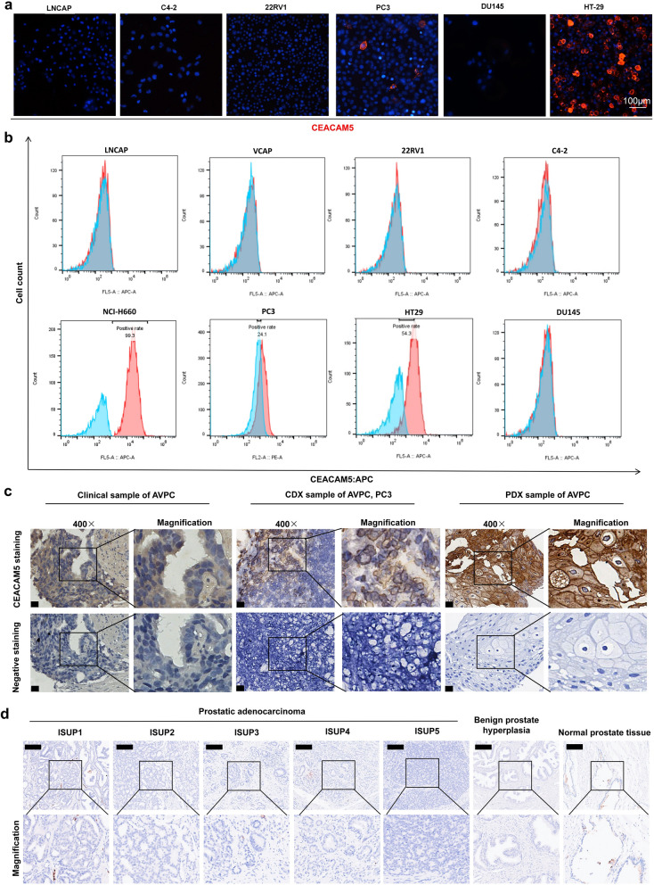
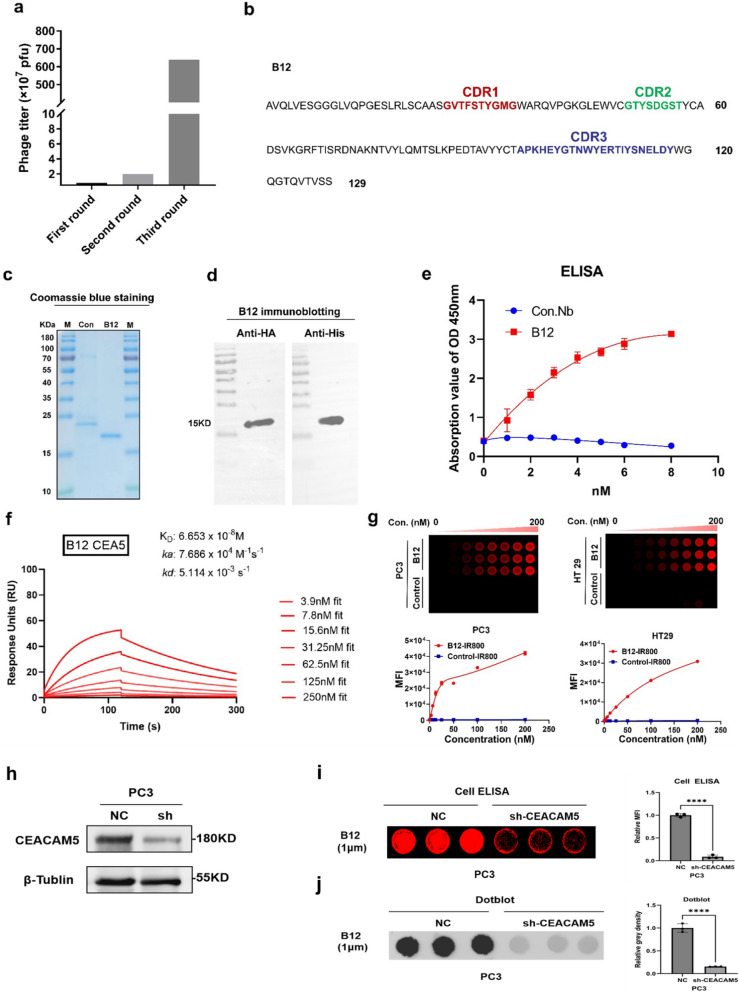
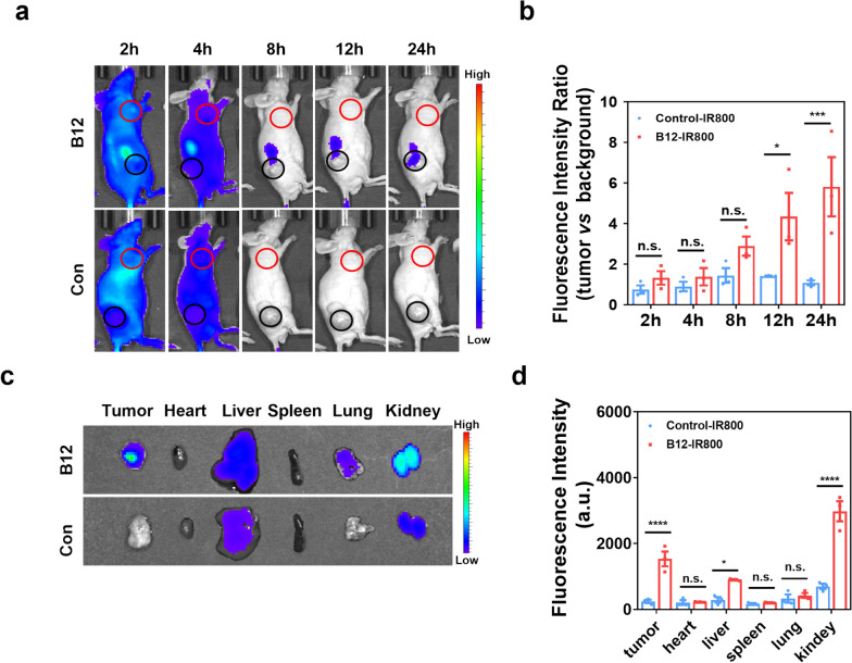
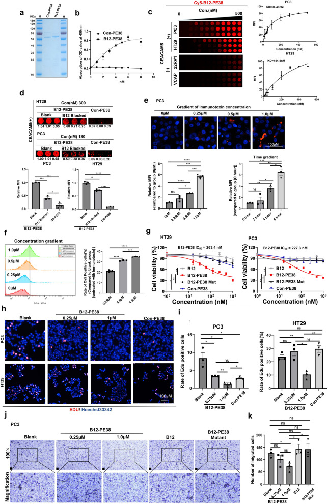
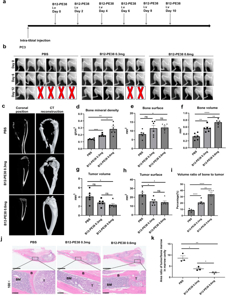
Aggressive variant prostate cancer (AVPC) originates from metastatic prostate cancer (mPCa) following androgen receptor-targeted therapies, leading to diverse pathological subtypes, notably castration-resistant prostate cancer (CRPC). Carcinoembryonic antigen-related cell adhesion molecule 5 (CEACAM5), is consistently expressed across AVPC phenotypes, including neuroendocrine prostate carcinoma (NEPC) and double-negative prostate carcinoma (DNPC), which are significant subtypes of CRPC, making it a promising therapeutic target. In this study, A high-affinity nanobody, B12, specific to CEACAM5, was discovered through phage library screening. B12 exhibited robust binding capabilities, enhanced tumor accumulation, and effective tissue penetration, facilitating rapid in vivo imaging of AVPC. The conjugation of B12 with PE38 to create the immunotoxin B12-PE38 showed significant anti-tumor activity in AVPC xenograft models, including one that mimics bone metastasis. When B12-PE38 was combined with docetaxel, it elicited enhanced tumor inhibitory effects, effectively inhibiting tumor progression. This study underscores CEACAM5 as a target for precise imaging and targeted therapy in AVPC, introducing novel diagnostic and therapeutic strategies for a disease that currently faces a dearth of effective treatment options due to the scarcity of well-defined targets.

Keywords: Aggressive variant prostate cancer; CEACAM5; Immunotoxin; Nanobodies; Phenotype transformation; Rapid imaging.


Conflict of interest statement

Declarations. Ethics approval and consent to participate: The research was approved by the Ethics Committee of the Fifth Affiliated Hospital of Southern Medical University (2024MNWK-K-001), the Shenzhen People’s hospital (AUP-230224-LZJ-543-01) and Nanchang University (BR/AF/SG-04/1.0-202112). Consent for publication: All subjects have written informed consent. Competing interests: The authors declare no competing interests.

Figures

Fig. 1
Fig. 1
Recapitulation of CEACAM5 expression profiles in PCa cell lines and tissues. a Immunofluorescence staining identified the membrane expression of CEACAM5 on DNPC derived PCa cell line PC3, but not in other PCa cell lines derived from prostatic adenocarcinoma, and HT-29 cells were used as a positive control. Scale bar, 100 μm. b Determination of CEACAM5 positivity in PCa derived cell lines by flow cytometry assay. The positive CEACAM5 staining was detected and confirmed in both AVPC derived PC3 and NCI-H660 cell lines, but not in cell lines derived from prostatic adenocarcinoma. The lower penal indicated CEACAM positive AVPC cell lines including NCI-H660 (100%) and PC3 (24.1%). The human colon carcinoma cell line HT29 was selected as the positive control (54.3%). The upper penal demonstrated negative CEACAM5 expression in the PCa cell lines. c Tissue CEACAM5 expression profiles in PCa tissue samples. Immunohistochemical assay verified the membrane expression of CEACAM5 in clinical samples (NEPC: 2/4) and PDX tissues, which were characterized by a consistent and diffuse CEACAM5 expression profile. In contrast, the expression profile of CEACAM5 in CDX samples of DNPC derived PC3 cell line was manifested as a focal and heterogenous expression profile. Scale bars, 20 μm. d CEACAM5 staining in prostatic adenocarcinoma (classified by ISUP grade), normal prostate tissue and benign prostatic hyperplasia. Notably, no positive staining of CEACAM5 was observed in these clinical samples. Scale bars, 250 μm
Fig. 2
Fig. 2
CEACAM5 nanobody isolation and characterization. a Output phage titer for three round biopanning against CEACAM5 protein. b Nanobody B12 sequence with three CDR domains. The whole amino acid sequence of B12 was presented and CDR domains 1, 2 and 3 were marked in red, green and blue, respectively. c, d The purification and confirmation of B12 nanobody by coomassie blue staining and immunoblotting with antibodies against HA and HIS tags. e The ELISA assay confirmed the superior binding activity of B12 over ConNb against CEACAM5 antigen (n = 3). Data are presented as mean ± SEM. f SPR assay to determine the binding dissociation constant (KD) of B12 to CEACAM5. The KD value of nanobody B12 against CEACAM5 antigen was 6.653 × 10–8 M. g The Cell ELISA assay to verify the binding ability of B12 to CEACAM5-expressing cells. Nanobodies were labelled with dye IR-800. Nanobody B12 was demonstrated the excellent binding ability against CEACAM5 expressing cell lines including PC3 and HT29, showing increased signals in a concentration-dependent manner over non-targeting ConNb (n = 3). Data are presented as mean ± SEM. h Western blot analysis of CEACAM5 knockdown efficiency in PC3 cells using targeted shRNA. i, j Cell ELISA and dot blot assays demonstrated reduced B12 fluorescence signals in CEACAM5-knockdown PC3 cells, confirming antibody specificity (n = 3). Data are analyzed by student t-test and represented with mean ± SEM; ****p < 0.0001
Fig. 3
Fig. 3
B12-IR800 effectively images AVPC tumors in vivo. a In vivo imaging exhibited the gradual accumulation of B12-IR800 in PC3 tumors over ConNb-IR800. The most significant difference occurred at 24 h post injection. The red circles indicate the selected background areas used as fluorescence intensity references in non-tumor regions, and the black circles mark the tumor regions. b Quantification analysis of fluorescence intensity with tumor-to-background ratio (TBR, tumor vs background) (n = 3). Data are expressed as mean ± SEM. Statistical analysis was conducted with Student’s t test. ****p < 0.0001; ns, not significant. TBR for B12-IR800 was significantly higher than ConNb-IR800 at 12 and 24 h post injection. c Ex vivo imaging on dissected various organs collected at 24 h post injection. B12-IR800 was primarily identified in tumor, liver and kidneys and Con Nb-IR800 was only observed in liver and kidneys. d Quantification analysis for fluorescence intensity from collected organs (n = 3). Data are expressed as mean ± SEM. *p < 0.05; **** < 0.0001; ns, not significant; Student’s t test
Fig. 4
Fig. 4
The B12-PE38 immunotoxin exhibits superb in vitro binding activity and anti-tumor efficacy against CEACAM5+ AVPC cells. a The purification of B12-PE38 and control non-targeting immunotoxin (ConNb-PE38) demonstrated by the Coomassie blue staining. b ELISA assay confirmed the better binding activity of B12-PE38 against CEACAM5 antigen over the non-targeting ConNb-PE38 (n = 3). Data are represented as mean ± SEM. c Cell ELISA assay validated the binding capability of B12-PE38 against CEACAM5 expressing cell lines including PC3 and HT29 (n = 3). The KD value of B12-PE38 against PC3 and HT29 cell lines was approximately 94.48 nM and 444.4 nM, respectively. Data are represented as mean ± SEM. d Pretreatment with B12 nanobodies blocked the binding activity of B12-PE38 against CEACAM5 expressing cell lines (n = 3). Data are represented as mean ± SEM. *p < 0.05; **p < 0.01; ***p < 0.001; ****p < 0.0001; One-way Anova. e, f Internalization and uptake assays for B12-PE38 in CEACAM5+ AVPC derived PC3 cells. Immunotoxins were labeled with dye Cy5. The drug uptake and internalization were determined by microscopy (e) and flow cytometry (f) (n = 3). Data are represented as mean ± SEM. One-way Anova was used to analyze the difference among difference groups. *p < 0.05; **p < 0.01; ***p < 0.001; ****p < 0.0001; ns, no significance. g In vitro cytotoxic assay by CCK8 revealed that the B12-PE38 immunotoxin significantly reduced the viability of cancer cells expressing CEACAM5, but not the B12 nanobody alone, inactive immunotoxin B12-PE38 mut or non-targeting ConNb-PE38 (n = 3). Data are represented as mean ± SEM. Two-way Anova was used to analyze the difference among difference groups. *p < 0.05; **p < 0.01. h, i EDU staining and quantification after various treatments, implying that B12-PE38 inhibits the proliferation in CEACAM5+ cell lines including PC3 and HT29 (n = 3). Data are represented as mean ± SEM. One-way Anova; *p < 0.05; **p < 0.01; ***p < 0.001; *p < 0.0001; ns, no significance. j, k In vitro migration assay indicated that B12-PE38 can efficiently suppress the migration of CEACAM5+ cell line PC3 (n = 3); scale bars: 50 μm. Data are represented as mean ± SEM. One-way Anova; *p < 0.05; ns, no significance
Fig. 5
Fig. 5
In vivo tumor targeting capability and anti-tumor efficacy of the immunotoxin B12-PE38 against CEACAM5+ xenografts. a, b The whole-body imaging and quantification of mutant immunotoxin B12-PE38 in AVPC derived PC3 xenografts. Approximately 100 μg of dye-labeled B12-PE38 mut and non-targeting ConNb-PE38 mut were systemically injected into tumor bearing mice and tumor images were collected in various time points (n = 3). The red circles indicate the selected background areas used as fluorescence intensity references in non-tumor regions, and the black circles mark the tumor regions. Data are expressed as mean ± SEM. Student’s t test; **p < 0.01; ****p < 0.0001; ns, not significant. c, d Ex vivo organ imaging and quantification for B12-PE38 mut and non-targeting ConNb-PE38 mut 24-h post-injection (n = 3). Data are expressed as mean ± SEM. Student’s t test; **p < 0.01; ****p < 0.0001; ns, not significant. e Treatment schedule of immunotoxin in PC3 tumor-bearing mice. f, g Tumor growth curve of PC3 tumors receiving the two different doses of immunotoxin B12-PE38 as well was B12 alone or PBS (n = 5). Data are expressed as mean ± SEM. Two-way Anova analysis was conducted to assess the difference among groups. **p < 0.01; ***p < 0.001; ****p < 0.0001; ns, not significant. The immunotoxin B12-PE38 significantly reduce the growth of PC3 xenograft, but no inhibition was observed in xenograft receiving B12. h Tumor weight from different treatment groups (n = 5). Data are expressed as mean ± SEM. One-way Anova; **p < 0.01. i The anti-tumor efficacy of B12-PE38 against HT29 xenografts (n = 5). Data are expressed as mean ± SEM. Two-way Anova analysis; ****p < 0.0001. j Survival analysis of mice bearing HT29 tumors receiving B12-PE38 treatment. The difference between two groups was assessed by Kaplan–Meier survival analysis (log-rank test); **p < 0.01
Fig. 6
Fig. 6
The combinational anti-tumor efficacy of the B12-PE38 immunotoxin with docetaxel against CEACAM5+ prostate cancers. a The treatment schedule for combination treatment of B12-PE38 with docetaxel in a PC3-derived CDX model. b Tumor growth curves among different groups to examine the combinational anti-tumor efficacy of the immunotoxin B12-PE38 with docetaxel against PC3 tumors (n = 5). Data are presented as mean ± SEM. Statistical analysis was performed by Two-way Anova test; **p < 0.01; ***p < 0.001; ns, not significant. c Survival study for the combination treatment of B12-PE38 with docetaxel in a PC3-derived CDX model. Statistical assessment for the survival curves was conducted with Kaplan–Meier survival analysis (log-rank test); *p < 0.05; **p < 0.01; ns, not significant. d The treatment schedule for combination treatment of B12-PE38 with docetaxel in a CEACAM5+ PDX model. e PDX tumor growth curves after the combination treatment (n = 5). Data are presented as mean ± SEM. Two-way Anova; ***p < 0.001; ****p < 0.0001; ns, not significant. f Kaplan–Meier survival analysis for the combination treatment (n = 5). **p < 0.01; ***p < 0.001; ns, no significance; log-rank test
Fig. 7
Fig. 7
The B12-PE38 immunotoxin effectively inhibits the progression of bone lesions induced by AVPC-derived cells. a Treatment schedule in PC3-induced tibial bone metastatic model. Drug administration started at 3 weeks after intra-tibial implantation of PC3 cells when X ray identified the local osteolytic lesions. Totally, six doses of B12-PE38 were administered every other day. b The X ray imaging for monitoring tibial bone lesions during the treatment (n = 6). The red cross indicated the visible signs of bone fracture and reached the endpoint of experiment. c CT scanning and bone reconstruction in three groups. di Multiple parameters to assess bone conditions, including bone mineral density (d), bone surface (e), bone volume (f), tumor volume (g), tumor surface (h), bone-to-tumor ratio (i) (n = 6). Data are presented as mean ± SEM (n = 6). *p < 0.05; **p < 0.01; ***p < 0.001; ****p < 0.0001; ns, not significant, One-way Anova. j Pathological examination by H&E staining to assess the area ratio of tumor (T) to bone marrow (BM) in tumor-bearing tibial (n = 3). B, bone tissues; scale bars, 250 μm. k Quantification analysis for area ratio of tumor (T) to bone marrow (BM) in J (n = 3). Data are expressed as mean ± SEM. *p < 0.05; **p < 0.01; One-way Anova

References

    1. Paolieri F, et al. Front-line therapeutic strategy in metastatic hormone sensitive prostate cancer: an updated therapeutic algorithm. Clin Genitourin Cancer. 2024;22(4):102096. - PubMed
    1. Belderbos BPS, et al. Novel treatment options in the management of metastatic castration-naïve prostate cancer; which treatment modality to choose? Ann Oncol. 2019;30(10):1591–600. - PubMed
    1. Davis ID, et al. Enzalutamide with standard first-line therapy in metastatic prostate cancer. N Engl J Med. 2019;381(2):121–31. - PubMed
    1. Yamada Y, Beltran H. The treatment landscape of metastatic prostate cancer. Cancer Lett. 2021;519:20–9. - PMC - PubMed
    1. Wang Y, et al. Mechanisms of enzalutamide resistance in castration-resistant prostate cancer and therapeutic strategies to overcome it. Br J Pharmacol. 2021;178(2):239–61. - PubMed

LinkOut - more resources