. 2025 Jul 18;24(1):295.
doi: 10.1186/s12933-025-02815-4.

C-C chemokine ligand 5 from women subcutaneous adipose tissue has a central role in vascular aging

Affiliations

C-C chemokine ligand 5 from women subcutaneous adipose tissue has a central role in vascular aging

Laura Le Pelletier et al. Cardiovasc Diabetol. .

Abstract

Background: Aging is associated with adipose tissue alterations, oxidative stress, and fibrosis and the onset of cardiometabolic complications. While it has been shown that perivascular adipose tissue (PVAT) contributes to vascular damage, the involvement of subcutaneous adipose tissue (SCAT) - particularly through its secretory activity - in vascular aging remains poorly understood. Previously, we have demonstrated that human adipose-derived stromal cells (ASCs) from the SCAT of aged women display senescence and oxidative stress. We hypothesized that the ASC secretome contributes to the onset of endothelial dysfunction, an early stage of vascular aging.

Methods: We prepared conditioned media from ASCs isolated from SCAT of healthy young (< 25y) or aged (> 60y) women. The ASCs secretome was analyzed and added on human coronary artery endothelial cells (HCAECs). Using clinical cohorts, we evaluated the expression of C-C-chemokine-ligand-5 (CCL5)/Regulated upon-Activation-Normally-T-expressed-and-secreted (RANTES) in adipose tissue of individuals with coronary heart disease.

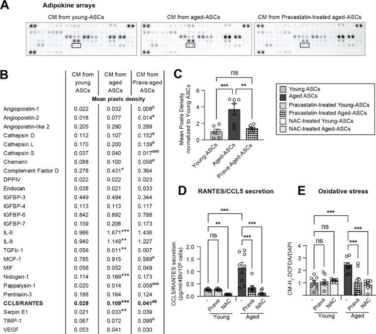
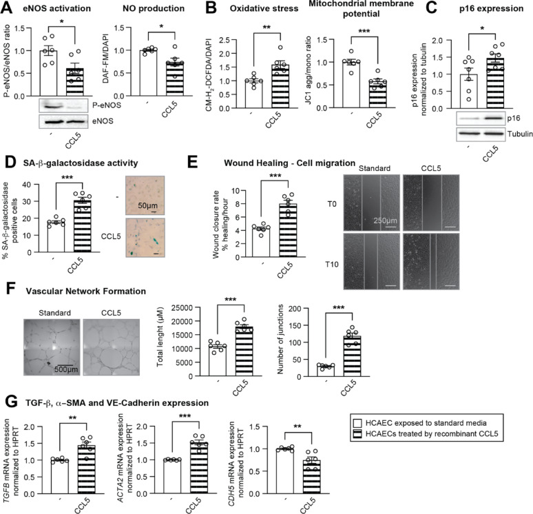
Results: The secretome of aged-donor ASCs induced endothelial cell dysfunction in HCAEC, as evidenced by lower nitric oxide production, higher oxidative stress, senescence, and a pro-adherent phenotype. Aged-donor ASCs also favored the endothelial-to-mesenchymal transition, characterized by the higher expression of mesenchymal markers, a pro-migratory profile and angiogenesis. We showed that the higher secretion of CCL5/RANTES in the secretome of aged- vs. young-donor ASCs and was responsible for these effects. Accordingly, CCL5/RANTES expression in SCAT, but not in epicardial adipose tissue, was associated with blood pressure in patients with coronary heart diseases, thus confirming the important role of SCAT in the onset of cardiometabolic disorders. CCL5’s ability to induce endothelial cell dysfunction and senescence was confirmed using a recombinant CCL5 and a CCL5/RANTES neutralizing antibody. Furthermore, we demonstrated that the CCL5/RANTES receptor antagonist drug maraviroc prevented the deleterious impact of CCL5/RANTES in both HCAECs and human cohorts. Thus, CCL5/RANTES secreted from SCAT during aging could contribute to endothelial dysfunction by exerting both local and systemic effects.

Conclusions: Our results highlighted the ability of the CCL5/RANTES released from aging SCAT and, specifically, from adipose stromal cells, to induce endothelial dysfunction and senescence - both of which are early steps in vascular aging - as well as a potential link between these phenomena and hypertension in particular.

Graphical abstract:

Supplementary Information: The online version contains supplementary material available at 10.1186/s12933-025-02815-4.

Keywords: Adipose stromal cells; Adipose tissue; Aging; CCL5/RANTES; Endothelial dysfunction; Vascular aging..


Conflict of interest statement

Declarations. Ethics approval and consent to participate: The ROCnRAL ANRS-157 was a single-arm study designed to evaluate a switch to a maraviroc plus raltegravir regimen in virologically suppressed HIV-1-infected patients. The Institutional Review Board of Pitie-Salpétrière Hospital approved the study protocol (ClinicalTrials.gov: NCT01420523). All patients provided written informed consent. The ANRS145 Marimuno study was a multicenter, noncomparative, prospective 36-week trial conducted in 16 “Agence nationale de recherche sur le sida et les hépatites” (ANRS) sites in France. The ethics committee in Toulouse, France approved the protocol (EudraCT: 2009-011171-76). All patients gave written informed consent. The PIECVIH ANRS EP52 study was a cross-sectional, study comparing the inflammatory state of epicardial adipose tissue (EAT) between people living with HIV and controls undergoing coronary artery bypass graft. The Institutional Review Board of Saint-Antoine Hospital approved the study protocol (ClinicalTrials.gov: NCT01899196). All patients provided written informed consent. Human adipose-derived stromal cells (ASCs). Adipose stromal cells were isolated from nine donors. All donors provided their written, informed consent to the use of their tissue specimens for research purposes. The study was performed in compliance with the principles of the Declaration of Helsinki and was approved by an institutional review board. The study was approved by the French regulatory authorities (CODECOH DC2023-5617). Competing interests: J.C. reports personal fees for lectures from ViiV Healthcare, Gilead and MSD outside the sub-mitted work. B.F. reports research grants from MSD, and personal fees for lectures for MSD, Amgen, Sanofi, NovoNordisk, and Lilly outside the submitted work. C.L. has received personal fees from MSD, outside the submitted work. F.B. reports research grants from Amgen; lecture fees from Gilead, ViiV Healthcare, Amgen, Sanofi, MSD, NovoNordisk, Novartis and Servier outside the submitted work. C. K. has received travel grants, consultancy fees, and honoraria or study grants from Gilead, Merck, Janssen, and ViiV Healthcare outside the submitted work. L.L., K.N.A., J.G., R.M., M.M., M.A., R.A., L.C., M.A., C.B., C.P., E.C. and V.B. declare no conflicts of interest.

Figures

Fig. 1
Fig. 1
The secretome of aged-donor ASCs induced HCAEC dysfunction, senescence, and endothelial-mesenchymal transition. HCAECs were incubated 48 hours with standard media, CMyoung or CMaged. (A) Representative immunoblots for Phospho-eNOS (phosphoSer1177) and eNOS are shown. Densitometry analysis results are expressed as the mean ± SEM ratio (Standard n=5, CMyoung, CMaged n=13). Nitric oxide (NO) production was evaluated by measuring the fluorescence of DAF-FM (normalized against DAPI) and expressed as the mean ± SEM, relative to standard media (Standard n=8, CMyoung, CMaged n=10) (B) The relative mRNA expression levels of InterCellular (ICAM1) and Vascular Adhesion Molecule (VCAM1) were normalized against that of HPRT, and expressed as the mean ± SEM (n=8). (C) ROS production was assessed by measuring the fluorescence of CM-H2-DCFDA (normalized against DAPI), and expressed as the mean ± SEM, relative to standard media (Standard n=8, CMyoung, CMaged n=10). Mitochondrial membrane potential was assessed using the cationic dye JC-1. Fluorescence results were normalized against MitoTracker and expressed as the mean ± SEM of aggregate/monomer, relative to standard media (Standard n=8, CMyoung, CMaged n=10). (D) Representative immunoblots for p16 and tubulin are shown. Densitometric analysis results are expressed as the mean ± SEM (Standard n=10, CMyoung, CMaged n=16). (E) Senescence was evaluated as the percentage of SA-β-galactosidase-positive cells (n=12). Representative images are shown. (F) Wound closure assay: Scratch wound closure rate was expressed as % of wound coverage/hour (n=5). Representative images of migration areas at 0 and 10 hours are shown. (G) The total tube lengths and number of junctions formed in the assay were measured and expressed as the mean ± SEM (n=8). Representative images are shown. (H) mRNA expression level of transforming growth factor β (TGFB), smooth muscle actin-α2 (ACTA2), and vascular-endothelial cadherin-5 (CDH5), were normalized against that of HPRT, and expressed as the mean ± SEM (n=8). Differences between conditions were determined by one-way ANOVA on data that passed the Brown-Forsythe or Bartlett’s test for heteroscedasticity and the Shapiro-Wilk test for normality of residuals. *P<0.05, **P<0.01, ***P<0.001
Fig. 2
Fig. 2
Pravastatin prevented the stress-induced senescence of aged-donor ASCs. Pravastatin (25 µmol/L) was added to the culture medium of aged-donor or young-donor ASCs for 7 days. (A) Senescence was evaluated with regard to SA-β-galactosidase activity and expressed as the percentage of SA-β-galactosidase-positive cells (n = 9). Representative images of SA-β-galactosidase-positive cells are shown. (B) Lysosomal accumulation was assessed by measuring Lysotracker fluorescence (normalized against DAPI). The results are expressed as the mean ± SEM, relative to young-donor ASCs (n = 9). (C) Representative immunoblots for p16 and tubulin are shown. Densitometric analysis results are expressed as the mean ± SEM (n = 9). (D) ROS production was assessed by measuring the fluorescence of CM-H2-DCFDA (normalized against DAPI), and expressed as the mean ± SEM, relative to young-donor ASCs (n = 9). Differences between conditions were determined by two-way ANOVA on raw data that passed Spearman’s test for heteroscedasticity and the Shapiro-Wilk test for normality of residuals. For B, data were not normally distributed, differences between conditions were determined by Kruskal-Wallis. *P < 0.05, **P < 0.01, ***P < 0.001
Fig. 3
Fig. 3
Pravastatin treatment of ASCs prevented the harmful effect of the aged-ASC secretome on endothelial cells. CM from young-ASCs treated with pravastatin (CMprava-young) or not (CMyoung) and CM from aged-ASCs treated with pravastatin (CMprava-aged) or not (CMaged). HCAECs were incubated 48 h with standard media or CM. (A) Representative immunoblots for Phospho-eNOS (phosphoSer1177) and eNOS are shown. Densitometry analysis results are expressed as the mean ± SEM ratio (n = 11). Nitric oxide (NO) production was evaluated by measuring the fluorescence of DAF-FM (normalized against DAPI) and expressed as the mean ± SEM, relative to standard media (n = 12). (B) The relative mRNA expression levels of ICAM1 and VCAM1 were normalized against that of HPRT, and expressed as the mean ± SEM (n = 8). (C) ROS production was assessed by measuring the fluorescence of CM-H2-DCFDA (normalized against DAPI), and expressed as the mean ± SEM, relative to standard media (n = 12). Mitochondrial membrane potential was assessed using the cationic dye JC-1. Fluorescence results were normalized against MitoTracker and expressed as the mean ± SEM of aggregate/monomer, relative to standard media (n = 12). (D) Representative immunoblots for p16 and tubulin are shown. Densitometric analysis results are expressed as the mean ± SEM (n = 8). (E) Senescence was evaluated as the percentage of SA-β-galactosidase-positive cells (n = 12). (F) Wound closure assay: Scratch wound closure rate was measured and expressed as % of wound coverage/hour (n = 3). Representative images of migration areas at 0 and 10 h are shown. (G) The total tube lengths and number of junctions formed in the assay were measured and expressed as the mean ± SEM (n = 6) Representative images are shown. (H) mRNA expression level of TGFB, ACTA2, and CDH5, were normalized against that of HPRT, and expressed as the mean ± SEM (n = 8). Differences between conditions were determined by two-way ANOVA on raw or transformed data that passed Spearman’s test for heteroscedasticity and the Shapiro-Wilk test for normality of residuals. For A right panel and C left panel, data were not normally distributed, differences between conditions were determined by Kruskal-Wallis. *P < 0.05, **P < 0.01, ***P < 0.001
Fig. 4
Fig. 4
Higher levels of CCL5/RANTES secretion by aged-donor than by young donor ASCs. (A) Representative adipokine arrays obtained for CM from either young-ASCs, aged-ASCs or pravastatin-treated-aged-ASCs. Black rectangle outlined CCL5/RANTES spot. (B) Densitometry analysis of adipokine arrays were performed and expressed as the mean ± SEM (n = 6). (C) Densitometry analysis for CCL5/RANTES were performed and expressed as the mean ± SEM, relative to young-donor ASCs (n = 6). (D) The levels of CCL5/RANTES in the culture medium from the last 48 h were determined with ELISA in young- or aged- donor ASCs treated or not by pravastatin or NAC for 7 days. The results are expressed as the mean ± SEM (n = 8). (E) ROS production was assessed by measuring the fluorescence of CM-H2-DCFDA (normalized against DAPI), and expressed as the mean ± SEM, relative to young-donor ASCs (n = 8). Differences between conditions were determined by two-way ANOVA on raw or transformed data that passed Spearman’s test for heteroscedasticity and the Shapiro-Wilk test for normality of residuals. For D, data were not normally distributed, differences between conditions were determined by Kruskal-Wallis. *P < 0.05, **P < 0.01, ***P < 0.001 young- vs. aged-donor ASCs. #P < 0.05, ##P < 0.01, ###P < 0.001 untreated vs. treated by pravastatin for B
Fig. 5
Fig. 5
CCL5/RANTES induced endothelial cell dysfunction, senescence and endothelial-mesenchymal transition. HCAECs were exposed to CCL5/RANTES (1 ng/mL) or not (untreated) for 48 h. (A) Representative immunoblots for Phospho-eNOS (phosphoSer1177) and eNOS are shown. Densitometry analysis results are expressed as the mean ± SEM ratio (n = 6). Nitric oxide (NO) production was evaluated by measuring the fluorescence of DAF-FM (normalized against DAPI) and expressed as the mean ± SEM, relative to standard media (n = 6) (B) ROS production was assessed by measuring the fluorescence of CM-H2-DCFDA (normalized against DAPI), and expressed as the mean ± SEM, relative to standard media (n = 6). Mitochondrial membrane potential was assessed using the cationic dye JC-1. Fluorescence results were normalized against MitoTracker and expressed as the mean ± SEM of aggregate/monomer, relative to standard media (n = 6). (C) Representative immunoblots for p16 and tubulin are shown. Densitometric analysis results are expressed as the mean ± SEM (n = 7). (D) Senescence was evaluated with regard to SA-β-galactosidase activity and expressed as the percentage of SA-β-galactosidase-positive cells (n = 6). Representative images are shown. (E) Wound closure assay: Scratch wound closure rate was measured and expressed as % of wound coverage/hour (n = 6). Representative images of migration areas at 0 and 10 h are shown. (F) Representative images of vascular network formation are shown. The total tube lengths and number of junctions formed in the assay were measured and expressed as the mean ± SEM (n = 6). (G) mRNA expression level TGFB, ACTA2, and CDH5, were normalized against that of HPRT, and expressed as the mean ± SEM (n = 6). Differences between the two groups were determined using the unpaired t-test, on data that passed the Shapiro-Wilk test for normality of residuals. *P < 0.05, **P < 0.01, ***P < 0.001
Fig. 6
Fig. 6
Maraviroc treatment of HCAECs prevented the harmful effects of the aged-donor ASC secretome. HCAECs were exposed to Maraviroc (MVC) (1 µM) 24 h prior to the exposure to CM or standard media. Maraviroc was then maintained through the 48 h exposure to CMaged. (A) Representative immunoblots for Phospho-eNOS (phosphoSer1177) and eNOS are shown. Densitometry analysis results are expressed as the mean ± SEM ratio (Standard n = 7, Standard + MVC n = 6, CMaged & CMaged+MVC n = 10). Nitric oxide (NO) production was evaluated by measuring the fluorescence of DAF-FM (normalized against DAPI) and expressed as the mean ± SEM, relative to standard media (n = 8). (B) The relative mRNA expression levels of ICAM1 and VCAM1 were normalized against that of HPRT, and expressed as the mean ± SEM (n = 8). (C) ROS production was assessed by measuring the fluorescence of CM-H2-DCFDA (normalized against DAPI), and expressed as the mean ± SEM, relative to standard media (n = 8). Mitochondrial membrane potential was assessed using the cationic dye JC-1. Fluorescence results were normalized against MitoTracker and expressed as the mean ± SEM of aggregate/monomer, relative to standard media (n = 8). (D) Representative immunoblots for p16 and tubulin are shown. Densitometric analysis results are expressed as the mean ± SEM (Standard n = 11, Standard + MVC n = 10, CMaged & CMaged+MVC n = 14). (E) Senescence was evaluated as the percentage of SA-β-galactosidase-positive cells (n = 8). Representative images are shown (F) Wound closure assay: Scratch wound closure rate was measured and expressed as % of wound coverage/hour (n = 5). Representative images of migration areas at 0 and 10 h are shown. (G) The total tube lengths and number of junctions formed in the assay were measured and expressed as the mean ± SEM (n = 6). Representative images are shown. (H) mRNA expression level of TGFB, ACTA2, and CDH5, were normalized against that of HPRT, and expressed as the mean ± SEM (n = 8). Differences between conditions were determined by two-way ANOVA on raw or transformed data that passed Spearman’s test for heteroscedasticity and the Shapiro-Wilk test for normality of residuals. *P < 0.05, **P < 0.01, ***P < 0.001
Fig. 7
Fig. 7
CCL5 mRNA expression in SCAT was correlated with blood pressure values. Subcutaneous thoracic and epicardial adipose tissue (EAT) were obtained from individuals from the PIECVIH study (n = 15). (A,C) Correlation between CCL5 mRNA expression and systolic blood pressure. (B, D) Correlation between CCL5 mRNA expression and diastolic blood pressure (r = Pearson’s correlation coefficient)
Fig. 8
Fig. 8
A switch to treatment with Maraviroc after conventional antiretroviral treatment reduced PBMCs senescence. Thirteen HIV-infected individuals from the ROCnRAL study were switched from conventional antiretroviral treatment to maraviroc-raltegravir combination therapy for a mean duration of 20 weeks. Senescence markers were evaluated in PBMCs sampled at inclusion (W0) and 20 weeks (W20). Representative immunoblots for p16 normalized against β-actin (A) and for Phospho-p53 (phosphoS15) and p53 (B) are shown. Densitometry analysis was performed and changes in levels of before (W0) and after (W20) initiation of maraviroc-raltegravir combination therapy were determined. Differences between conditions were determined using the paired t-test. ***P < 0.001

References

    1. Jia G, Aroor AR, Jia C, Sowers JR. Endothelial cell senescence in aging-related vascular dysfunction. Biochim Biophys Acta Mol Basis Dis. 2019;1865(7):1802–9. - PubMed
    1. Bu LL, Yuan HH, Xie LL, Guo MH, Liao DF, Zheng XL. New dawn for atherosclerosis: vascular endothelial cell senescence and death. Int J Mol Sci 2023, 24(20). - PMC - PubMed
    1. Donato AJ, Machin DR, Lesniewski LA. Mechanisms of dysfunction in the aging vasculature and role in Age-Related disease. Circ Res. 2018;123(7):825–48. - PMC - PubMed
    1. Koenen M, Hill MA, Cohen P, Sowers JR. Obesity, adipose tissue and vascular dysfunction. Circ Res. 2021;128(7):951–68. - PMC - PubMed
    1. Ungvari Z, Tarantini S, Donato AJ, Galvan V, Csiszar A. Mechanisms of vascular aging. Circ Res. 2018;123(7):849–67. - PMC - PubMed

LinkOut - more resources