. 2025 Jul 18;18(1):118.
doi: 10.1186/s12920-025-02187-4.

A recurrent ABCC2 c.2439 + 5G > A variant disturbs mRNA splicing and causes Dubin-Johnson syndrome

Affiliations

A recurrent ABCC2 c.2439 + 5G > A variant disturbs mRNA splicing and causes Dubin-Johnson syndrome

Rongyue Sun et al. BMC Med Genomics. .

Abstract

Background: Hyperbilirubinemia is the main clinical manifestation of Dubin-Johnson syndrome (DJS), of which most cases can be attributed to the variants in the ABCC2 gene. This study aimed to characterize the mechanism of a splicing variant of the ABCC2 gene and interrogate the variant pathogenicity.

Methods: Whole exome sequencing (WES) was performed to identify potential genetic causes. Bioinformatics analysis was performed to predict the variant pathogenicity. Minigene assays were performed to investigate the effects of the identified variant on mRNA splicing. Western blot (WB) experiments were performed to verify the impact of the variant on protein expression. Protein subcellular localization was analyzed by indirect immunofluorescence (IF).

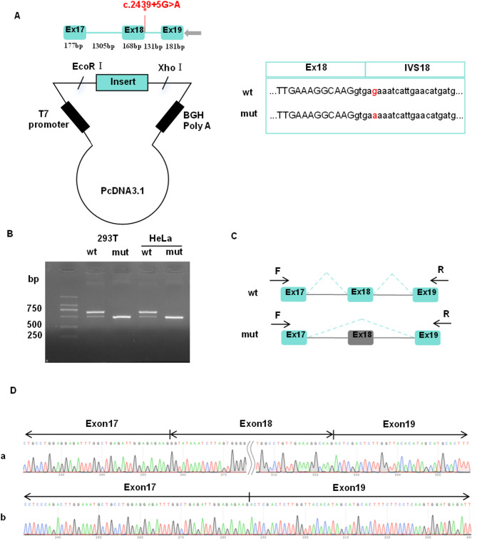
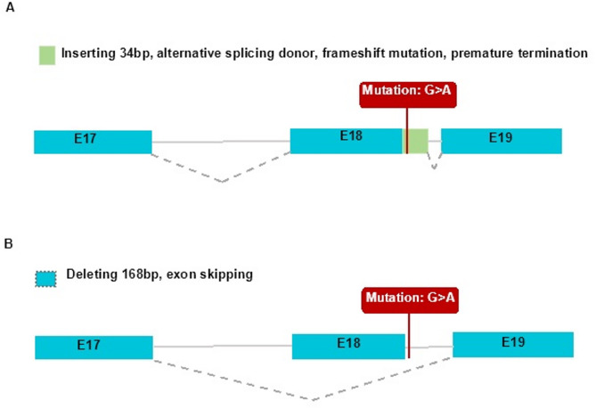
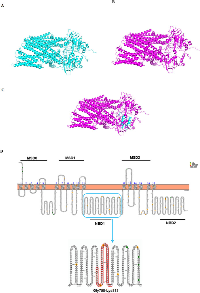
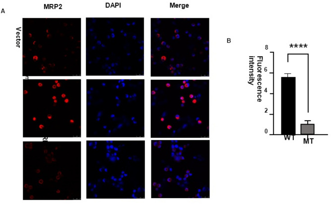
Results: Genetic analysis revealed compound heterozygous variants in ABCC2 gene: the splice-site variant c.2439 + 5G > A inherited from the mother and the nonsense variant c.3825 C > G (p.Y1275X) inherited from the father. The recurrent ABCC2 c.2439 + 5G > A variant can affect mRNA splicing which leads to the skipping of exon 18 and induces the loss of 56 native amino acids, resulting in a shortened MRP2 protein (p.Gly758_Lys813del). The c.2439 + 5G > A variant may cause mislocalization of the mutant protein and significantly reduces its expression.

Conclusion: Our study identifies c.2439 + 5G > A in ABCC2 as a pathogenic variant underlying DJS through aberrant splicing. Critically, the proband's phenotype resulted from compound heterozygous variants leading to biallelic loss of functional protein. These findings highlight the necessity of comprehensive ABCC2 genetic testing for DJS, facilitating accurate diagnosis and personalized patient management.

Keywords: ABCC2; Dubin-johnson syndrome; Heterozygous intronic variant; Minigene assays.


Conflict of interest statement

Declarations. Ethics approval and consent to participate: The study was conducted by the Declaration of Helsinki and approved by the ethics committee of The First Affiliated Hospital of Wenzhou Medical University (No. YS2024-268). Informed consent to participate was obtained from all of the participants and parents of the minor participant in the study. Consent for publication: Not applicable. Competing interests: The authors declare no competing interests.

Figures

Fig. 1
Fig. 1
Functional analysis of the ABCC2 variant effect on mRNA splicing. (A) Construction strategy of pcDNA3.1 minigene vector, * representative of the variant site, the sequencing of wild-type ABCC2 gene vector and c.2439 + 5G > A ABCC2 variant gene vector. (B) Gel electrophoresis of reverse transcription polymerase chain reaction products displayed a single band a (estimated 670 bp) from the wild-type (wt) and a smaller band b (estimated 502 bp) in the mutant type (mut). (C) The splicing pattern corresponding to the band a and band b. (D) Illustration of the sequencing of band a (wild-type in HEK293T and Hela cells) and band b (variant c.2439 + 5G > A in HEK293T and Hela cells) products lead to a shorter transcript with deletion of exon 18 including 168 bp
Fig. 2
Fig. 2
Pedigree of the family. Circles and squares represent females and males, respectively. The black-filled symbols represent the proband
Fig. 3
Fig. 3
The sequencing results of the family. The rectangular symbol indicates the variant site. The variant c.2439 + 5G > A is inherited from the mother
Fig. 4
Fig. 4
The sequencing results of the family. The rectangular symbol indicates the variant site. The variant c.3825 C > G is inherited from the father
Fig. 5
Fig. 5
Two patterns of RNA splicing prediction. (A). Pattern one: Inserting 34 bp, alternative splicing donor, frameshift mutation, premature termination. (B). Pattern two: Deleting 168 bp, exon skipping. E17: exon 17; E18: exon 18; E19: exon 19
Fig. 6
Fig. 6
Prediction of MRP2 structure and domain. (A) Wild-type MRP2 protein. (B) Mutant MRP2 (p.Gly758_Lys813del). (C) Compared wild-type and mutant MRP2 protein. RMSD: 32.8021 Å. (D) Predicted MRP2 protein domains showed the loss of 56aa (p.Gly758_Lys813del) located in nucleotide-binding domain 1 (NBD1) domain
Fig. 7
Fig. 7
The protein expression of MRP2 p.Gly758_Lys813del was decreased in cell lines. (A, D) Western blot bands demonstrated that the protein levels of over-expressed MRP2MT were significantly lower than that of MRP2WT. (B, E) The quantification found that the protein levels of MRP2MT were only 57 ± 2.39% and 50 ± 2.39% (both p < 0.0001, n = 3) that of wild-type MRP2WT in HuH-7 and HepG2 cell lines, respectively. (C, F) The co-expressed Flag tag showed that the protein levels were only 59 ± 2.39% and 55 ± 2.39% (both p < 0.001, n = 3) within these groups, respectively. All data are expressed as Mean ± SEM. Asterisks (*) represent statistically significant differences (***P < 0.001, ****P < 0.0001, n = 3). WT: wild-type MRP2; MT: MRP2 p.Gly758_Lys813del
Fig. 8
Fig. 8
The subcellular localization of MRP2 p.Gly758_Lys813del. (A, B) Immunofluorescence assay showed the decreased MRP2 p.Gly758_Lys813del expression both in cell membrane and cytoplasm. All data are expressed as Mean ± SEM. Asterisks (*) represent statistically significant differences (****P < 0.0001, n = 3). WT: wild-type MRP2; MT: MRP2 p.Gly758_Lys813del
Fig. 9
Fig. 9
MRP2 p.Gly758_Lys813del mutant exhibited decreased organic anion transport activity. Compared to wild-type MRP2, the MRP2 p.Gly758_Lys813del mutant exhibited decreased organic anion transport activity in Huh-7 and HepG2 cell lines. (*) represent statistically significant differences (***P < 0.001, n = 5). Each bar represents the Mean ± SD

Similar articles

References

    1. Mao YX, et al. Transport mechanism of human bilirubin transporter ABCC2 tuned by the inter-module regulatory domain. Nat Commun. 2024;15(1):1061. - PMC - PubMed
    1. Strassburg CP. Hyperbilirubinemia syndromes (Gilbert-Meulengracht, Crigler-Najjar, Dubin-Johnson, and rotor syndrome). Best Pract Res Clin Gastroenterol. 2010;24(5):555–71. - PubMed
    1. Memon N, et al. Inherited disorders of bilirubin clearance. Pediatr Res. 2016;79(3):378–86. - PMC - PubMed
    1. Dubin IN, Johnson FB. Chronic idiopathic jaundice with unidentified pigment in liver cells; a new clinicopathologic entity with a report of 12 cases. Med (Baltim). 1954;33(3):155–97. - PubMed
    1. Paulusma CC, et al. A mutation in the human canalicular multispecific organic anion transporter gene causes the Dubin-Johnson syndrome. Hepatology. 1997;25(6):1539–42. - PubMed

Substances

LinkOut - more resources