Review
. 2025 Jul 18;23(1):347.
doi: 10.1186/s12964-025-02335-4.

The gut-heart axis: a correlation between Paneth cells' dysfunction, microbiome dysbiosis, and cardiovascular diseases

Affiliations
Review

The gut-heart axis: a correlation between Paneth cells' dysfunction, microbiome dysbiosis, and cardiovascular diseases

Aysa Rezabakhsh et al. Cell Commun Signal. .

Abstract

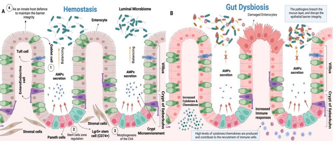
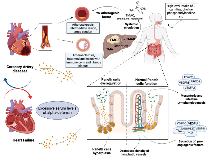
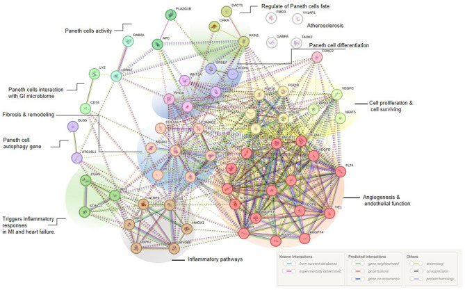
Gut microbiota dysbiosis is characterized by an imbalance in the core microbial equilibrium, leading to changes in the homeostasis of the gastrointestinal tract (GIT) environment. As guardians of the gut microbiota, Paneth cells (PCs) secrete antimicrobial peptides (AMPs) and play a crucial role in maintaining gut integrity and innate immunity in the small intestine. The gut-heart axis has emerged as a critical mediator in cardiovascular disease (CVD) pathogenesis and has drawn significant attention. In this regard, the reciprocal relationship between gut dysbiosis and PC dysfunction has been proposed, which may contribute to a compromised gut barrier and increased systemic inflammation, one of the main drivers of CVD development. It is also well-established that dysfunctional PCs disrupt gut homeostasis and subsequently permit the translocation of pro-inflammatory metabolites like trimethylamine N-oxide (TMAO) while reducing protective short-chain fatty acids (SCFAs), which correlates with atherosclerosis, hypertension, and heart failure.

A better understanding of the underlying mechanisms linking gut health, PCs function, and cardiovascular outcomes is warranted for developing novel gut-target therapies against major CVD risks. This review aimed to comprehensively discuss the predominant role of PCs in the gut-heart axis, some effective compounds on PC function and AMP modulation, and finally, a possible correlation between PC dysfunction and CVD pathogenesis, encouraging future research to further elucidate this crosstalk.

Keywords: Bioinformatics analysis; Cardiovascular disease; Gut barrier; Gut microbiota; Paneth cells.


Conflict of interest statement

Declarations. Ethics approval and consent to participate: N/A. Consent for publication: N/A. Competing interests: The authors declare no competing interests.

Figures

Fig. 1
Fig. 1
The impacts of PCs on Gut Microbiota. The PCs, strategically located at the base of the intestinal crypts of Lieberkühn, play a substantial role in maintaining a healthy gut ecosystem. (A) These specialized epithelial cells are defined as the first line of defense against pathogens and further contribute to intestinal hemostasis and promote microbial diversity— by establishing a balance in the gut microbiota composition. PCs prevent gut dysbiosis and contribute to the host’s well-being through (1) AMPs secretion and immune regulatory activities, (2) Lgr5 + stem cell support (niche maintenance), (3) CVA morphogenesis, and (4) Maintenance of gut barrier integrity. (B) There is a close cross-link between gut dysbiosis and PCs dysfunction in which the reduced AMP secretion allows pathogenic bacteria to overgrow, leading to the microbial imbalance. Also, disruption in gut barrier integrity due to PCs dysfunction can result in bacterial translocation, recruitment of immune cells, and excessive cytokine/chemokine production and further exacerbating dysbiosis and systemic inflammation. Abbreviations: AMPs, anti-microbial peptides; CVA, crypt-villus axis; CD74+, cluster of differentiation 74+; Lgr5+, Leucine-rich repeat-containing G-protein coupled receptor 5+
Fig. 2
Fig. 2
Comprehensive signaling pathways in PCs. (A) Autophagy plays a critical role in the function of PCs. The coordinated action of ATG5, ATG7, and ATG16, as key genes in the autophagy process, leads to the formation of double-membrane structures known as autophagosomes. Autophagy machinery especially contributes to the regulation of AMPs secretion and the degradation of intracellular pathogens beyond the recycling of cellular components. (B) This part illustrates the canonical Wnt signaling pathway in PCs. In the absence of Wnt ligands to stimulate Frizzled receptors, the inhibitory complex, composed of APC, Axin, GSK-3β, and CK1, targets β-catenin for phosphorylation. Following Wnt signaling activation, stabilized β-catenin (phosphorylated form) translocates into the nucleus and forms a complex of TCF/LEF transcription factors, which in turn, activate the transcription of Wnt target genes. (C) NOD2, a key pattern recognition receptor, plays a crucial role in directing both gene expression and proper sorting and secretion of AMPs i.e., lysozyme, as a key enzyme for degrading bacterial cell walls (D) The presence of some TLRs (TLR-9, TLR-3, TLR-4) in regulating AMPs expression, highlighting the key role of TLRs, and related downstream transcription factors named TRIF, MyD88 in this regard. Abbreviations: Akt-1, Akt serine-threonine protein kinase-1; AMPs, antimicrobial peptides; APC, antigen-presenting cell; ATG, autophagy-related gene; CK1, Casein Kinase 1; c-Kit, c-receptor tyrosine kinase; CRP, C-reactive protein; DCVs, dense core vesicles; ER, Endoplasmic reticulum; GSK-3β, glycogen synthase kinase 3β; IRF3/7, IFN regulatory factors 3 and 7; LEF, lymphoid enhancer factor; LRRK2, leucine-rich repeat kinase 2; NF-κB, nuclear factor ‘kappa-light-chain-enhancer of activated B-cells; PI3K, phosphatidylinositol-3 kinase; Rab2a, Ras-related protein Rab-2A; NOD2, nucleotide-binding oligomerization domain-containing protein 2; Reg3 β/γ, regenerating islet-derived protein 3 beta/gamma; SCF, Stem cell factor; TCF, T-cell factor; TLR, Toll-like receptor; Trif, TIR domain-containing adaptor inducing interferon
Fig. 3
Fig. 3
Immunomodulatory impact of Paneth cells. Abbreviations: AMPs, Antimicrobial peptides; Dact1, dishevelled binding antagonist of beta-catenin 1; EGF, Epidermal growth factor; FGF-R3, fibroblast growth factor receptor 3; FXR, farnesoid X receptor; Gfi-1, growth factor independence-1; ICS, intestine stem cell; INF, interferon; HNF4α, hepatocyte nuclear factor 4α; IL-17 A, interleukin-17 A, IL-17 A R, interleukin-17 A receptor; Lgr5+, Leucine-rich repeat-containing G-protein coupled receptor 5; MAPK, mitogen-activated protein kinase; Nur77, Nuclear receptor 77; Shp2, Src homology 2-conatining protein tyrosine phosphatase 2; Wnt3a, wingless-type MMTV integration site family 3a, DII4, Delta-like 4; Math 1, mouse atonal homolog 1; Spdef, SAM pointed domain-containing Ets transcription factor; SOX-9, SRY-box transcription factor 9; Th17, T-helper 17
Fig. 4
Fig. 4
A correlation between Paneth cells regulation and cardiovascular health. Following the gut dysbiosis, serum levels of toxic metabolite, TMAO, which plays a crucial role in atherosclerotic plaque formation, increase. Positive impacts likely refer to the pro-angiogenic factors secreted by PCs contributing to the development and maintenance of the intricate vascular network and lymphoid system (right side). However, negative impacts have also been reported following PCs’ dysregulation and excessive release of AMPs in the serum of patients with CVD, such as heart failure (left side). Abbreviations: ANGPT2, angiopoietin 2; FMO3, flavin-containing dimethylaniline monooxygenases 3; FOXC2, Forkhead box protein C2; PROX-1, Prospero homeobox protein 1; TMA, Trimethylamine; TMAO, Trimethylamine N-oxide; VEGF, vascular endothelial growth factor
Fig. 5
Fig. 5
Protein–protein interaction network obtained using STRING software for prediction of Hub genes and signaling pathways in cardiovascular pathology and Paneth cells dysfunction. The images show the confidence view (http://string-db.org/). Stronger associations are represented by thicker lines 10

Similar articles

References

    1. Nakamura K, Sakuragi N, Takakuwa A, Ayabe T. Paneth cell α-defensins and enteric microbiota in health and disease. Biosci Microbiota Food Health. 2016;35:57–67. - PMC - PubMed
    1. Ferrari AJ, Santomauro DF, Aali A, Abate YH, Abbafati C, Abbastabar H, Abd ElHafeez S, Abdelmasseh M, Abd-Elsalam S, Abdollahi A, et al. Global incidence, prevalence, years lived with disability (YLDs), disability-adjusted life-years (DALYs), and healthy life expectancy (HALE) for 371 diseases and injuries in 204 countries and territories and 811 subnational locations, 1990–2021: a systematic analysis for the Global Burden of Disease Study 2021. The Lancet. 2024;403:2133–2161. - PMC - PubMed
    1. Gaziano TA, Bitton A, Anand S, Abrahams-Gessel S, Murphy A. Growing epidemic of coronary heart disease in low- and middle-income countries. Curr Probl Cardiol. 2010;35:72–115. - PMC - PubMed
    1. Mocumbi AO. Cardiovascular health care in Low- and Middle-Income countries. Circulation. 2024;149:557–9. - PubMed
    1. Frioux C, Ansorge R, Özkurt E, Ghassemi Nedjad C, Fritscher J, Quince C, Waszak SM, Hildebrand F. Enterosignatures define common bacterial guilds in the human gut microbiome. Cell Host Microbe. 2023;31:1111–e11251116. - PubMed

LinkOut - more resources