. 2025 Jul 19;15(1):26217.
doi: 10.1038/s41598-025-12268-0.

TFEB overexpression alleviates autophagy-lysosomal deficits caused by progranulin insufficiency

Affiliations

TFEB overexpression alleviates autophagy-lysosomal deficits caused by progranulin insufficiency

Wren O Nader et al. Sci Rep. .

Abstract

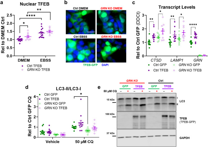
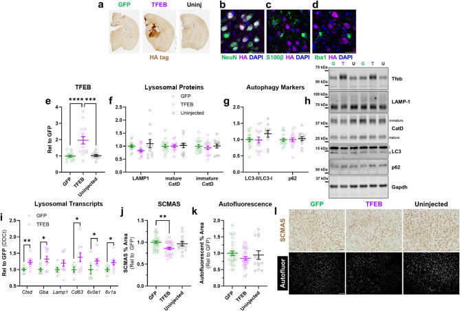
Progranulin is a pro-protein that is necessary for maintaining lysosomal function. Loss-of-function progranulin (GRN) mutations are a dominant cause of frontotemporal dementia (FTD). Brains of people with FTD due to GRN mutations accumulate lysosomal storage material and exhibit increased expression of lysosomal transcripts, which may be driven by TFEB and related transcription factors. While this may be a compensatory response to lysosomal impairment, overproduction of lysosomal proteins may also contribute to FTD pathogenesis. To investigate how TFEB may contribute to disease in people with GRN mutations, we analyzed the effects of TFEB overexpression in progranulin-insufficient cells and mice. We generated GRN knockout HEK-293 cells (GRN KO cells), which exhibited increased nuclear localization of TFEB and expression of lysosomal transcripts, but impaired autophagy. TFEB overexpression in GRN KO cells further increased lysosomal transcripts and partially normalized autophagy. We next injected an AAV vector expressing mouse Tfeb (AAV-TFEB) into the thalamus of Grn-/- mice, which accumulates lysosomal storage material. AAV-TFEB increased lysosomal transcripts and reduced immunoreactivity for SCMAS, a marker of lysosomal storage material, in Grn-/- thalamus. These data show that TFEB activity alleviates some autophagy-lysosomal deficits caused by progranulin insufficiency, suggesting potential utility of lysosome-based therapies for GRN-associated diseases.

Keywords: Autophagy; Lysosomes; Progranulin; TFEB.


Conflict of interest statement

Declarations. Competing interests: The authors declare no competing interests.

Figures

Fig. 1
Fig. 1
GRN Knockout HEK-293 Cells Exhibit Autophagy-Lysosomal Abnormalities. We deleted the entire GRN coding sequence from HEK-293 cells using CRISPR-Cas9, resulting in complete loss of progranulin. (a, b), Lysates of GRN knockout (GRN KO) cells had elevated levels of mature cathepsin D (CatD, t test, p = 0.0013, n = 9 per group). (c, d), GRN KO cells also exhibited a deficit in autophagy, defined as a reduced LC3-II/LC3-I ratio after incubating for one hour with 50 µM chloroquine (CQ) (ANOVA effect of CQ, p < 0.0001, * = p = 0.0406 by Holm-Sidak post-hoc test, n = 9 per group). (eg), Fluorescent imaging of cells transfected with GFP-LC3-RFP-LC3ΔG confirmed a reduction in autophagic flux in GRN KO cells. The ratio of GFP/RFP fluorescence was significantly increased in GRN KO cells versus controls (e, linear mixed effects model effect of GRN, p = 0.0002, *** = p < 0.001 by Tukey’s post-hoc test, n = 528–594 cells per group from 4 independent cultures). Additionally, incubation with 50 µM chloroquine for 3 h increased the GFP/RFP ratio in control cells (e, linear mixed effects model effect of CQ, p < 0.0001, *** = p < 0.001 by Tukey’s post-hoc test) but not in GRN KO cells (p = 0.534 by Tukey’s post-hoc test). Data in (e) are shown as the median fluorescent intensity normalized to control vehicle-treated cells from each culture, with violin plots showing the distribution of all cells measured and symbols showing the median value for each culture. (f), Similar results were obtained by analyzing the frequency distribution of the GFP/RFP ratio in cells transfected with GFP-LC3-RFP-LC3ΔG (f, Kolmogorov-Smirnov test, control vehicle vs. GRN KO vehicle, p = 0.0147, control vehicle vs. control CQ, p = 0.0472, GRN KO vehicle vs. GRN KO CQ, p = 0.7649). Scale bars in (g) represent 20 μm. (h, i), Restoration of progranulin (PGRN) to GRN KO cells reduced levels of mature CatD, though CatD still remained elevated compared to control cells (ANOVA effect of GRN KO, p < 0.0001, effect of PGRN expression, p < 0.0001, ** = p < 0.01, *** = p < 0.001, **** = p < 0.0001 by Holm-Sidak post-hoc test, n = 6 per group). (g, h), Restoration of PGRN to GRN KO cells also normalized the LC3-II/LC3-I ratio (ANOVA effect of PGRN expression, p = 0.0328, n = 7–9 per group), as GFP-transfected GRN KO cells had lower LC3-II/LC-I after chloroquine treatment than control cells (** = p = 0.0092 by Holm-Sidak test) while PGRN-transfected GRN KO cells did not differ from controls (p = 0.3058 by Holm-Sidak test). Full images of immunoblots are shown in Fig. S2.
Fig. 2
Fig. 2
TFEB Overexpression Increases Expression of Lysosomal Transcripts and Normalizes Autophagy in GRN Knockout HEK-293 Cells. (a, b), When transfected with a TFEB-GFP construct, a higher proportion of GRN KO cells than controls exhibited nuclear TFEB localization under standard culture conditions (ANOVA effect of GRN, p = 0.0133, * = p = 0.0357 by Holm-Sidak test, n = 9–11/group). However, both control and GRN KO cells exhibited an increase in nuclear TFEB localization after nutrient starvation by incubating for one hour in EBSS (ANOVA effect of EBSS, p < 0.0001, ** = p = 0.005 and **** = p < 0.0001 by Holm-Sidak test). (c), GRN KO cells exhibited higher expression of lysosomal transcripts such as CTSD than controls, with a similar trend for LAMP1 (MANOVA effect of GRN, p < 0.001, CTSD ANOVA effect of GRN, p = 0.0087, LAMP1 ANOVA effect of GRN, p = 0.0895). Transfection with TFEB-GFP increased lysosomal transcripts in both control and GRN KO cells versus transfection with a GFP control plasmid (MANOVA effect of TFEB, p < 0.001, * = p < 0.05, ** = p < 0.01, **** = p < 0.0001 by Holm-Sidak post-hoc test, n = 9–10 samples/group). (d, e), TFEB overexpression also normalized the autophagy deficits of GRN KO cells (3-way ANOVA effect of CQ, p < 0.0001, effect of GRN, p < 0.0001, CQ x TFEB, p < 0.0282, n = 10–12/group), as GFP-transfected GRN KO cells had lower LC3-II/LC-I after chloroquine treatment than control cells (* = p = 0.0266 by Holm-Sidak test), while TFEB-transfected GRN KO cells did not (p = 0.3923 by Holm-Sidak test). The scale bar in b represents 20 μm. Full images of immunoblots are shown in Fig. S3.
Fig. 3
Fig. 3
TFEB Overexpression in Wild-type Mouse Brain Increases Levels of Lysosomal Proteins. (a), We injected AAV2 vectors expressing either GFP or HA-tagged codon-optimized mouse TFEB into the ventroposteromedial/lateral nuclei of the thalamus (VPM/VPL) of wild-type mice. After 8 weeks, we collected brains for analysis of TFEB expression and lysosomal proteins. (b–d), AAV-TFEB primarily transduced neurons, based on immunostaining for the HA tag and markers of neurons (NeuN), astrocytes (S100β), and microglia (Iba1). (e), TFEB overexpression in thalamus was further confirmed by immunoblot (t test, p = 0.0373). (f), Immunoblot of thalamic tissue confirmed increases in lysosomal proteins in TFEB-treated mice (MANOVA effect of TFEB, p = 0.006), with increases in LAMP-1 (t test, p = 0.0141), mature cathepsin D (t test, p = 0.0017), and immature cathepsin D (t test, p = 0.0003). (g), However, analysis of the LC3-II/LC3-I ratio (t test, p = 0.6163) and p62 (t test, p = 0.5056) did not reveal signs of increased autophagy. n = 6–7 mice per group. Scale bars in (b–d) represent 25 μm. Panel (a) created using Biorender.com. (Arrant, A. (2025) https://BioRender.com/flnev93 and https://BioRender.com/6cia20y). Full images of immunoblots are shown in Fig. S4.
Fig. 4
Fig. 4
TFEB Overexpression Reduces Lysosomal Storage Material in Grn–/– Mice. (a), AAV-TFEB expressed HA-tagged TFEB throughout the ventroposteromedial/lateral thalamic nuclei of Grn–/– mice. (bd), Similar to wild-type mice, AAV-TFEB primarily transduced neurons and increased TFEB levels in the thalamus (e, ANOVA effect of TFEB, p < 0.0001, *** = p = 0.0001 and **** = p < 0.0001 by Holm-Sidak post-hoc test). (f), In contrast to wild-type mice, AAV-TFEB did not increase LAMP-1 or cathepsin D (CatD) in Grn–/– mice (MANOVA effect of TFEB, p = 0.467). AAV-TFEB also did not alter the LC3-II/LC3-I ratio (g, ANOVA effect of TFEB, p = 0.2413) or p62 levels (g, ANOVA effect of TFEB, p = 0.9191) in Grn–/– mice. For (eg), n = 10–15 mice/group. i, However, AAV-TFEB did increase levels of multiple lysosomal transcripts as measured by qPCR (MANOVA effect of TFEB, p = 0.04, * = p < 0.05, ** = p < 0.01 by t test, n = 6–10 samples/group). (j), AAV-TFEB reduced immunoreactivity for SCMAS, a marker of lysosomal storage material that is elevated in Grn–/– thalamus (ANOVA effect of TFEB, p = 0.0063, * = p = 0.0039 by Holm-Sidak post-hoc test, n = 13–27 mice/group). (k), AAV-TFEB produced a similar trend for autofluorescent lipofuscin (ANOVA effect of TFEB, p = 0.2730), which exhibited greater variability, especially in the uninjected group. Representative 10X images of SCMAS immunostaining and autofluorescence are shown in l with 100 μm scale bars. SCMAS = subunit C of mitochondrial ATP synthase. Full images of immunoblots are shown in Fig. S5.

References

    1. Bateman, A. & Bennett, H. P. The granulin gene family: from cancer to dementia. Bioessays31, 1245–1254. 10.1002/bies.200900086 (2009). - PubMed
    1. Cenik, B., Sephton, C. F., Cenik, K., Herz, B., Yu, G. & J. & Progranulin: a proteolytically processed protein at the crossroads of inflammation and neurodegeneration. J. Biol. Chem.287, 32298–32306. 10.1074/jbc.R112.399170 (2012). - PMC - PubMed
    1. Nguyen, A. D., Nguyen, T. A., Martens, L. H., Mitic, L. L. & Farese, R. V. Jr. Progranulin: at the interface of neurodegenerative and metabolic diseases. Trends Endocrinol. Metab.24, 597–606. 10.1016/j.tem.2013.08.003 (2013). - PMC - PubMed
    1. Baker, M. et al. Mutations in progranulin cause tau-negative frontotemporal dementia linked to chromosome 17. Nature442, 916–919 (2006). - PubMed
    1. Cruts, M. et al. Null mutations in progranulin cause ubiquitin-positive frontotemporal dementia linked to chromosome 17q21. Nature442, 920–924 (2006). - PubMed

MeSH terms

Substances