. 2025 Jul 19;8(1):1076.
doi: 10.1038/s42003-025-08491-0.

OICR-41103 as a chemical probe for the DCAF1 WD40 domain

Affiliations

OICR-41103 as a chemical probe for the DCAF1 WD40 domain

Serah W Kimani et al. Commun Biol. .

Abstract

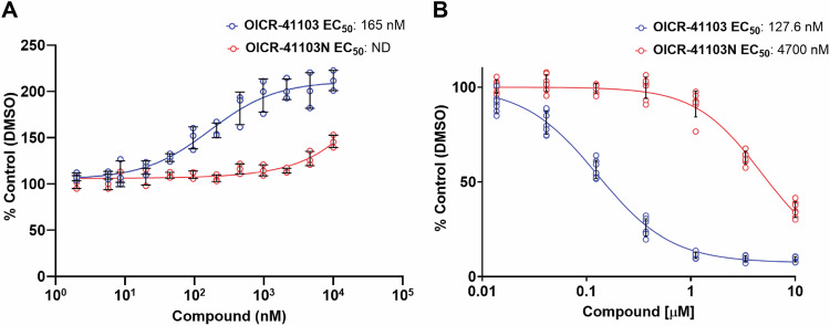
Human DCAF1 is a multidomain protein that plays a critical role in protein homeostasis. Its WDR domain functions as a substrate recruitment module for RING-type CRL4 and HECT family EDVP E3 ubiquitin ligases, enabling the ubiquitination and proteasomal degradation of specific substrates. DCAF1's activity has been implicated in cell proliferation and is documented to promote tumorigenesis. Additionally, the DCAF1 WDR domain is hijacked by lentiviral accessory proteins to induce the degradation of host antiviral factors, such as SAMHD1 and UNG2. These diverse roles make DCAF1 an attractive target for therapeutic development in oncology and antiviral strategies. It is also a promising candidate for use in targeted protein degradation. We previously reported a novel ligand, OICR-8268, that targets the DCAF1 WDR domain. In this study, we present the development of OICR-41103, a potent, selective, and cell-active small molecule chemical probe for DCAF1, derived from OICR-8268. The co-crystal structure of the DCAF1-OICR-41103 complex reveals the ligand's binding mode within the WDR central pocket, demonstrating its potential for PROTAC design and development. Notably, OICR-41103 effectively displaces the lentiviral Vpr protein from DCAF1 in both biochemical and cellular settings, highlighting its potential for the development of HIV therapeutics.


Conflict of interest statement

Competing interests: The authors declare no competing interests.

Figures

Fig. 1
Fig. 1. Development of pyrroles OICR-41103 and OICR-41103N from pyrazole OICR-8268.
OICR-8268 is a previously reported DCAF1 ligand. OICR-41103 (1) represents the active chemical probe, while OICR-41103N serves as the corresponding negative control.
Fig. 2
Fig. 2. Binding of OICR-41103 and OICR-41103N to the DCAF1 WDR domain.
Characterization of OICR-41103 (A) and OICR-41103N (B) by SPR. C Thermal stabilization of. DCAF1 WDR domain at 20 µM by OICR-41103 with a ΔTm of 23.0 ± 0.1 °C (blue dashed trace), and OICR-41103N with a ΔTm of 8.8 ± 0.3 °C (red trace) compared to the DCAF1 WDR domain with no compound (black trace), as measured by DSF. D The WDR domain stabilization was concentration dependent. E A close-up view of the OICR-41103 (deep teal sticks) binding site within the DCAF1 WDR central pocket. Coordinating water molecules are shown as red spheres. Residues interacting with the ligands are rendered as thin sticks, and hydrogen bonds are shown in black dashes. F A 2D diagram of the protein-compound interactions. The most important interaction classes, including water-mediated hydrogen bonds, are represented. All protein residues within 5.0 Å from ligand are displayed. The picture was generated using the LID tool from Maestro software (Schrodinger) from the prepared X-ray structure (PDB: 9D4E).
Fig. 3
Fig. 3. OICR-41103 engages and binds to the WDR domain of DCAF1 in cells.
A Cellular Thermal Shift Assay (CETSA) was used to assess OICR-41103 binding to the WDR domain of DCAF1 in NCI-H460 cells expressing HiBiT-tagged WDR domain of DCAF1. OICR-41103 thermally stabilized the WDR domain of DCAF1 in a dose dependent manner with an EC50 of 167 nM. Results are shown as an average +/−SD (n = 3 biologically independent samples). B NanoBRET assay with fluorescently labeled DCAF1 tracer was used to confirm cellular target engagement. OICR-41103 decreases the NanoBRET ratio between DCAF1 tracer and N-terminally NL-tagged DCAF1(WD40) in a dose-dependent manner. The negative control compound OICR-41103N was over 35-fold less potent. HEK 293T cells were transfected with N-terminally NL-tagged DCAF1(WD40) for 24 h and treated with tracer in presence or absence of compounds for 1 h. Results are shown as an average +/−SD (n = 7, 2 biologically independent samples).
Fig. 4
Fig. 4. Displacement of Vpr by OICR-41103.
A A transparent surface representation of the DCAF1 WDR domain with the bound OICR-41103 ligand (deep teal spheres), superimposed with the DCAF1-Vpr-SAMHD1 ternary structure (PDB: 5AJA). B A close-up view of the OICR-41103 (deep teal sticks) binding site, compared to the binding site of the parent compound, OICR-8268, (magenta sticks, PDB: 8F8E) and the Vpr binding site in the DCAF1-Vpr-SAMHD1 structure (PDB: 5AJA). OICR-41103 creates a steric clash with Vpr. C An HTRF assay demonstrating that OICR-41103 potently disrupts the interaction of the DCAF1 WDR domain and full-length Vpr protein. OICR-41103 displaces Vpr from DCAF1 with a Kdisp below 100 nM, demonstrating competition for the same binding site as Vpr. The opposite enantiomer, OICR-41103N, was used as a negative control and demonstrates substantially weaker potency (~180 × difference in Kdisp) in displacing the interaction, ±SD n = 3 incubations performed in separate wells at given compound concentrations. D The effect of OICR-41103 on disrupting the WDR-DCAF1/Vpr interaction in cells was assessed using NanoBit complementation assay. HEK293EMT cells were transfected with the WDR-DCAF1 tagged to smBiT and Vpr tagged to LgBiT. OICR-41103 disrupted WDR-DCAF1/Vpr interaction in a dose-dependent manner, with an EC50 of 1 μM, and had no effect on the negative control pair, p53/MDM2 interaction. Results are shown as an average +/−SD (n = 3 biologically independent samples).
Fig. 5
Fig. 5. DCAF1 knockdown results in growth suppression of NCIH-1703, NCIH-2170, and NCIH-1915 cell lines, with no toxicity observed upon OICR-41103 treatment.
A Western-blot analysis showing DCAF1 knockdown after 4 days of doxycycline induction (−/+DOX treatment) in NCI-H1703, NCI-H2170, and NCI-H1915 cells. B Trypan blue exclusion assay was used to count the number of cells in -DOX and +DOX treated cells. The percentage of remaining cells was calculated (+DOX/−DOX). shRNAs for Polo-like kinase (PLK1) and a non-targeting control (CTR2) were used as positive and negative growth suppression controls, respectively. C ATPlite assay was used to assess the effect of OICR-41103 in NCI-H1703, NCI-H2170, and NCI-H1915 cells after 7 days of compound treatment. Results are shown as an average +/−SD (n = 3 biologically independent samples). shDCAF1 cells are compared to shCTR2 cells using unpaired t-test, and *P-value < 0.05.

References

    1. Maddika, S. & Chen, J. Protein kinase DYRK2 is a scaffold that facilitates assembly of an E3 ligase. Nat. Cell Biol.11, 409–419 (2009). - PMC - PubMed
    1. Angers, S. et al. Molecular architecture and assembly of the DDB1-CUL4A ubiquitin ligase machinery. Nature443, 590–593 (2006). - PubMed
    1. He, Y. J., McCall, C. M., Hu, J., Zeng, Y. & Xiong, Y. DDB1 functions as a linker to recruit receptor WD40 proteins to CUL4-ROC1 ubiquitin ligases. Genes Dev.20, 2949–2954 (2006). - PMC - PubMed
    1. Kim, K. et al. VprBP has intrinsic kinase activity targeting histone H2A and represses gene transcription. Mol. Cell52, 459–467 (2013). - PMC - PubMed
    1. Mohamed, W. I. et al. The CRL4(DCAF1) cullin-RING ubiquitin ligase is activated following a switch in oligomerization state. EMBO J.40, e108008 (2021). - PMC - PubMed

LinkOut - more resources