. 2025 Jul 21;23(1):532.
doi: 10.1186/s12951-025-03567-9.

Prunus mume derived extracellular vesicle-like particles alleviate experimental colitis via disrupting NEK7-NLRP3 interaction and inflammasome activation

Affiliations

Prunus mume derived extracellular vesicle-like particles alleviate experimental colitis via disrupting NEK7-NLRP3 interaction and inflammasome activation

Qi Lv et al. J Nanobiotechnology. .

Abstract

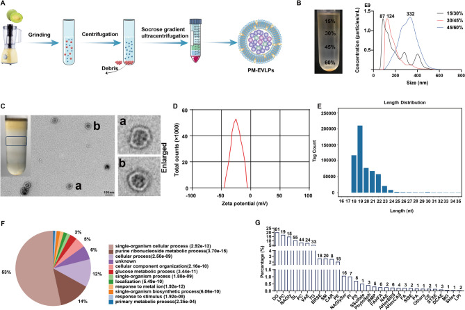
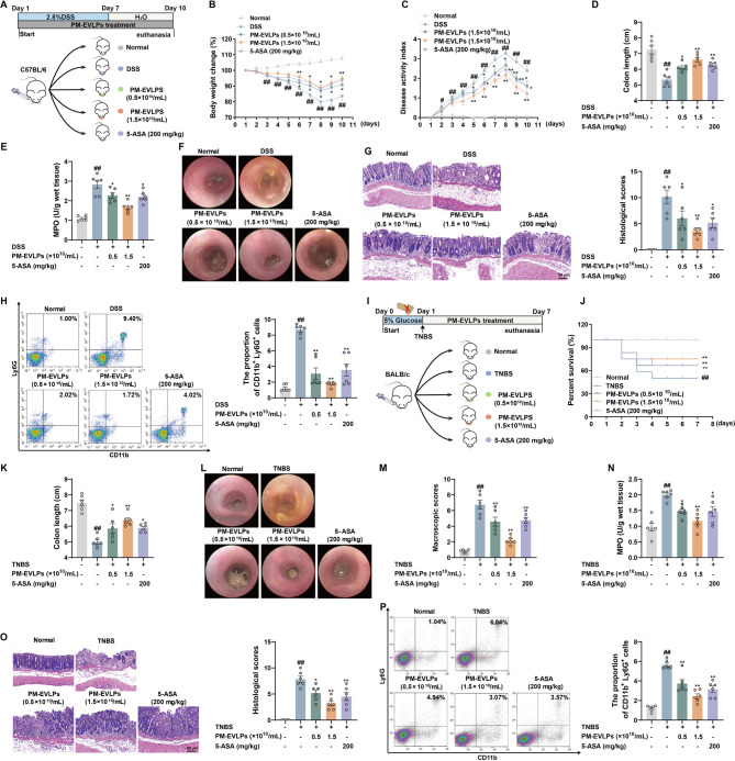
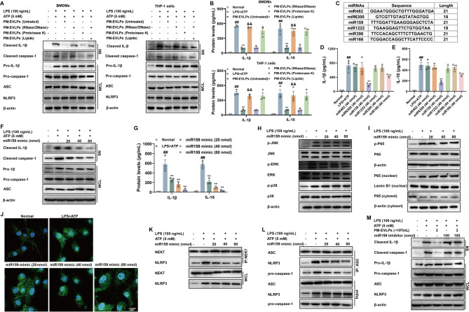
Edible plant derived extracellular vesicle-like particles (EVLPs) have garnered attention as potential therapeutic agents for chronic inflammatory diseases. Prunus mume (PM) is a functional fruit known for its gastrointestinal benefits, yet the detail material basis and potential mechanism remain unclear. Here, we reported that oral administration of prunus mume derived EVLPs (PM-EVLPs) substantially mitigated experimental colitis in mice. The in vivo bio-distribution analysis revealed that PM-EVLPs specifically targeted inflamed colon of colitis mice. Further in vitro studies demonstrated that PM-EVLPs were predominantly internalized by macrophages. The combined treatment with clodronate liposomes confirmed that macrophage was the target cell for PM-EVLPs-mediated anti-colitis activity. Mechanistically, PM-EVLPs selectively inhibited caspase-1 auto-cleavage and IL-1β secretion caused by NLRP3 inflammasome activation, while exerting minimal impact on AIM2, NLRP1 or NLRC4 inflammasome activation. Excluding the effects on mitochondrial ROS generation, K+ efflux or Ca2+ influx, PM-EVLPs disrupted the NEK7-NLRP3 interaction, thereby preventing NLRP3 inflammasome assembly. Notably, the inhibitory activity was attributed to RNAs rather than lipids or proteins within PM-EVLPs. Deep RNA sequencing, coupled with the application of miRNA mimics/inhibitors identified miR159 as the material basis for PM-EVLPs' inhibition of NLRP3 inflammasome activation and anti-colitis efficacy. Collectively, these findings suggest that PM-EVLPs represent a promising nanomedicine with potential as a novel therapeutic strategy for colitis and deserves further investigation and development.

Keywords: NEK7-NLRP3 interaction; NLRP3 inflammasome; PM-EVLPs; Ulcerative colitis; miR159.


Conflict of interest statement

Declarations. Ethical approval: The animal experimental protocols were approved by the Experimental Animal Ethics Center of Nanjing University of Chinese Medicine (Approved number: 202012A017; 202103A029; 202207A012; 202304A069). Consent for publication: All the authors agree on the final submission. Competing interests: The authors declare no competing interests.

Figures

Fig. 1
Fig. 1
The characterization of PM-EVLPs. (A) PM juice was purified through sucrose density gradient ultracentrifugation. (B) The particle size distribution of vesicles collected form the 15/30%, 30/45% and 45/60% sucrose gradient interfaces following ultracentrifugation. The size profiles were determined using the Nanoparticle Tracking Analysis. (C) The sucrose-gradient band, highlighted by the frame was collected for transmission electron microscopy examination. (D) The surface charge of the particles was assessed using a Zetasizer. (E-G) The miRNA, protein and lipid compositions of PM-EVLPs were detected by RNA deep sequencing, proteomic profiling and lipidomic assays, respectively. All analyses were derived from three biological replicates
Fig. 2
Fig. 2
Oral administration of PM-EVLPs significantly alleviates experimental colitis in mice. (A) The female C57BL/6 mice were subjected to DSS-induced colitis model, PM-EVLPs (0.5, 1.5 × 1010/mL) and 5-ASA (200 mg/kg) were orally administrated for 10 consecutive days. (B) Body weight loss. (C) DAI scores. (D) Colon length. (E) MPO activity. (F) High-resolution mini-endoscopy pictures of colons. (G) Histopathological damage in colons. (H) The infiltration of neutrophils in colonic lamina propria. (I) The male BALB/c mice were subjected to TNBS-induced colitis model, PM-EVLPs (0.5, 1.5 × 1010/mL) and the 5-ASA (200 mg/kg) were orally administrated for consecutive 7 days. (J) Survival rate. (K) Colon length. (L) High-resolution mini-endoscopy pictures of colons. (M) The macroscopic scores. (N) MPO activity in colons. (O) Histopathological damage in colons. (P) The infiltration of neutrophils in colonic lamina propria. The data was expressed as means ± S.E.M. of six mice in each group. ##P < 0.01 vs. normal group; *P < 0.05 and **P < 0.01 vs. DSS or TNBS group
Fig. 3
Fig. 3
Oral administration of PM-EVLPs targets the colonic macrophage. (A) The healthy and DSS-induced colitis mice was starved for 12 h, followed by orally administration of DiR-labeled PM-EVLPs at various time points. The heart, liver, spleen, lung, kidney, stomach, small intestine and colon were collected, and the distribution of PM-EVLPs was analyzed using the IVIS Lumina III imaging system. (B) The healthy and DSS-induced colitis mice were orally administered DiR-labeled PM-EVLPs. The localization of PM-EVLPs in the mucosa, submucosa, muscularis propria and serosa layer was examined using immunofluorescence assay stained with β-actin for cytoskeleton visualization. (C, D) T cells, BMDMs and PMA-differentiated THP-1 cells were incubated with DiO-labeled PM-EVLPs at 37 ℃ for 0.5, 1, 2 and 4 h, the cellular uptake was detected using immunofluorescence and flow cytometry assays. (E) The DSS-induced colitis mice were orally administered DiR-labeled PM-EVLPs, and the co-localization between DiR-labeled PM-EVLPs and F4/80 macrophages in colonic lamina propria was detected. The data was expressed as means ± S.E.M. of six mice in each group or three independent in vitro experiments
Fig. 4
Fig. 4
The macrophage was responsible for PM-EVLPs-mediated alleviation of colitis. (A) The female C57BL/6 mice were subjected to DSS-induced colitis model, PM-EVLPs (1.5 × 1010/mL) and 5-ASA (200 mg/kg) were orally administrated for 10 consecutive days, the clodronate and control liposomes were intraperitoneally injection 12 h prior to the onset of DSS treatment and followed by injection every other day. (B) DAI scores. (C) Colon length. (D) High-resolution mini-endoscopy pictures of colons. (E) Histopathological damage in colons. (F) MPO activity. (G) Infiltration of neutrophils in colonic lamina propria. (H-N) The male BALB/c mice were subjected to TNBS-induced acute colitis model, PM-EVLPs (0.5, 1.5 × 1010/mL) and the 5-ASA (200 mg/kg) were orally administrated for consecutive 7 days, clodronate and control liposomes were intraperitoneally injection 12 h prior to the onset of TNBS treatment and followed by injection every other day. Survival rate (H); colon length (I); high-resolution mini-endoscopy pictures of colons (J); histopathological damage in colons (K); infiltration of neutrophils in colonic lamina propria (L); macroscopic scores of colons (M); quantification of MPO activity in colons (N). Data was expressed as means ± S.E.M. of six mice in each group. ##P < 0.01 vs. normal group; *P < 0.05 and **P < 0.01 vs. DSS or TNBS group; &&P < 0.01 vs. DSS or TNBS + control liposomes + PM-EVLPs group
Fig. 5
Fig. 5
PM-EVLPs specifically inhibits NLRP3 inflammasome activation in colonic macrophages. (A-D) The mice were subjected to DSS-induced colitis, and protein expression of cleaved caspase-1, IL-1β and IL-18 in colonic macrophages and epithelial cells was examined by western blotting and ELISA, respectively. (E-J) BMDMs and PMA-stimulated THP-1 cells were cultured with LPS (100 ng/mL) for 3 h, and then treated with PM-EVLPs for 1 h, followed by addition of ATP (5 mM) for 45 min, nigericin (4 µM) for 3 h or monosodium urate crystal (150 µg/mL) for 6 h. The protein expressions of cleaved caspase-1 and cleaved IL-1β in supernatants were examined by western blotting assay (E, G, I); secretion of IL-1β and IL-18 in supernatants were quantified by ELISA (F, H, J). Data was expressed as means ± S.E.M. of six mice in each group or three independent in vitro experiments. ##P < 0.01 vs. Normal group; *P < 0.05 and **P < 0.01 vs. DSS or LPS + ATP/MSU/Nigericin group
Fig. 6
Fig. 6
PM-EVLPs blocks the NEK7-NLRP3 interaction to disrupted NLRP3 inflammasome assembly. (A, B) BMDMs and PMA-stimulated THP-1 cells were cultured with LPS (100 ng/mL) for 3 h, and then treated with PM-EVLPs for 1 h, followed by addition of ATP (5 mM) for 45 min. The ASC oligomerization was determined by western blotting assay (A); formation of NLRP3/ASC/pro-caspase-1 complex was detected by Co-IP assay (B). (C, D) BMDMs and PMA-stimulated THP-1 cells were incubated with PBS or PM-EVLPs for 1 h, and the whole cell lysis was suffered from CETSA and SIP assays. (E, F) Immunoprecipitation analysis of interaction between NLRP3 and NEK7 in HEK-293T cells transfected with indicated plasmid and treated with PM-EVLPs for 24 h. (G) The BMDMs and PMA-stimulated THP-1 cells were subjected to NLRP3 inflammasome activation model, and the formation of NEK7-NLRP3 complex was detected by Co-IP assay. (H) The schematic diagram of in situ proximity ligation assay (NEK7-NLRP3 PLA). (I) The interaction between NEK7 and NLRP3 (red color) was analyzed by PLA in BMDMs. (J) The schematic diagram of NEK7 and NLRP3 fusion proteins used for bimolecular fluorescence complementation (BiFC) analysis. (K) BiFC signals were detected in BMDMs transfected with NEK7-VC155 and NLRP3-VN173 plasmids. (L) The BMDMs were subjected to NLRP3 inflammasome activation model, and the interaction of NEK7-NLRP3 was detected using laser scanning confocal microscope. Data was expressed as means ± S.E.M. of three independent in vitro experiments
Fig. 7
Fig. 7
The miR159 in PM-EVLPs was identified in inhibiting NLRP3 inflammasome activity. (A, B) PM-EVLPs were either untreated or treated with proteinase to digest protein, DNase I/RNase A to remove the majority of RNAs, or chloroform/methanol to extract total lipids. BMDMs and PMA-stimulated THP-1 cells were subjected to NLRP3 inflammasome activation model in the presence of treated PM-EVLPs, protein expressions of cleaved caspase-1, cleaved IL-1β and generation of IL-1β, IL-18 were examined by western blotting assay and ELISA, respectively. (C) The six most abundant miRNAs in PM-EVLPs were listed. (D, E) BMDMs were cultured with LPS for 3 h, and then treated with synthetic miR159, miR482, miR6300, miR1222, miR396 and miR166 mimics for 1 h, followed by addition of ATP for 45 min. Secretion of IL-1β and IL-18 were quantified by ELISA. (F, G) BMDMs were subjected to NLRP3 inflammasome activation model in the presence of miR159 mimic, protein expressions of cleaved caspase-1, cleaved IL-1β and secretion of IL-1β, IL-18 in supernatants were examined by western blotting and ELISA, respectively. (H-J) BMDMs were treated with miR159 mimic and LPS (100 ng/mL) for 3 h, the phosphorylation modification of p38, ERK, JNK, p65 and nuclear translocation of p65 were detected by western blotting and immunofluorescence assays. (K, L) BMDMs were subjected to NLRP3 inflammation activation model in the presence of miR159 mimic, formation of NEK7-NLRP3 and NLRP3/ASC/pro-caspase-1 complex was detected by Co-IP assay. (M) BMDMs were subjected to NLRP3 inflammation activation model in the presence of miR159 inhibitor and PM-EVLPs, protein expressions of cleaved caspase-1, cleaved IL-1β were examined by western blotting assay. The data was expressed as means ± S.E.M. of three independent in vitro experiments. ##P < 0.01 vs. Normal group; **P < 0.01 vs. LPS + ATP group; &&P < 0.01 vs. PM-EVLPs (untreated) group
Fig. 8
Fig. 8
The miR159 in PM-EVLPs alleviates experimental colitis. (A) Female C57BL/6 mice were subjected to the DSS-induced colitis model, miR159 agomir (400, 800 pmol) and 5-ASA (200 mg/kg) were orally administrated for 10 consecutive days. (B) Body weight change. (C) DAI scores. (D) Colon length. (E) High-resolution mini-endoscopy pictures indicating degree of intestinal inflammation. (F) Histopathological damage in colons. (G) Quantification of MPO activity in colons. (H) Proportion of neutrophils in colonic lamina propria. (I-O) The male BALB/c mice were subjected to TNBS-induced acute colitis model, miR159 agomir (400, 800 pmol) and 5-ASA (200 mg/kg) were orally administrated for 7 consecutive days. The survival rate (I); colon length (J); high-resolution mini-endoscopy pictures of colonic tissues (K); histopathological damage in colons (L); infiltration of neutrophils in colonic lamina propria (M); macroscopic scores of colons (N); MPO activity in colons (O). The data was expressed as means ± S.E.M. of six mice in each group. ##P < 0.01 vs. normal group; *P < 0.05 and **P < 0.01 vs. DSS or TNBS group
None

References

    1. Zeeshan M, Ain QU, Weigmann B, Story D, Smith BR, Ali H. Dual pH and microbial-sensitive galactosylated polymeric nanocargoes for multi-level targeting to combat ulcerative colitis. Asian J Pharm Sci. 2023;18:100831. - PMC - PubMed
    1. Le Berre C, Honap S, Peyrin-Biroulet L. Ulcerative colitis. Lancet. 2023;402:571–84. - PubMed
    1. Rogler G, Singh A, Kavanaugh A, Rubin DT. Extraintestinal manifestations of inflammatory bowel disease: current concepts, treatment, and implications for disease management. Gastroenterology. 2021;161:1118–32. - PMC - PubMed
    1. Shin AE, Tesfagiorgis Y, Larsen F, Derouet M, Zeng PYF, Good HJ, et al. F4/80 + Ly6Chigh macrophages lead to cell plasticity and cancer initiation in colitis. Gastroenterology. 2023;164:593–609. - PMC - PubMed
    1. Neurath MF. Strategies for targeting cytokines in inflammatory bowel disease. Nat Rev Immunol. 2024;24:559–76. - PubMed

LinkOut - more resources