. 2025 Jul 4:19:1538956.
doi: 10.3389/fnins.2025.1538956. eCollection 2025.

Phase contrast-derived cerebral blood flow is associated with neurodegeneration and cerebrovascular injury in older adults

Affiliations

Phase contrast-derived cerebral blood flow is associated with neurodegeneration and cerebrovascular injury in older adults

Jeffrey D Pyne et al. Front Neurosci. .

Abstract

Global cerebral blood flow and the local delivery of blood through the vascular network are essential to maintain brain and cognitive health throughout the lifespan. In this cross-sectional study, we examined the association of extracranial blood flow into the brain, measured with phase contrast magnetic resonance imaging, with regional brain volumes, cortical thickness, white matter tract integrity, white matter hyperintensity volume, and cerebral microbleeds. Our study included 311 older adults (mean age: 77 years, standard deviation: 5.6) from the Washington Heights Inwood Columbia Aging Project (WHICAP), a community-based study in northern Manhattan. We found that lower extracranial cerebral blood flow is associated with lower cortical regional volumes, lower white matter tract integrity, and higher white matter hyperintensity volume. We observed that lower extracranial cerebral blood flow, quantified by total, anterior, and posterior circulations, is associated with lower white matter tract integrity in the forceps minor, cingulum cingulate gyrus, and inferior fronto-occipital fasciculus. Additionally, lower total extracranial cerebral blood flow is associated with higher white matter hyperintensity volume, a marker of small vessel cerebrovascular disease. These findings support our hypothesis that lower extracranial cerebral blood flow is associated with a greater degree of vascular brain injury and indicators of neurodegeneration and are consistent with the guiding conceptual framework that diminished extracranial blood flow could be a factor that promotes or exacerbates neurodegeneration and cerebrovascular injury in older adults. Future longitudinal studies are needed to establish causality and temporality.

Keywords: aging; cerebral blood flow; cerebrovascular; hypoperfusion; phase contrast MRI; white matter.


Conflict of interest statement

AMB is an inventor of a patent for white matter hyperintensity quantification (US Patent US9867566B2). The remaining authors declare that the research was conducted in the absence of any commercial or financial relationships that could be construed as a potential conflict of interest.

Figures

Figure 1
Figure 1
Visualization of internal carotid and vertebral artery flow measurements, representing anterior (green) and posterior (orange) cerebral blood flow, by phase contrast MRI. (A) The internal carotid arteries (green), representing the anterior circulation, and the vertebral arteries (orange), representing the posterior circulation are measured using phase contrast. Example 2D phase contrast plane perpendicular alignments are shown for the internal carotid arteries (green plane) and vertebral arteries (orange plane). The blue lines indicate the approximate target location for blood vessel measurement. (B) 2D phase contrast MRI produces cardiac-gated magnitude (right) and phase (left) cross-sectional slices. (C) Representation of typical internal carotid and vertebral blood flow profiles. (D) The sum of internal carotid and vertebral blood flow profiles characterizes extracranial total cerebral blood flow.
Figure 2
Figure 2
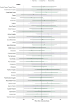
Summary forest plot showing the effect sizes of the relationship of extracranial total, anterior, and posterior cerebral blood flow with cortical thickness. Analyses are adjusted for age, vascular risk factors, intracranial volume, and sex, with corrections applied for multiple comparisons. Each bar represents the results of a separate linear model. Only regions that are perfused by the supplying artery are tested for associations (Liu et al., 2023). Filled and open circles represent significant and non-significant associations, respectively.
Figure 3
Figure 3
Illustration showing the significant associations of extracranial total (yellow), anterior (green), and posterior (purple) cerebral blood flow with cortical, subcortical, and ventricular system regional volume ROIs. Regional volume ROIs with nonsignificant associations with extracranial blood flow are shown in gray. Analyses are adjusted for age, vascular risk factors, intracranial volume, and sex, with corrections applied for multiple comparisons.
Figure 4
Figure 4
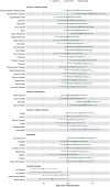
Summary forest plot showing the effect sizes of the relationship of extracranial total, anterior, and posterior cerebral blood flow with regional volumes. Analyses are adjusted for age, vascular risk factors, intracranial volume, and sex, with corrections applied for multiple comparisons. Each bar represents the results of a separate linear model. Organized by cortical, subcortical, and ventricular system regions. Only regions that are perfused by the supplying artery are tested for associations (Liu et al., 2023). Filled and open circles represent significant and non-significant associations, respectively.
Figure 5
Figure 5
Summary forest plot showing the effect sizes of the relationship of extracranial total, anterior, and posterior cerebral blood flow with white matter tract integrity. Analyses are adjusted for age, vascular risk factors, intracranial volume, and sex, with corrections applied for multiple comparisons. Each bar represents the results of a separate linear model. Organized by commissural, projection, limbic, and association tracts. Filled and open circles represent significant and non-significant associations, respectively.
Figure 6
Figure 6
Scatter plots displaying the association between extracranial total, anterior, and posterior cerebral blood flow and total WMH volume. Linear regression curves, with 95% confidence intervals, are adjusted for age, vascular risk factors, intracranial volume, and sex. Scatter plot datapoints are visualized on unadjusted values.

References

    1. Alsop D. C., Detre J. A., Golay X., Günther M., Hendrikse J., Hernandez-Garcia L., et al. (2015). Recommended implementation of arterial spin-labeled perfusion MRI for clinical applications: a consensus of the ISMRM perfusion study group and the European consortium for ASL in dementia. Magn. Reson. Med. 73, 102–116. doi: 10.1002/mrm.25197, PMID: - DOI - PMC - PubMed
    1. Amin-Hanjani S., Du X. J., Pandey D. K., Thulborn K. R., Charbel F. T. (2015). Effect of age and vascular anatomy on blood flow in major cerebral vessels. J. Cereb. Blood Flow Metab. 35, 312–318. doi: 10.1038/jcbfm.2014.203, PMID: - DOI - PMC - PubMed
    1. Appelman A. P. A., Van der Graaf Y., Vincken K. L., Tiehuis A. M., Witkamp T. D., Mali W. P. T. M., et al. (2008). Total cerebral blood flow, white matter lesions and brain atrophy: the SMART-MR study. J. Cerebr. Blood Flow. Metab. 28, 633–639. doi: 10.1038/sj.jcbfm.9600563, PMID: - DOI - PubMed
    1. Aslan S., Xu F., Wang P. L., Uh J., Yezhuvath U. S., van Osch M., et al. (2010). Estimation of labeling efficiency in pseudocontinuous arterial spin labeling. Magn. Reson. Med. 63, 765–771. doi: 10.1002/mrm.22245, PMID: - DOI - PMC - PubMed
    1. Avila J. F., Renteria M. A., Jones R. N., Vonk J. M. J., Turney I., Sol K., et al. (2021). Education differentially contributes to cognitive reserve across racial/ethnic groups. Alzheimers Dement. 17, 70–80. doi: 10.1002/alz.12176, PMID: - DOI - PMC - PubMed

LinkOut - more resources