. 2025 Jul 4:23:e00673.
doi: 10.1016/j.ohx.2025.e00673. eCollection 2025 Sep.

Open-source tubing-free impeller pump platform for controlled recirculating fluid flow for microfluidics and organs-on-chip

Affiliations

Open-source tubing-free impeller pump platform for controlled recirculating fluid flow for microfluidics and organs-on-chip

Sophie R Cook et al. HardwareX. .

Abstract

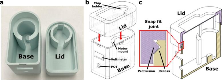
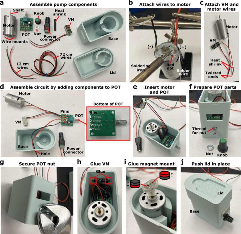
Fluid flow is utilized in many microscale technologies, including microfluidic chemical reactors, diagnostics, and organs-on-chip (OOCs). In particular, OOCs may rely on fluid flow for nutrient delivery, cellular communication, and application of shear stress. In order for microscale flow systems to be readily adopted by non-experts, a tubing-free, user-friendly pump would be useful, particularly one that is simple to use, affordable, and compatible with cell culture incubators. To address these needs, here we share the design and fabrication of an impeller pump platform that provides recirculating fluid flow through a microfluidic loop without the need for tubing connections. Flow is driven by rotating a magnetic stir bar or 3D-printed impeller in a pump well, using magnets mounted on a DC motor. The DC motors used produce negligible heat output in a compact system, making it compatible with cell culture incubators. The pump platform accommodates user-defined microfluidic or OOC device geometries, which may be easily customized by 3D printing. Furthermore, the system is easily assembled from low-cost materials and simple circuitry by someone with no prior training. We demonstrate the ability of the platform to drive recirculating fluid flow in a microfluidic device at well-characterized flow velocities ranging from µm/s to mm/s for use with microfluidic technologies. Though designed with OOCs in mind, we envision that this platform will enable users from ranging disciplines to incorporate fluid flow in customized microscale technologies.

Keywords: 3D printing; Bioreactor; Micropump; Multi-organ-on-chip; Stir bar.


Conflict of interest statement

The authors declare that they have no known competing financial interests or personal relationships that could have appeared to influence the work reported in this paper.

Figures

None
Graphical abstract
Fig. 1
Fig. 1
Overview of the tubing-free impeller pump platform. (a) The impeller pump drives recirculating fluid flow on-chip by rotating a magnetic stir bar within a cylindrical pump well within the device. (b) An image of a device loaded on to the impeller pump. The chip was filled with food dye and a 10 mm stir bar was added to the pump well. (c) An image of the interior of the pump platform, showing the motor, mounted magnets, voltmeter, and POT within the 3D-printed base. (d,e) An image of six impeller pumps resting on a laser-cut acrylic pump holder on the bench (d) and inside a standard cell culture incubator (e). (f) An image of the chip cover being added to an impeller pump to limit evaporation.
Fig. 2
Fig. 2
Features of the Impeller Pump Housing. (a) An image of the FDM 3D-printed Impeller Pump Base (left) and Impeller Pump Lid (right). (b) A schematic from Fusion 360 showing the assembly of the pump housing. The red arrows mark the snap fit joints. (c) A schematic from Fusion 360 of a central cut plane of the assembled pump housing. The snap fit joint consisted of a protruding feature on the lid (purple) and a corresponding recess on the base (yellow).
Fig. 3
Fig. 3
Adjustable features of the 3D-printed device. These features include (i) pump well dimensions, (ii) channel dimensions and corners, (iii) channel/well intersection, and (iv) addition of functional features (e.g. well for tissue slice culture).
Fig. 4
Fig. 4
Motor circuit diagram. Schematic showing how wires from the (i) DC motor, (ii) voltmeter, and (iii) power connector are attached to the positive and negative terminals of the (iv) POT. A (v) power adapter is used to connect the (iii) power connector to an outlet.
Fig. 5
Fig. 5
Assembly of impeller pump external housing. (a) Components required for pump assembly, (b) wires attached to the DC motor, (c) heat shrink used to hold twisted voltmeter and motor wires together, (d) wires inserted into the POT and power connector, (e) the motor and POT secured in the Impeller Pump Base, (f) components to secure the POT in place, (g) POT nut tightened with a wrench, (h) voltmeter glued into place, (i) disk magnets glued to the Magnet on the motor shaft, and (j) Impeller Pump Lid snapped into place to finish pump assembly.
Fig. 6
Fig. 6
Characterization and stability of impeller. (a) As the voltage increased, the impeller RPMs increased for pumps 1–6. Note pump #1 was slower than the others and failed the quality control test. (b) RPM of the impeller remained stable across 3 pumps for 5 days at 1.7 V.
Fig. 7
Fig. 7
Experimental maximum channel velocity quantification using the microscale impeller pump. (a) Time-lapse images of dye movement in the channel of the demo chip (1270 RPM, 1.8 V) at different time points (0.000 s, 3.027 s, 8.998 s). The blue arrow notes the flow direction. The solid red line (arrow) indicates the distance from the tip of the parabola to the reference line, used to quantify the dye movement. The red dashed line marks the reference line used to measure dye movement over time, demonstrating the velocity measurements. (b) Experimentally measured maximum channel velocity within the demo chip as a function of RPM, showing an increase with higher RPM values. Dots and error bars represent mean and standard deviation (n = 4); some error bars are too small to see.
Fig. 8
Fig. 8
Fluid recirculation using the impeller pump. Schematic depicting flow direction and stir bar size alongside still images from video s of recirculating fluid flow using in the demo chip. Using a (a) 5 mm and (b) 10 mm stir bar at 1.7 V (1255 RPM), purple dye was added to the pump well and moved through the channel over time. The dye front is marked with a red arrow, and the stir bar rotation direction is noted with a white arrow. Scale bar = 5 mm. Stills are taken from Movie 4 and Movie 5 included in supplementary files.

References

    1. Leung C.M., de Haan P., Ronaldson-Bouchard K., Kim G.-A., Ko J., Rho H.S., Chen Z., Habibovic P., Jeon N.L., Takayama S., Shuler M.L., Vunjak-Novakovic G., Frey O., Verpoorte E., Toh Y.-C. Nat. Rev; Methods Primer: 2022. A guide to the organ-on-a-chip.
    1. Ronaldson-Bouchard K., Vunjak-Novakovic G. Organs-on-a-Chip: A fast Track for engineered human tissues in drug development. Cell Stem Cell. 2018;22:310–324. doi: 10.1016/j.stem.2018.02.011. - DOI - PMC - PubMed
    1. Griffith L.G., Swartz M.A. Capturing complex 3D tissue physiology in vitro. Nat. Rev. Mol. Cell Biol. 2006;7:211–224. doi: 10.1038/nrm1858. - DOI - PubMed
    1. Shim S., Belanger M.C., Harris A.R., Munson J.M., Pompano R.R. Two-way communication between ex vivo tissues on a microfluidic chip: application to tumor–lymph node interaction. Lab. Chip. 2019;19:1013–1026. doi: 10.1039/C8LC00957K. - DOI - PMC - PubMed
    1. Ortiz-Cárdenas J.E., Zatorski J.M., Arneja A., Montalbine A.N., Munson J.M., Luckey C.J., Pompano R.R. Towards spatially-organized organs-on-chip: photopatterning cell-laden thiol-ene and methacryloyl hydrogels in a microfluidic device. Organs–-Chip 4. 2022 doi: 10.1016/j.ooc.2022.100018. - DOI - PMC - PubMed

LinkOut - more resources