. 2025 Jul 11:30:371-383.
doi: 10.1016/j.reth.2025.06.014. eCollection 2025 Dec.

Fabrication of Piezo1 protein encapsulated pressure-sensitive multifunctional hydrogel in modulating cellular response and wound healing in pressure ulcer conditions

Affiliations

Fabrication of Piezo1 protein encapsulated pressure-sensitive multifunctional hydrogel in modulating cellular response and wound healing in pressure ulcer conditions

Jing Ning et al. Regen Ther. .

Abstract

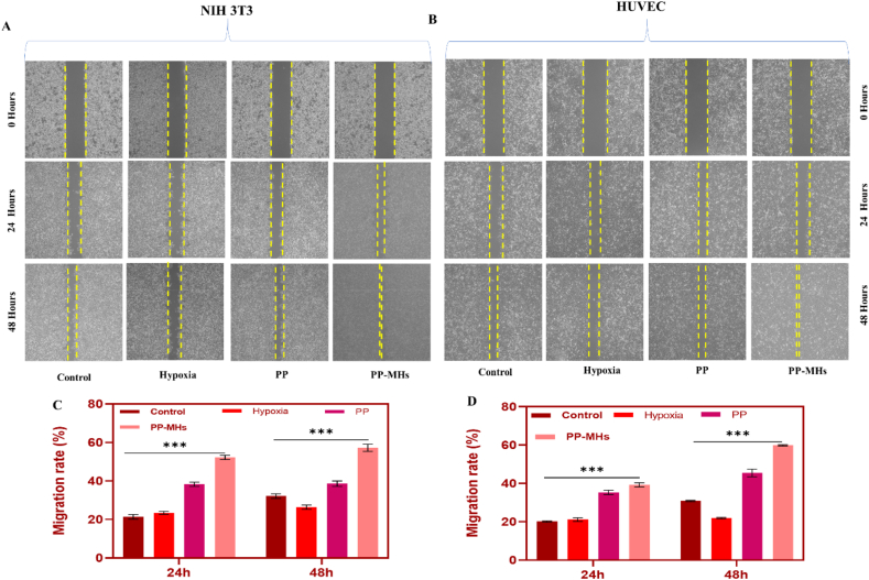
Pressure ulcers (PUs) are prevalent skin lesions characterized by significant morbidity, susceptibility to infection, and a complex healing process. This study aims at the synthesis of piezo1 protein-encapsulated, pressure-sensitive multifunctional hydrogel to modulate cellular response and promote wound healing in PUW conditions. The hydrogel synthesized from carboxyl methyl cellulose hydrogel exhibits the optimal swelling ratio and is found to have a high storage modulus (G'). This shows the mechanical strength and viscoelastic nature of the synthesized hydrogel. The PP encapsulation and releasing efficiency has been analyzed, and this proves the prolonged activation of mechanotransduction properties. In vitro analysis on 3T3 and HUVEC proves a high proliferation rate and proves to have an enhanced cell migration rate in hypoxia-induced cell lines. The angiogenesis was also found to be increased, which is indicated by tube formation that enhances the wound healing rate. The pressure ulcer animal model was analyzed for 3, 7, 10, and 14 days, and the wound healing rate. The reduction in inflammatory cytokine expression and the collagen deposition rate has been analyzed. By day 14, the wound closure reached above 91%, significantly higher than the untreated group. These findings demonstrate that PP-MH enhances cell proliferation and angiogenesis, thereby acts as a promising strategy for advanced pressure ulcer management.

Keywords: Multifunctional hydrogel; Piezo1 protein; Pressure ulcer; Wound healing.


Conflict of interest statement

The authors declare that they have no known competing financial interests or personal relationships that could have appeared to influence the work reported in this paper.

Figures

Fig. 1
Fig. 1
Characterization analysis: (A) FT-IR Analysis, (B) 13C NMR spectrum of PP-MHs, (C) SEM analysis of the hydrogel.
Fig. 2
Fig. 2
Characterization properties of the PP-MHs: (A&B) Indicate the water absorbance rate and selling time that denote the hydration capacity of the hydrogel; (C) Degradation rate analysis; (D) Protein absorption analysis; and (E) Indicate the protein release from the hydrogel and show that the composition 1 was effective for absorption and releasing analysis; (F) swelling ratio analysis over various time intervals for MHs and PP-MHs that indicates the hydrogel expansion and retention properties. The data in each bar represents the mean ± SD of three independent samples. They were statistically significant, as indicated by the symbols ∗ for (p < 0.05), ∗∗ for (p < 0.01), and ∗∗∗ for (p < 0.001). Comparative analysis of hydrogel formulations prepared with a constant amount of CMC (5 g) and varying concentrations of PEGDE: Entry 1 (0.5 g), Entry 2 (1.0 g), Entry 3 (1.5 g), Entry 4 (2.0 g), and Entry 5 (2.5 g).
Fig. 3
Fig. 3
The effect of PP-MHs on NHI 3T3 and HUVEC was analyzed: (A&B) Cell viability analysis, (C–E) Cell proliferation analysis changes were observed in hypoxic conditions and when treated with PP and PP-MHs. The data in each bar represents the mean ± SD of three independent samples. They were statistically significant, as indicated by the symbols ∗ for (p < 0.05), ∗∗ for (p < 0.01), and ∗∗∗ for (p < 0.001).
Fig. 4
Fig. 4
(A–D) Wound healing for assessing cell migration in NIH 3T3 and HUVEC cell under different treatment condition. The changes were observed at 0, 24, 48 h show the migration of cells in control, Hypoxia, PP, and PP-MHs where the yellow lines indicate the migration rate. The quantification of migration rate of 3T3 and HUVEC cell. The data in each bar represents the mean ± SD of three independent samples. They were statistically significant, as indicated by the symbols ∗∗ for (p < 0.01), and ∗∗∗ for (p < 0.001).
Fig. 5
Fig. 5
Determination of the angiogenic potential and gene expression of NHI 3T3 and HUVEC cell lines. (A–C) Denotes the number of nodes and segments of 3T3 cell lines and the representative images for tube formation assay. (D–F) Indicates the tube formation changes of HUVEC when treated with PP-MHs. (G & H) Represents the relative mRNA expressions of angiogenic genes (VEGF, FGE2, ADAM19, MM9) in NHI 3T3 and HUVEC cell lines. The data in each bar represents the mean ± SD of three independent samples. They were statistically significant, as indicated by the symbols ∗∗ for (p < 0.01), and ∗∗∗ for (p < 0.001).
Fig. 6
Fig. 6
The in vivo analysis: (A) Wound healing rate of different treatment group under various time intervals, (B) Denotes the wound healing rate at day 3, 7, 10 and 14 and found that PP-MHs were effective in treating PUW when compared to the control group, (C) Indicates the changes observation in PUW animal model at various days (0,3,7,10,14) and the found PP-MHs was effective in treating PUW. The data in each bar represents the mean ± SD of three independent samples. They were statistically significant, as indicated by the symbols ∗∗∗ for (p < 0.001).
Fig. 7
Fig. 7
(A) Histological examination of wound healing was observed in PP-MHs treated group over various time intervals, (B–E) Denotes the scar length, Epidermis thickness was found to increase when treated with PP-MHs, collagen content and no of appendix organs in tissue sections suggesting enhanced tissue remodelling. The data in each bar represents the mean ± SD of three independent samples. They were statistically significant, as indicated by the symbols ∗∗ for (p < 0.01), and ∗∗∗ for (p < 0.001).
Fig. 8
Fig. 8
Analysis of inflammatory and remodelling markers in wound healing under PP-MHs treatment conditions: (A–C) The mRNA expression of inflammatory genes (IL-1β, IL-6 and IL-10), (D–I) Quantification of MPO positive neutrophils at day 7&14, The relative expression of elastase/β-actin as control denotes the changes in protease activity, The analysis of F4/80-Positive macrophage at various days 7&14, The expression of MMP-12/β-actin that denotes the extracellular matrix modelling, The expression of TGF-β1/TβR1 level, MCP-1 expression analysis, (J) Denotes the western blotting analysis of elastase, MCP-1, MMP-12, TGF- β with β-actin as a control. The data in each bar represents the mean ± SD of three independent samples. They were statistically significant, as indicated by the symbols ∗ for (p < 0.05), ∗∗ for (p < 0.01), and ∗∗∗ for (p < 0.001).

Similar articles

  • Dressings and topical agents for treating pressure ulcers.
    Westby MJ, Dumville JC, Soares MO, Stubbs N, Norman G. Westby MJ, et al. Cochrane Database Syst Rev. 2017 Jun 22;6(6):CD011947. doi: 10.1002/14651858.CD011947.pub2. Cochrane Database Syst Rev. 2017. PMID: 28639707 Free PMC article.
  • Hydrogel dressings for venous leg ulcers.
    Ribeiro CT, Dias FA, Fregonezi GA. Ribeiro CT, et al. Cochrane Database Syst Rev. 2022 Aug 5;8(8):CD010738. doi: 10.1002/14651858.CD010738.pub2. Cochrane Database Syst Rev. 2022. PMID: 35930364 Free PMC article.
  • Foam dressings for treating pressure ulcers.
    Walker RM, Gillespie BM, Thalib L, Higgins NS, Whitty JA. Walker RM, et al. Cochrane Database Syst Rev. 2017 Oct 12;10(10):CD011332. doi: 10.1002/14651858.CD011332.pub2. Cochrane Database Syst Rev. 2017. PMID: 29025198 Free PMC article.
  • Endovenous ablation for venous leg ulcers.
    Cai PL, Hitchman LH, Mohamed AH, Smith GE, Chetter I, Carradice D. Cai PL, et al. Cochrane Database Syst Rev. 2023 Jul 27;7(7):CD009494. doi: 10.1002/14651858.CD009494.pub3. Cochrane Database Syst Rev. 2023. PMID: 37497816 Free PMC article.
  • Debridement for venous leg ulcers.
    Gethin G, Cowman S, Kolbach DN. Gethin G, et al. Cochrane Database Syst Rev. 2015 Sep 14;2015(9):CD008599. doi: 10.1002/14651858.CD008599.pub2. Cochrane Database Syst Rev. 2015. PMID: 26368002 Free PMC article.

References

    1. Lupiáñez-Pérez I., Morilla-Herrera J.C., Ginel-Mendoza L., Martín-Santos F.J., Navarro-Moya F.J., Sepúlveda-Guerra R.P., et al. Effectiveness of olive oil for the prevention of pressure ulcers caused in immobilized patients within the scope of primary health care: study protocol for a randomized controlled trial. Trials. 2013;14:1–7. http://www.trialsjournal.com/content/14/1/348 - PMC - PubMed
    1. Hajhosseini B., Longaker M.T., Gurtner G.C. Lippincott Williams and Wilkins; Apr. 01, 2020. Pressure injury. - DOI - PubMed
    1. Assis De Brito T.L., Monte-Alto-Costa A., Romana-Souza B. Propranolol impairs the closure of pressure ulcers in mice. Life Sci. Apr. 2014;100(2):138–146. doi: 10.1016/j.lfs.2014.02.007. - DOI - PubMed
    1. Kottner Jan, Cuddigan Janet, Carville Keryln, Balzer Katrin, Berlowitz Dan, Law Susan, et al. Pressure ulcer/injury classification today: An international perspective. J Tissue Viability. 2020;29(3):197–203. doi: 10.1016/j.jtv.2020.04.003. - DOI - PubMed
    1. Gray R.J., Voegeli D., Bader D.L. Features of lymphatic dysfunction in compressed skin tissues - implications in pressure ulcer aetiology. J Tissue Viability. Feb. 2016;25(1):26–31. doi: 10.1016/j.jtv.2015.12.005. - DOI - PubMed

LinkOut - more resources