. 2025 Aug 1;24(8):4139-4153.
doi: 10.1021/acs.jproteome.5c00273. Epub 2025 Jul 21.

OCIAD2 Promotes Cancer Progression via Metabolic Reprogramming in Lung Adenocarcinoma

Affiliations

OCIAD2 Promotes Cancer Progression via Metabolic Reprogramming in Lung Adenocarcinoma

Yi-Hui Huang et al. J Proteome Res. .

Abstract

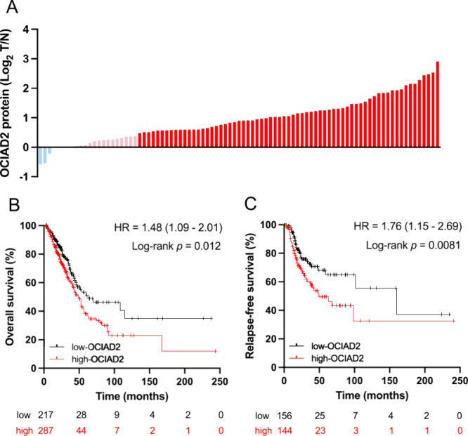
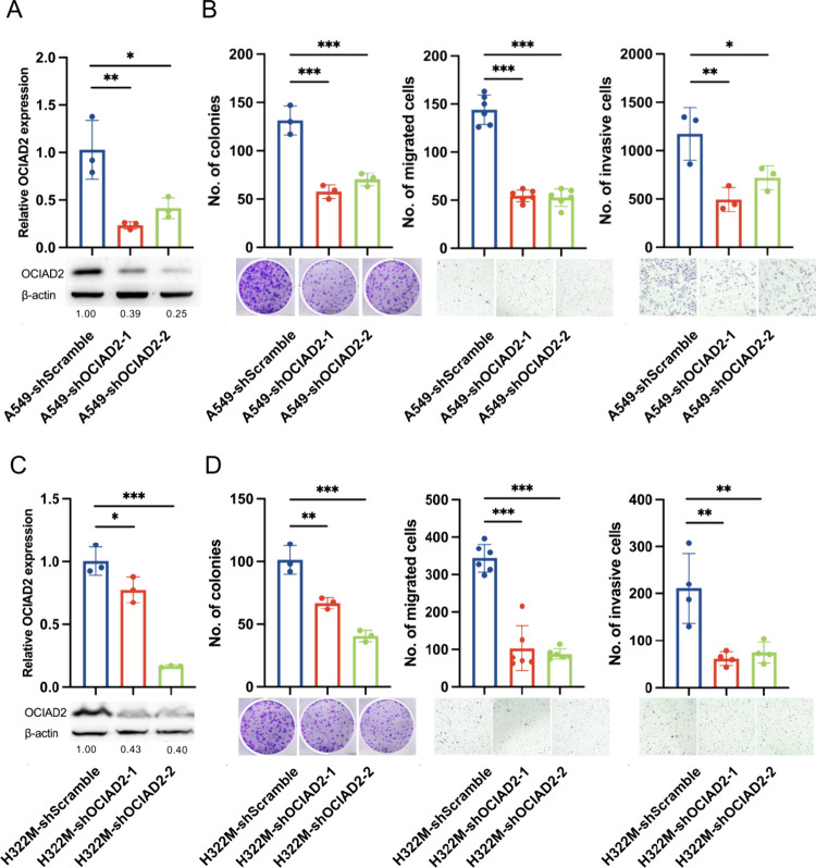
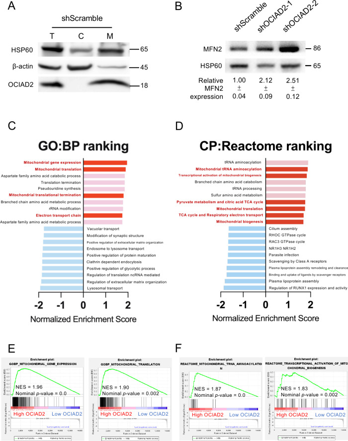
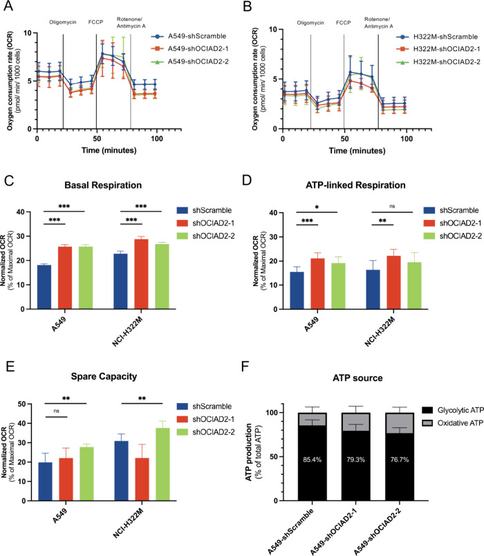
Given the limited proteomic insights and high incidence of lung adenocarcinoma, further investigation of uncharacterized proteins in cancer progression remains crucial. In this study, a poorly characterized protein, OCIA domain-containing 2 (OCIAD2), encoded by chromosome 4 was identified as being upregulated in lung adenocarcinoma from our previous proteogenomics data using the Taiwan Cancer Moonshot cohort. OCIAD2 was highly expressed in tumor tissues in 95.5% of lung adenocarcinoma patients in our cohort, with elevated expression correlating with worse survival. Functional studies revealed that the silencing of the OCIAD2 decreased cell migration, invasion, and colony-forming abilities. Gene Set Enrichment Analysis (GSEA) indicated the involvement of OCIAD2 in oxidative phosphorylation (OXPHOS). Subsequently, mitochondrial metabolic assay demonstrated that OCIAD2 impairs OXPHOS function, accompanied by a metabolic shift toward glycolysis. These findings suggest that OCIAD2 promotes cancer progression through metabolic reprogramming, highlighting the role of OCIAD2 as a potential biomarker and therapeutic target for lung adenocarcinoma.

Keywords: Chromosome-centric Human Proteome Project; Lung adenocarcinoma; Ovarian Cancer Immunoreactive Antigen Domain Containing 2; Uncharacterized protein existence level 1.


Figures

1
1
Clinical relevance of OCIAD2 in lung adenocarcinoma. (A) Relative expression levels of the OCIAD2 protein from tumors (T) and adjacent normal tissues (N) of 89 lung adenocarcinoma patients in the Log2 scale. Dark red: patients with Log2 T/N ratio ≥ 0.38. Light red: patients with 0.38 > Log2 T/N > 0. Light blue: patients with Log2 T/N ≤ 0. (B and C) Kaplan–Meier survival curves of overall survival (OS) (B) and relapse-free survival (RFS) (C) in lung adenocarcinoma patients grouped by high-OCIAD2 and low-OCIAD2 mRNA expression were analyzed using the public database. Red line = high-OCIAD2 mRNA group; black line = low-OCIAD2 mRNA group.
2
2
Pathway analysis of OCIAD2-associated in lung adenocarcinoma progression. (A and B) Volcano plots display the coexpressed genes of OCIAD2 in LUAD obtained from LinkedOmics (A) and cBioPortal (B) database which identified with a p value <0.05 by Welch’s t test (y-axis) and | Spearman correlation coefficient (R) | (x-axis) > 0.25. The positively correlated genes with R > 0.25 (x-axis) are in red and the negatively correlated genes with R < −0.25 (x-axis) are in blue. (C and D) The top 10 enriched pathways were analyzed by Metacore using the data assessed from LinkedOmics (C) and cBioPortal (D) database. Cell motility-related pathways were highlighted in red. (E) Differentially expressed proteins derived from Taiwan Cancer Moonshot cohort were identified with a p value < 0.05 by Welch’s t test (y-axis) and | Spearman correlation coefficient (R) | (x-axis) > 0.25. Of the 1031 differentially expressed proteins, 508 up-regulated proteins were colored red, while 523 downregulated proteins were marked blue in the volcano plot. (F) The top 10 enriched pathways were analyzed using the Taiwan Cancer Moonshot cohort data with Metacore. Red-colored pathways were associated with cell motility.
3
3
Functional assessment of OCIAD2 in lung cancer cells. (A and C) OCIAD2 in A549 (A) and NCI-H322 M (C) cells was evaluated through real-time quantitative RT-PCR (mean ± SD, n = 3), using TATA-box binding protein (TBP) as the internal control. Western blotting revealed the OCIAD2 protein expression level in shScramble control cells and two OCIAD2-silenced cells (shOCIAD2–1 and shOCIAD2–2). Numbers below the Western blot images represent the levels of expression of the β-actin for OCIAD2 quantified by ImageJ and normalized to β-actin as a loading control. (B and D) Three functional assays were conducted on A549 (B) and NCI-H322 M (D) cells, including colony formation assay (left), transwell migration assay (middle), and transwell invasion assay (right). All data are presented as the mean ± SD. Statistical significance was determined as *p < 0.05, **p < 0.01, and ***p < 0.001 by one-way ANOVA and Tukey’s multiple comparisons tests, compared to the shScramble control cells.
4
4
Investigation of the role of OCIAD2 in mitochondria-associated pathways in lung cancer cells. (A) The OCIAD2 protein in cytosolic (C) and mitochondrial (M) fractions extracted from A549-shScramble cells was detected by Western blotting. The fractionations of mitochondria and cytosol were validated by the specific markers HSP60 (heat shock protein 60) and β-actin, respectively. T: total cell lysate; C: cytosolic fraction; M: mitochondrial fraction. (B) Western blot analysis was performed on mitochondrial fractions in OCIAD2-silenced and scramble control A549 cells to determine the expression level of Mitofusin-2 (MFN2) protein. The relative expression of MFN2 was quantified by ImageJ software and normalized to mitochondrial loading control HSP60 (mean ± SD, n = 3). (C and D) Normalized Enrichment Score (NES) of the top 10 positively (red) and negatively (blue) enriched gene sets resulted from GSEA analysis of high-OCIAD2 group (Log2 T/N ≥ 0.38) vs low-OCIAD2 group (Log2 T/N < 0.38). Of note, the mitochondrial-related pathways are shown in dark red columns. (E and F) Enrichment plots of 2 top-ranked pathways enriched in (C) GO-BP and (D) CP-Reactome gene sets show the normalized enrichment score (NES) and ranking metric score.
5
5
Influence of OCIAD2 on mitochondrial metabolic functions in lung cancer cells. (A and B) The oxygen consumption rate (OCR) was measured in real-time in A549 (A) and NCI-H322 M (B) cells transduced with shScramble or shOCIAD2 constructs following sequential injections of Oligomycin, FCCP, and Rotenone/Antimycin A. (C–E) Quantification of Basal respiration (C), ATP-linked respiration (D), and Spare capacity (E) in the A549 and NCI-H322 M cells. Data in (A-E) represent the mean ± SD (n = 9, 3 technical replicates × 3 biological replicates). Statistical significance for (CE) was determined as *p < 0.05, **p < 0.01, and *** p < 0.001 by one-way ANOVA and Tukey’s multiple comparisons tests, compared to the shScramble control cells. (F) The relative luminescence unit (RLU) values were used to determine the proportional contributions of ATP generated by glycolysis and OXPHOS in A549 shScramble and A549 shOCIAD2 cells. Glycolytic ATP was indicated by the decreased ATP level under 2-DG treatment, while oxidative ATP was calculated by subtracting glycolytic ATP from the total ATP (represented by ATP levels in the nontreated group). All data were displayed as the mean ± SD (n = 3) and normalized to the total ATP.

Similar articles

References

    1. Legrain P., Aebersold R., Archakov A., Bairoch A., Bala K., Beretta L., Bergeron J., Borchers C. H., Corthals G. L., Costello C. E.. The human proteome project: current state and future direction. Mol. Cell. Proteomics. 2011;10(7):M111.009993. doi: 10.1074/mcp.M111.009993. - DOI - PMC - PubMed
    1. Omenn G. S., Orchard S., Lane L., Lindskog C., Pineau C., Overall C. M., Budnik B., Mudge J. M., Packer N. H., Weintraub S. T.. et al. The 2024 Report on the Human Proteome from the HUPO Human Proteome Project. J. Proteome Res. 2024;23(12):5296–5311. doi: 10.1021/acs.jproteome.4c00776. - DOI - PMC - PubMed
    1. Paik Y. K., Jeong S. K., Omenn G. S., Uhlen M., Hanash S., Cho S. Y., Lee H. J., Na K., Choi E. Y., Yan F.. et al. The Chromosome-Centric Human Proteome Project for cataloging proteins encoded in the genome. Nat. Biotechnol. 2012;30(3):221–223. doi: 10.1038/nbt.2152. - DOI - PubMed
    1. Paik Y. K., Lane L., Kawamura T., Chen Y. J., Cho J. Y., LaBaer J., Yoo J. S., Domont G., Corrales F., Omenn G. S.. et al. Launching the C-HPP neXt-CP50 Pilot Project for Functional Characterization of Identified Proteins with No Known Function. J. Proteome Res. 2018;17(12):4042–4050. doi: 10.1021/acs.jproteome.8b00383. - DOI - PMC - PubMed
    1. Zhang Y., Vaccarella S., Morgan E., Li M., Etxeberria J., Chokunonga E., Manraj S. S., Kamate B., Omonisi A., Bray F.. Global variations in lung cancer incidence by histological subtype in 2020: a population-based study. Lancet Oncol. 2023;24(11):1206–1218. doi: 10.1016/S1470-2045(23)00444-8. - DOI - PubMed

MeSH terms

Substances