. 2025 Jul;29(14):e70720.
doi: 10.1111/jcmm.70720.

CD44 Participates to Extramedullary Haematopoiesis Onset by Mediating the Interplay Between Monocytes and Haematopoietic Stem Cells in Myelofibrosis

Affiliations

CD44 Participates to Extramedullary Haematopoiesis Onset by Mediating the Interplay Between Monocytes and Haematopoietic Stem Cells in Myelofibrosis

Margherita Mirabile et al. J Cell Mol Med. 2025 Jul.

Abstract

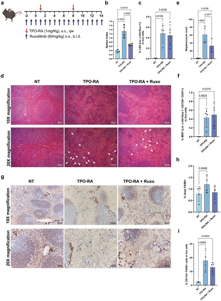
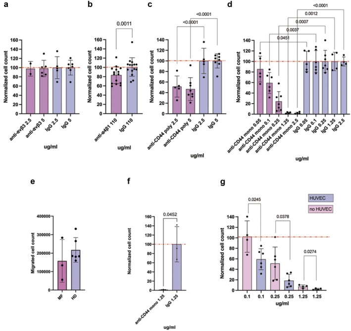
Extramedullary haematopoiesis (EMH) refers to blood generation outside of the bone marrow (BM). In Myelofibrosis (MF), a myeloproliferative neoplasm, the disruption of BM microenvironment promotes haematopoietic stem and progenitor cells (HSPCs) mobilisation, resulting in the onset of EMH in the spleen, and then in splenomegaly. Although JAK2 inhibitors have a good efficacy in reducing splenomegaly, the presence of a significant proportion of non-responder patients underlines the need to explore the cellular mechanisms responsible for the EMH onset. In a MF mouse model, Ruxolitinib induces a reduction in spleen volume but does not affect EMH. CD44 inhibition successfully reduces monocyte and HSPC migration in an in vitro extravasation model. Strikingly, MF monocytes are more effective in promoting HSPC migration through the production of hyaluronic acid. Collectively, our results demonstrate that CD44 regulates the migration of monocytes that are crucial for the onset of EMH in MF patients, as they produce CD44 ligands recruiting HSPCs from the BM.

Keywords: CD44; cell migration; extramedullary haematopoiesis; hyaluronic acid; myelofibrosis; osteopontin.


Conflict of interest statement

The authors declare no conflicts of interest.

Figures

FIGURE 1
FIGURE 1
EMH takes place in the spleen of TPO‐RA mice. (a) Schematic outline for the generation of MF mice. MF was induced by sub‐cutaneous injection of a TPO‐RA (Romiplostim, 1 mg/kg, once weekly). Mice were given Ruxolitinib 60 mg/kg through oral gavage twice daily starting 3 days before the first TPO‐RA injection. Mice were sacrificed after 15 days of TPO‐RA treatment. (b) Spleen index at sacrifice of not treated (NT), TPO‐RA treated and TPO‐RA + Ruxolitinib treated mice (n = 4 for each group). (c) Flow cytometry evaluation of Lineage Sca1+ and c‐Kit+ (LSK) cells in the spleen of NT (n = 2), TPO‐RA (n = 4) and TPO‐RA + Ruxolitinib (n = 5) treated mice. Each dot represents a mouse. (d) Representative images of haematoxylin–eosin stained spleen sections from controls, TPO‐RA treated mice and MF mice receiving Ruxolitinib. Megakaryocytes are indicated using white arrows in 20X images and represent large cells with multilobed nuclei and abundant cytoplasm. (e) The megakaryocyte count for NT (n = 4), TPO‐RA treated (n = 4) and TPO‐RA + Ruxolitinib treated mice (n = 3). Each dot represents the mean count of megakaryocytes in at least 3 fields of the same section at 10X magnification (n = 4 for each group). (f) Flow cytometry evaluation of Lineage Sca1 c‐Kit+ CD150+ CD41+ megakaryocyte progenitors in the spleen of NT (n = 2), TPO‐RA (n = 4) and TPO‐RA + Ruxolitinib (n = 5) treated mice. Each dot represents a mouse. (g) Immunohistochemistry for F4/80 of NT, TPO‐RA treated and TPO‐RA and Ruxolitinib treated mice. (h) Immunohistochemistry quantification of spleen sections from NT, TPO‐RA treated and TPO‐RA and Ruxolitinib treated mice. Each dot represents the percentage of area covered by F4/80 signal in a representative field (n = 4 for each group). (i) Flow cytometry evaluation of CD11b+ F4/80+ macrophages in the spleen of NT (n = 2), TPO‐RA (n = 4) and TPO‐RA + Ruxolitinib (n = 5) treated mice. Each dot represents a mouse. In graphs each dot represents a mouse, each column represents group mean ± standard deviation. Comparisons were performed by means of one‐way ANOVA. p‐values are reported only if < 0.05. Scale bars of 100 and 50 μm are shown for 10× and 20× magnification images, respectively.
FIGURE 2
FIGURE 2
The effect of Ruxolitinib on splenomegaly is not mediated by cell migration but it is correlated with OPN. Scheme in panel (a) represents the workflow of our in vitro extravasation system based on Transwell with HUVECs. Each Transwell received 80,000 HUVEC cells treated with 100 μg/mL of TNF‐α. HUVECs were seeded upside down for an overnight incubation. The next day the Transwell modified with HUVECs was loaded with immunopurified CD14+ or CD34+ cells. After an overnight incubation, the cells in the down side of the system were collected and counted with flow cytometry. Panels (b–e) show the effect of Ruxolitinib treatment on in vitro migration of, respectively, HD CD14+ cells (n = 3), MF CD14+ cells (n = 3), HD CD34+ cells (n = 3) and MF CD34+ cells (n = 3). The red dashed line represents 100% migration. On X axis are reported Ruxolitinib doses, on Y axis the normalised cell count (see Section 2). (f) The OPN plasma levels for NT, TPO‐RA treated and TPO‐RA + Ruxolitinib treated mice. In graphs each dot represents a replicate for migration experiments or a mouse for ELISA assay, each column represents group mean ± standard deviation. Comparisons in mice were performed by means of one‐way ANOVA for panels (b, d, f), and unpaired T‐test for panels (c, e). p‐values are reported only when < 0.05.
FIGURE 3
FIGURE 3
CD44 is highly expressed in MF monocytes and HSPCs and its expression is increased in the spleen of MF mice. (a) Immunophenotypic evaluation of α4β1, αvβ3 and CD44 in circulating CD14 + monocytes in HDs (n = 15) and MF patients (n = 8). (b) Immunophenotypic evaluation of α4β1, αvβ3 and CD44 in circulating CD34 + cells in HDs (n = 15) and MF patients (n = 8). (c, d) Flow cytometry evaluation of CD44, α4β1 and αvβ3 expression in macrophages (c) and LSK cells (d) in the spleen of NT (n = 2) and TPORA (n = 4) treated mice. Each dot represents a mouse. (e, f) Flow cytometry evaluation of the frequency of CD44 + macrophages (e) and LSK cells (f) in the spleen of NT (n = 2), TPORA (n = 4) and TPORA + Ruxolitinib (n = 5) treated mice. Each dot represents a mouse. In graphs, each dot represents a patient or a mouse, and each column represents group mean ± standard deviation. Comparisons were performed by means of unpaired T‐test for panels (a–d) and one‐way ANOVA for panels (e, f). p‐values are reported only when < 0.05.
FIGURE 4
FIGURE 4
CD44 regulates HD and MF CD14+ monocyte migration. Panel (a) shows the effect of anti‐αvβ3 antibody on migration of HD CD14+ monocyte in vitro in presence of HUVEC coating while panel (b) displays results for the anti‐α4β1 antibody treatment. Panel (c) shows the effect of the polyclonal anti‐CD44 antibody on migration of HD CD14+ monocyte in vitro in presence of HUVEC endothelium while panel (d) displays results for the monoclonal anti‐CD44 antibodies. (e) Graph displays the difference in migration between HD (n = 6) and MF (n = 3) CD14+ monocytes in the established in vitro extravasation model. (f) The effect of anti‐CD44 antibody on migration of MF CD14+ monocyte in vitro (n = 3). (g) Bar plot represents results of HD CD14+ monocyte migration assays with and without HUVEC for the monoclonal anti‐CD44 antibody; Transwell samples with HUVEC are represented with purple bars while Transwell samples without HUVEC are illustrated with pink bars; the percentage of migration is normalised considering control IgG samples, not shown in this graph. In all bar plots the red dashed line represents 100% migration. On X axis are reported antibodies doses, Y axis reports the normalised cell count (see Section 2). In graphs each dot represents a replicate, each column represents group mean ± standard deviation. Comparisons between samples treated with the specific antibody or control IgG were performed by means of paired T‐test. p‐values are reported only if < 0.05.
FIGURE 5
FIGURE 5
CD44 regulates CD14+ monocytes migration through the interaction with OPN and HA. (a) The graph displays the concentration of HA (ng/mL) in HD (n = 8) and MF (n = 13) plasma samples. Comparison between HD and MF was performed by means of unpaired T‐test. Panels (b, c) show, respectively, the effect of neutralising antibody against OPN and HA inhibitor on HD CD14+ monocyte migration. Panels (d, e) show the effect on HD CD14+ monocytes migration of the combination of neutralising antibody against OPN (d) or HA inhibitor (e) with the monoclonal anti‐CD44 antibody. The upper red dashed line represents 100% migration while the down red dashed line represents the migration inhibition induced by the anti‐CD44 0.1 μg/mL treatment. X axis display antibodies and inhibitor doses, Y axis shows the normalised cell count (see Section 2). In graphs each dot represents a replicate, each column represents group mean ± standard deviation. Comparisons between samples treated with the specific antibody or control IgG were performed by means of paired T‐test. p‐values are reported only if < 0.05.
FIGURE 6
FIGURE 6
CD44 regulates CD34+ HSPC migration in vitro through the binding with HA. (a) Graph displays the difference in migration between HD (n = 22) and MF (n = 10) CD34+HSPCs in our in vitro extravasation model. (b) The plot shows the inhibition of migration induced by the anti‐CD44 monoclonal antibody on HD (n = 22) and MF (n = 10) CD34+ HSPCs. The red dashed line represents 100% migration. On X axis are reported the antibodies at different doses, on Y axis the normalised cell count (see Section 2). Each dot represents a replicate. Panels (c, d) show the OPN and HA produced by HD (n = 11) and MF (n = 13 for panel c and n = 14 for panel d) CD14+ monocytes in vitro, respectively. The number of live monocytes was determined at time of medium collection, 96 h after plating, and it was used to normalise ligand concentration to obtain a measure of OPN and HA produced by single monocytes in culture. (e) The effect of HD (n = 11) and MF (n = 10) CD14+ supernatants on HD CD34+ cell migration; the number of migrated CD34+ cells was normalised based on the number of remaining monocytes at time of supernatants harvesting. Each dot represents a replicate. Panel (f) shows the effect of anti‐CD44 monoclonal antibody on HD CD34+ cells migration induced by MF monocytes conditioned medium (n = 3). (g) The effect of anti‐OPN antibody and HA inhibitor, alone or in combination, on HD CD34+ cells migration induced by MF monocytes conditioned medium (n = 3). Panels (h, i) show the correlation between log transformed CD34+ normalised migrated cells and HA (panel h, n = 14) and OPN (panel i, n = 19) concentration. In graphs each dot represents a replicate, each column represents group mean ± standard deviation. Comparisons were performed by means of unpaired T‐test for panels (a, c, d, e), paired T‐test for panels (b, f), and one‐way ANOVA for panel (g). Correlation analysis was performed by simple linear regression. p‐values are reported only if < 0.05.

References

    1. Cenariu D., Iluta S., Zimta A.‐A., et al., “Extramedullary Hematopoiesis of the Liver and Spleen,” Journal of Clinical Medicine 10 (2021): 5831, 10.3390/jcm10245831. - DOI - PMC - PubMed
    1. Sohawon D., Lau K. K., Lau T., and Bowden D. K., “Extra‐Medullary Haematopoiesis: A Pictorial Review of Its Typical and Atypical Locations,” Journal of Medical Imaging and Radiation Oncology 56 (2012): 538–544, 10.1111/j.1754-9485.2012.02397.x. - DOI - PubMed
    1. Wright D. E., Wagers A. J., Gulati A. P., Johnson F. L., and Weissman I. L., “Physiological Migration of Hematopoietic Stem and Progenitor Cells,” Science 294 (2001): 1933–1936, 10.1126/science.1064081. - DOI - PubMed
    1. Yang X., Chen D., Long H., and Zhu B., “The Mechanisms of Pathological Extramedullary Hematopoiesis in Diseases,” Cellular and Molecular Life Sciences 77 (2020): 2723–2738, 10.1007/s00018-020-03450-w. - DOI - PMC - PubMed
    1. Grabek J., Straube J., Bywater M., and Lane S. W., “MPN: The Molecular Drivers of Disease Initiation, Progression and Transformation and Their Effect on Treatment,” Cells 9 (2020): 1901, 10.3390/cells9081901. - DOI - PMC - PubMed

MeSH terms