. 2025 Jul 29;122(30):e2407224122.
doi: 10.1073/pnas.2407224122. Epub 2025 Jul 21.

RACK1A positively regulates opening of the apical hook in Arabidopsis thaliana via suppression of its auxin response gradient

Affiliations

RACK1A positively regulates opening of the apical hook in Arabidopsis thaliana via suppression of its auxin response gradient

Qian Ma et al. Proc Natl Acad Sci U S A. .

Abstract

Apical hook development is an ideal model for studying differential growth in plants and is controlled by complex phytohormonal crosstalk, with auxin being the major player. Here, we identified a bioactive small molecule that decelerates apical hook opening in Arabidopsis thaliana. Our genetic studies suggest that this molecule enhances or maintains the auxin maximum found in the inner hook side and requires certain auxin signaling components to modulate apical hook opening. Using biochemical approaches, we then revealed the WD40 repeat scaffold protein RECEPTOR FOR ACTIVATED C KINASE 1A (RACK1A) as a direct target of this compound. We present data in support of RACK1A playing a positive role in apical hook opening by activating specific auxin signaling mechanisms and negatively regulating the differential auxin response gradient across the hook, thereby adjusting differential cell growth, an essential process for organ structure and function in plants.

Keywords: Arabidopsis; apical hook; auxin; differential cell growth.


Conflict of interest statement

Competing interests statement:The authors declare no competing interest.

Figures

Fig. 1.
Fig. 1.
The compound DAPIA decelerates apical hook opening in Arabidopsis etiolated seedlings. (A) Chemical structure of DAPIA. (B and C) DAPIA dose–response analysis – representative images of apical hook phenotypes (B) and quantification of average hook angle (C) after 4 d of growth (including germination time) of Col-0 and axr1-30 in darkness on medium supplemented with DMSO (mock) or DAPIA. Values in square brackets represent concentrations in µM. (Scale bar, 5 mm.) Data are shown as box plots and different letters indicate significantly different means of N = 8 to 17 seedlings at P < 0.05 (one-way ANOVA, Tukey’s multiple comparisons test) (C). (D) Kinematics of apical hook angle in Col-0 and axr1-30 as measured every 4 h for 10 d of growth starting from germination (0 h) in darkness on medium supplemented with DMSO (mock) or 10 µM DAPIA. Error bars represent SEM; N = 23 to 61 seedlings. Color-coded values beside the curves represent the rate of early hook opening, expressed as the mean slope angle in degrees of the late maintenance-opening phase (from the maximum mean hook angle to the first mean hook angle below 30% of the maximum), for which asterisks indicate significant differences (Wilcoxon rank-sum test; **P < 0.01; ***P < 0.001).
Fig. 2.
Fig. 2.
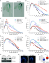
DAPIA affects the auxin response in the apical hook and requires the auxin signaling components AXR2, ARF7, and ARF19 to decelerate hook opening. (A) Representative images of apical hooks of GUS-stained DR5::GUS seedlings after 4 d of growth (including germination time) in darkness on medium supplemented with DMSO (mock) or DAPIA. Values in square brackets represent concentrations in µM. (Scale bar, 150 µm.) (BF) Kinematics of apical hook angle in tir1-1afb2-3 (B), axr2-1 (C), shy2-2 (D), arf2-8 (E), and arf7-1arf19-1 (F), together with the relevant WT, as measured every 4 h for 10 d of growth starting from germination (0 h) in darkness on medium supplemented with DMSO (mock) or 10 µM DAPIA. Error bars represent SEM; N = 16 to 61 seedlings. Color-coded values beside the curves represent the rate of early hook opening, expressed as the mean slope angle in degrees of the late maintenance-opening phase (from the maximum mean hook angle to the first mean hook angle below 30% of the maximum), for which asterisks indicate significant differences (Wilcoxon rank-sum test; ns–not significantly different; **P < 0.01; ***P < 0.001). (G) Immunoblot analysis of tissue samples from ARF19::ARF19-Venus and ARF7::ARF7-Venus seedlings, grown for 4 d in darkness on medium supplemented with DMSO (mock) or 10 µM DAPIA, probed with anti-YFP antibody. Ponceau stain was used as loading control. (H and I) Representative confocal images (H) and quantification of Venus fluorescence gradient (inner:outer side average fluorescence intensity ratio) (I) of apical hooks of dark-grown ARF19::ARF19-Venus seedlings at the late maintenance-opening phase. Seedlings were grown on medium supplemented with DMSO (mock) or 10 µM DAPIA. (Scale bar, 100 µm.) Low-to-high Venus signal intensity is represented as a blue-to-white color gradient according to the inset, and maximum intensity projections of z-stacks are shown (H). Data are shown as box plots and asterisks indicate significantly different means of N = 11 to 18 seedlings (Wilcoxon rank-sum test; *P < 0.05) (I). Values in square brackets represent concentrations in µM.
Fig. 3.
Fig. 3.
RACK1A is the direct target of DAPIA. (AC) Direct binding between DAPIA and RACK1A validated by proteolytic analysis (A) and molecular docking (B and C). (A) Total protein extracts from 4-d-old etiolated seedlings of axr1-30, as well as Col-0 and rack1a-3 as controls, were incubated with DMSO (mock) or DAPIA and subjected to various dilutions of pronase (expressed as total enzyme:total protein ratios). RACK1A protein levels were detected through protein gel blot analysis using anti-RACK1A antibody (Upper blot) and anti-α-Tubulin antibody (Lower blot) as a reference protein control. Each blot has its own reference ladder, viewed with a different mode and therefore saved as a separate image. Values in square brackets represent concentrations in µM. (B) Top (Left panel) and bottom (Right panel) views of RACK1A 3D structure with the DAPIA binding site predicted by the docking simulation. RACK1A, in the ribbon diagram, adopts a β-propeller conformation. The seven blades of the β-propeller are numbered 1-7 from the N terminus (N-ter, blue) to the C terminus (C-ter, red) in rainbow mode. The four antiparallel β-strands in each blade are labeled A–D, as exemplified in blade 2. The small molecule DAPIA is bound in a cavity in the central channel and is shown in stick-and-ball and surface mesh modes in salmon color. (C) A close-up view of DAPIA in the predicted binding pocket of RACK1A. The thirteen RACK1A residues involved in the interaction with DAPIA are shown in sticks, with the same color codes as used in B. Hydrogen bonds are shown as black dashed lines. (D) Multiple sequence alignment of Arabidopsis, human, and Drosophila RACK1 in relationship with the 3D structure of Arabidopsis RACK1A docked with DAPIA. Protein sequences were obtained from UniProt using the following accession numbers: Arabidopsis thaliana RACK1A: O24456, Homo sapiens (human) RACK1: P63244, Drosophila melanogaster RACK1: O18640. Sequences are aligned using Clustal Omega. Secondary structural elements of Arabidopsis RACK1A (four β-strands in each of the seven blades) are indicated above the sequences, with the same alphanumeric and color codes as used in B. Invariant amino acids are denoted by asterisks. The DAPIA-interacting residues are indicated by salmon circles and those constituting the conserved region 2 by purple squares.
Fig. 4.
Fig. 4.
RACK1A regulates apical hook opening at the level of auxin response. (A) Kinematics of apical hook angle in Col-0 and rack1a-3 as measured every 4 h for 10 d of growth starting from germination (0 h) in darkness. (B) Representative confocal image of the apical hook of dark-grown RACK1A::RACK1A-GFP seedling at the late maintenance-opening phase. (C) Kinematics of apical hook angle in Col-0, axr1-30, rack1a-3, and axr1-30rack1a-3 as measured every 4 h for 10 d of growth starting from germination (0 h) in darkness. Error bars represent SEM; N = 11 to 46 seedlings (A and C). Color-coded values beside the curves represent the rate of early hook opening, expressed as the mean slope angle in degrees of the late maintenance-opening phase (from the maximum mean hook angle to the first mean hook angle below 30% of the maximum), for which asterisks (A) or different letters (C) indicate significant differences (Wilcoxon rank-sum test; ***P < 0.001; different letters: P < 0.05). (D and E) Representative confocal images (D) and quantification of auxin response gradient (inner:outer side average nuclear GFP fluorescence intensity ratio) (E) of apical hooks of dark-grown DR5::n3GFP seedlings in Col-0, axr1-30, rack1a-3, and axr1-30rack1a-3 backgrounds at time points representing the maintenance phase and late maintenance-opening phase of the WT. [Scale bar, 100 µm (B) and 200 µm (D).] Low-to-high GFP signal intensity is represented as a blue-to-yellow color gradient according to the insets and maximum intensity projections of z-stacks are shown (B and D). Data are shown as box plots and asterisks indicate significantly different means of N = 17 to 29 seedlings (Wilcoxon rank-sum test; ns–not significantly different; ***P < 0.001) (E).

References

    1. Žádníková P., et al. , A model of differential growth-guided apical hook formation in plants. Plant Cell 28, 2464–2477 (2016). - PMC - PubMed
    1. Raz V., Ecker J. R., Regulation of differential growth in the apical hook of Arabidopsis. Development 126, 3661–3668 (1999). - PubMed
    1. Silk W. K., Erickson R. O., Kinematics of hypocotyl curvature. Am. J. Bot. 65, 310–319 (1978).
    1. Abbas M., Alabadí D., Blázquez M., Differential growth at the apical hook: All roads lead to auxin. Front. Plant Sci. 4, 441 (2013). - PMC - PubMed
    1. Mazzella M. A., Casal J. J., Muschietti J. P., Fox A. R., Hormonal networks involved in apical hook development in darkness and their response to light. Front. Plant Sci. 5, 52 (2014). - PMC - PubMed

MeSH terms

LinkOut - more resources