. 2025 Jul 21;16(1):6685.
doi: 10.1038/s41467-025-61811-0.

Mechanism of Rad51 filament formation by Rad52 and Rad55-Rad57 in homologous recombination

Affiliations

Mechanism of Rad51 filament formation by Rad52 and Rad55-Rad57 in homologous recombination

Jaigeeth Deveryshetty et al. Nat Commun. .

Abstract

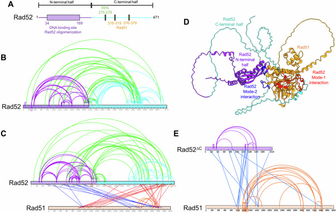
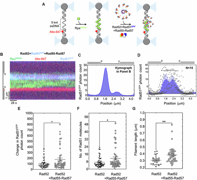
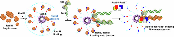
Homologous recombination (HR) repairs double-stranded DNA breaks (DSBs) by generating single-stranded DNA (ssDNA), which is initially coated by Replication Protein A (Rpa). Rad51, a recombinase, catalyzes strand invasion but binds ssDNA with lower affinity than Rpa, necessitating mediator proteins like Rad52 (yeast) or BRCA2 (humans) for Rad51 loading. The mechanisms of this exchange remain unclear. We show that Saccharomyces cerevisiae Rad52 uses its disordered C-terminus to sort polydisperse Rad51 into discrete monomers. Using fluorescent-Rad51 and single-molecule optical tweezers, we visualize Rad52-mediated Rad51 filament formation on Rpa-coated ssDNA, preferentially at ssDNA-dsDNA junctions. Deleting the C-terminus of Rad52 disrupts Rad51 sorting and loading. Addition of the Rad51 paralog Rad55-Rad57 enhances Rad51 binding by ~60%. Despite structural differences, Rad52 and BRCA2 share conserved functional features. We propose a unified "Sort, Stack & Extend" (SSE) mechanism by which mediator proteins and paralogs coordinate Rad51 filament assembly during HR.


Conflict of interest statement

Competing interests: The authors declare no competing interests.

Figures

Fig. 1
Fig. 1. S. cerevisiae Rad51 is polydisperse in solution.
A Schematic of Rad51 denoting the positions of the Walker A and B motifs for ATP-binding and hydrolysis. The FVTA motif promotes Rad51 oligomerization. Loops 1 and 2 in site 1 and residues in site 2 are critical for DNA binding. The N-terminal disordered region (NDR), N-terminal lobe domain (NLD), and RecA-like domains are also denoted. B Structure of the Rad51 filament in the absence of DNA (PDB:1SZP). The monomeric units are colored gray and orange. Phe-144 and Ala-147 dock into defined pockets of the adjacent Rad51 protomer to promote oligomerization. C Mass photometry analysis of Rad51 (100 nM) shows a wide range of oligomers ranging from monomers through decamers, with dimers being the predominant species. D Analytical ultracentrifugation sedimentation velocity analysis of Rad51 at a higher concentration (10 µM) also shows a similar distribution of oligomers. E Crosslinking mass spectrometry (XL-MS) analysis of Rad51 using BS3 crosslinker shows extensive intra- and inter-Rad51 crosslinks. Source data are provided as a Source Data file.
Fig. 2
Fig. 2. The C-terminus of Rad52 sorts Rad51 into monomers.
A Schematic of Rad52 showing the ordered N-terminal and disordered C-terminal halves of Rad52. The Rpa and Rad51 binding motifs reside in the C-terminal half. B Crosslinking mass spectrometry (XL-MS) analysis of Rad52 reveals extensive crosslinks within the N-terminal (purple) half, the C-terminal (cyan) half, and between the two halves (green). C XL-MS analysis of the Rad52-Rad51 complex reveals intra-Rad52 crosslinks as shown in (B), in addition to two sets of crosslinks between Rad51 and Rad52. Crosslinks between the N-terminal half of Rad52 and Rad51 are shown in blue, while crosslinks between the C-terminal half of Rad52 and Rad51 are shown in red. Interestingly, almost all of the inter- and intra-Rad51 crosslinks observed in the Rad51 alone XL-MS analysis are absent when in complex with Rad52. Only three crosslinks within Rad51 are captured (orange). D AlphaFold3 prediction of the complex between one Rad52 subunit and Rad51. Residues in Rad51 that are proposed to mediate mode-1 interactions with Rad52 are shown in red. Residues that mediate mode-2 interactions are shown in blue and reside in the L1 and L2 loops of Rad51. E Rad52ΔC, lacking the C-terminal half of Rad52, retains interactions with Rad51. XL-MS analysis of this complex reveals the reappearance of the inter- and intra-Rad51 crosslinks (orange), suggesting that Rad51 forms oligomers when bound to Rad52ΔC. Thus, the Rad51 sorting properties arise from interactions with the C-terminus of Rad52 (mode-1). Intra-Rad52ΔC crosslinks are denoted in purple, and those with Rad51 are shown in blue. Source data are provided as a Source Data file.
Fig. 3
Fig. 3. The Rad51-sorting function is mediated by mode-1 interactions and facilitated through the disordered C-terminus of Rad52.
A Schematic of the two Rad51 interaction motifs in the disordered C-terminus of Rad52. Rad52ΔN* is a construct that possesses both these motifs, and B is able to sort Rad51 from a polydisperse to a monodispersed species in solution. Mass photometry analysis of C Rad51, D Rad52ΔN*, and E the Rad51-Rad52ΔN* complex. Rad51 is polydisperse, with monomeric to decameric species observed in solution. Rad52ΔN* is a single lower molecular weight species in solution. It should be noted that the predicted mass is below the detectable limit of the mass photometry methodology. The Rad51-Rad52ΔN* complex shows a loss of the higher order Rad51 species and accumulation of a predominantly single species that corresponds to one Rad51 molecule bound per Rad52ΔN*. Source data are provided as a Source Data file.
Fig. 4
Fig. 4. Direct visualization of Rad51 binding events on single-stranded DNA.
A Schematic of the sfGFP-Rad51 (Rad51GFP) construct. The sfGFP is engineered at position 55 in the non-conserved N-terminal region of S. cerevisiae Rad51. Two 16 aa flexible linkers flank the sfGFP and are required for maintaining Rad51 functionality. B An AF-model of the Rad51GFP protein. sfGFP and Rad51 are colored green and orange, respectively. The two 16 aa linkers are colored gray. C Rad51 and Rad51-variants support the formation of D-loops. Cy5-labeled ssDNA was incubated with Rad51/Rad51-variant, followed by the addition of Rpa and Rad54+dsDNA, and incubated as depicted. D Image of the gel showing D-loop formation. The smear observed in the lanes corresponding to the Rad51Cy5 sample arises from the fluorescence signal of the protein (Supplementary Fig. 5c). E Quantification of the percent D-loops formed shows all Rad51 proteins retaining similar levels of D-loop formation activity. F Design of the optical trap assay to visualize Rad51GFP binding to ssDNA. A ~ 48.5 knt ssDNA was tethered to biotin handles, bound to streptavidin beads, and captured by two optical traps. The trapped DNA is then sequentially moved to channels containing Rad51GFP in the absence or presence of ATP. No Rad51 binding is observed in the absence of ATP, but robust Rad51GFP binding to ssDNA is observed in the presence of ATP. G Rad51GFP binding to ssDNA is dependent on ATP concentration. Data points from multiple Rad51 binding events measured across several tethers (N) for each ATP concentration are shown, and the SEM are plotted as error bars. N = 13, 5, 13, and 16 tethers for experiments with 0.1, 0.34, 1, and 2 mM ATP, respectively. Source data are provided as a Source Data file.
Fig. 5
Fig. 5. Rad52 enhances Rad51 binding to ssDNA.
A Schematic of the optical trap experiment showing Rad51GFP binding to a ~ 48.5 knt ssDNA substrate in the absence or presence of Rad52. Kymographs of optical trap data in the B absence or C presence of Rad52 shows stimulation of Rad51GFP binding to ssDNA. D Kymograph showing no binding of Rad51GFP to an Rpa-coated ssDNA in the absence of Rad52. E Rad52ΔC, which lacks the C-terminal Rad51 and Rpa interaction domains, poorly promotes Rad51GFP binding to ssDNA. F Under these single-molecule experimental conditions (100 nM Rad51), low levels of Rad51GFP binding to dsDNA are observed. G Quantitation of Rad51GFP filaments formed under the denoted conditions. Representative data from a minimum of n = 5 tethers per condition is shown, and the SEM is plotted as error bars. An unpaired t test was performed between the ±Rad52 conditions, and ** denotes p = 0.0098. Source data are provided as a Source Data file.
Fig. 6
Fig. 6. Rad51 preferentially localizes to the recessed Rpa-coated ss-dsDNA junctions in the presence of Rad52.
A Schematic of the optical trap experiment where the DNA is first preincubated with MB543-labeled-Rpa (RpaMB543; 0.25 nM) and then moved to a channel containing fluorescent Rad51 (either Rad51Cy5 or Rad51GFP; 50 nM) in the presence of Rad52 (5 nM decamer) and ATP (5 mM). An Atto-647 fluorophore is embedded within one arm of the dsDNA handle (red) to help define polarity. Stacks of frames (kymograph) were recorded by continuous confocal scanning along the DNA axis. The kymographs show binding of either B Rad51Cy5 (magenta) or C Rad51GFP (blue) onto the RpaMB543-coated ssDNA gap (green) in the presence of Rad52 and ATP. The scale bars for the length of the DNA (y axis) and time (x axis) are denoted for each kymograph. D Photon counts for Rad51Cy5 (magenta) and RpaMB543 (green) were quantified and plotted versus the position of DNA in the kymograph. Rad51Cy5 preferentially binds to the 5′-recessed junction when in complex with Rad52. In addition, Rad51 and Rpa binding events are not mutually exclusive, suggesting higher-order complex formation between Rpa, Rad52, and Rad51 at this junction. E Similar junction-preferential binding of Rad51GFP (blue) is observed under these conditions. Compilation of photon count data from experiments using either F Rad51Cy5 (n = 9 independent tethers) or G Rad51GFP (n = 13 independent tethers) shows binding events at both junctions and internal regions on the ssDNA gap, but with a strong bias for the 5′-recessed junction. The SEM is plotted as error bars. Source data are provided as a Source Data file.
Fig. 7
Fig. 7. Rad55-Rad57, the Rad51-paralog, increases the number of Rad51 molecules bound during nucleation and an increase in filament length.
A Design of optical trap experiments to investigate the effect of Rad55-Rad57 on Rad51 filament formation. RpaMB543-coated (green; 0.25 nM) gapped ssDNA were moved to a channel containing Rad51GFP (blue; 100 nM), Rad52 (5 nM decamer), and Rad55-Rad57 (50 nM) in the presence of ATP (5 mM). B Kymograph from a single tether shows preferential binding of Rad51GFP close to the 5′-recessed junction. C Photon count of Rad51GFP binding events along the DNA in the kymograph shown in B and D, across multiple individual tethers (n = 15 tethers) shows broader coverage of the ssDNA gap with Rad51GFP. E Quantitation of the change in Rad51GFP count (15 tethers, 46-50 traces), or F the number of Rad51GFP molecules bound, shows an increase in Rad51 molecules in the presence of Rad55-Rad57 compared to Rad52 alone (15 tethers, 46-50 traces). G Binding of more Rad51 molecules in the presence of Rad55-Rad57 results in an increase in the length of the filament by ~0.1 μm (15 tethers, 28-40 traces). The SEM is plotted as error bars. Two-tailed unpaired t-tests were performed and * and ** denote p-values of 0.0314 (E), 0.0296 (F), and 0.0026 (G), respectively. Source data are provided as a Source Data file.
Fig. 8
Fig. 8. A sort, stack, and extend (SSE) model for mediator and Rad51-paralog-promoted Rad51 filament formation on Rpa-coated ssDNA during HR.
A sort, stack, and extend model for events in presynapsis is depicted. Rad51 is polydisperse in solution and is sorted into defined monomeric units by Rad52. There are two modes of interaction between Rad52 and Rad51. Mode-1 sorts Rad51 into monomers, whereas mode-2 is asymmetric and occurs at one position in the Rad52 ring. The Rad52-Rad51 complex engages the Rpa-coated resected ssDNA during pre-synapsis, and preferential binding to the ss-dsDNA junction is proposed. However, Rad52 is not sufficient to promote Rad51 filament growth. Rad51-paralogs (Rad55-Rad57) promote filament growth/extension. The binding positions of Rad52 and Rad55-Rad57 are speculative and need to be experimentally established.

Similar articles

References

    1. Negrini, S., Gorgoulis, V. G. & Halazonetis, T. D. Genomic instability-an evolving hallmark of cancer. Nat. Rev. Mol. Cell Biol.11, 220–228 (2010). - PubMed
    1. Pfeiffer, P., Goedecke, W. & Obe, G. Mechanisms of DNA double-strand break repair and their potential to induce chromosomal aberrations. Mutagenesis15, 289–302 (2000). - PubMed
    1. Wright, W. D., Shah, S. S. & Heyer, W. D. Homologous recombination and the repair of DNA double-strand breaks. J. Biol. Chem.293, 10524–10535 (2018). - PMC - PubMed
    1. Heyer, W. D., Ehmsen, K. T. & Liu, J. Regulation of homologous recombination in eukaryotes. Annu. Rev. Genet.44, 113–139 (2010). - PMC - PubMed
    1. San Filippo, J., Sung, P. & Klein, H. Mechanism of eukaryotic homologous recombination. Annu. Rev. Biochem.77, 229–257 (2008). - PubMed

MeSH terms

LinkOut - more resources