. 2025 Jul 21;16(1):6677.
doi: 10.1038/s41467-025-62061-w.

E2F activity determines mitosis versus whole-genome duplication in G2-arrested cells

Affiliations

E2F activity determines mitosis versus whole-genome duplication in G2-arrested cells

Kibum Kim et al. Nat Commun. .

Abstract

While mitogenic signaling is known to regulate cell-cycle entry during the G1 phase, its function in the G2 phase remains elusive. Here we show that mitogenic signaling controls whether G2-arrested cells proceed through mitosis or undergo whole-genome duplication. Although mitogenic signaling is not required for the G2/M transition under normal conditions, it modulates E2F transcriptional activity via c-Myc. When G2 arrest occurs due to CDK4/6 and CDK2 suppression, E2F activity levels determine the status of APC/C inactivation and the CDK2-Rb feedback loop. Upon release from G2 arrest, cells maintaining APC/C inactivation promptly induce CDK2 activation and FoxM1 phosphorylation, driving mitotic entry. Conversely, APC/C reactivation degrades cyclin A and abolishes the CDK2-Rb loop, necessitating CDK4/6 activation for cell-cycle re-entry. This regulatory mechanism mirrors the G1-phase process, resulting in whole-genome duplication. In cancer cells, this process promotes genome instability and oncogene amplification, contributing to aggressive behavior. These findings reveal a previously unrecognized mitogen-dependent checkpoint that governs cell fate in the G2 phase.


Conflict of interest statement

Competing interests: The authors declare no competing interests.

Figures

Fig. 1
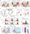
Fig. 1. G2-arrested cells need CDK4/6 activity to re-enter the cell cycle, resulting in WGD.
a Density scatterplot showing DNA content versus EdU staining in primary MEFs (n = 3000 cells). b Histogram of phosphorylated Rb (p-Rb, S807/811) normalized to total Rb (t-Rb) levels in S and G2 phases in MEFs. Dotted lines represent the threshold used to classify cells into p-Rb negative and positive populations (n > 1000 cells/condition). c Representative images of Hoechst, γH2AX, and 53BP1 in MCF-10A cells 48 h after mitogen removal and sorted by DNA content. G1- and G2-arrested cells were classified based on DNA content. Scale bar is 20 μm. d Single-cell violin plots of γH2AX and 53BP1 puncta area in G1 and G2-arrested cells (n > 2000 cells/condition). Asterisks indicate significant differences in the two-tailed unpaired t-test (*** p ≤ 0.0001). e Representative images of Hoechst, p-Rb (S807/811), and E2F1 mRNA FISH in mitogen-starved MCF-10A cells stimulated with mitogens for 12 h. Scale bar is 20 μm. f Percentage of cells positive for p-Rb (S807/811) and averaged levels of E2F1 and Cdc25A mRNA as a function of time since mitogen stimulation. Solid lines represent sigmoidal best-fit curves. g, h Density scatterplot showing DNA content versus EdU staining (n > 1000 cells) (g) and percentage of p-Rb-positive cells in G2-arrested cells treated with DMSO or palbociclib (1 µM) for 15 min (h). Mitogen-starved cells were stimulated with mitogens for 12 h before fixation. Data are shown as mean ± SD (n = 3 biological replicates). Asterisks indicate significant differences in a two-tailed unpaired t-test (*p ≤ 0.05). i Schematic illustrating live-cell sensors for APC/C activity (top) and cell-cycle phase (bottom). j, k Single-cell traces of nuclear area (left), Geminin-degron (middle), and Cdt1-degron (right) levels in G1- (j) and G2-arrested (k) cells. Cells were synchronized by mitogen removal for 48 h and classified as G1- or G2-arrested based on the nuclear area (n = 69 cells/condition). l Histogram showing DNA content in G1- and G2-arrested cells 25 hr after mitogen stimulation (G1-arrested: n = 642 cells; G2-arrested: n = 69 cells).
Fig. 2
Fig. 2. APC/C reactivation in G2-arrested cells is associated with low E2F activity and WGD.
a Histogram showing the time to mitosis in G2-phase cells treated with DMSO or PF-06873600 (500 nM) for 24 h (DMSO: n = 409 cells; CDK2/4/6i: n = 1984 cells). b Single-cell traces of Geminin-degron and Cdt1-degron levels. Circles mark the first mitosis after PF-06873600 (500 nM) treatment. Cells were classified based on APC/C reactivation status 24 h after treatment (n = 100 cells). c Histogram of DNA content 32 h after CDK2/4/6i withdrawal (APC/C reactivation: n = 847 cells; no APC/C reactivation: n = 284 cells). d Single-cell traces of Geminin-degron levels (left), CDK4/6 (middle), and CDK2 (right) activities. G2-phase cells were treated with NCS (1 µg/mL) and classified by APC/C status 24 h after treatment (n = 100 cells). e Percentage of cells with APC/C reactivation in wild-type (WT) and p53-knockout cells. G2-phase cells were treated with NCS (1 µg/mL), Etoposide (10 µM), or Zeocin (500 µg/mL) for 24 h and classified based on APC/C reactivation status. Data are shown as mean ± SD (n = 3 biological replicates). Asterisks indicate significant differences in the two-tailed unpaired t-test (*p ≤ 0.05; **p ≤ 0.001). fg Single-cell traces of Geminin-degron levels (left), CDK4/6 (middle), and CDK2 (right) activities in WT (f) and p53-knockout (g) cells. G2-phase cells were treated with Zeocin (500 µg/mL) and classified by APC/C status 24 h after treatment (n = 100 cells).
Fig. 3
Fig. 3. Rb/E2F pathway determines APC/C reactivation in G2-arrested cells.
a Single-cell traces of Geminin degron in G2-phase cells treated with PF-06873600 (500 nM) and classified by APC/C reactivation status 24 h after treatment. Circles mark the time of fixation and staining (n = 50 cells). b Single-cell violin plot showing ACTB (left), E2F1 (middle), and Cdc25A (right) mRNA levels (n > 250 cells/condition). Asterisks indicate significant differences in the two-tailed unpaired t-test (***p ≤ 0.0001). c Histogram showing the time to mitosis in WT and Rb-knockout cells (WT DMSO, n = 352 cells; WT CDK2/4/6i, n = 939 cells; Rb-knockout DMSO, n = 410 cells; Rb-knockout CDK2/4/6i, n = 658 cells). d Single-cell traces of Geminin degron in WT and Rb-knockout cells treated with PF-06873600 (500 nM) and classified by APC/C reactivation status (n = 100 cells/condition). eg Percentage of cells with APC/C reactivation in WT and Rb-knockout cells (e), after Emi1 or Cdh1 siRNA knockdown (f), and with/without Emi1AxxA (R322A, L325A) induction (g). G2-phase cells were treated with PF-06873600 (500 nM) and −/+ doxycycline (1 µM) (g) and classified by APC/C reactivation status. Data are shown as mean ± SD (n = 3 biological replicates). Asterisks indicate significant differences in the two-tailed unpaired t-test (*p ≤ 0.05; **p ≤ 0.001).
Fig. 4
Fig. 4. APC/C reactivation disrupts the CDK2-Rb feedback loop.
a Single-cell traces of Geminin-degron levels (left) and CDK4/6 (middle) and CDK2 (right) activities in G2-phase cells treated with PF-06873600 (500 nM), classified by their APC/C reactivation status 24 h after treatment (n = 100 cells). b Phase plot of CDK activities (y-axis) versus Geminin-degron levels (x-axis), with cell trajectories classified by APC/C reactivation status and color-coded to represent the time since CDK2/4/6i treatment. Distinct color schemes are applied to highlight differences based on APC/C reactivation (n > 300 cells/condition). Single-cell violin plot showing levels of cyclin A (c) and p21 (d) in G2-phase cells treated with PF-06873600 (500 nM) for 24 h (n = 1200 cells/condition). Asterisks indicate significant differences in the two-tailed unpaired t-test (***p ≤ 0.0001). ef Histogram of p-Rb (S807/811) normalized to t-Rb. MCF-10A (top) and RPE1 (bottom) cells were treated with DMSO, palbociclib (1 µM), or PF-06873600 (500 nM) for 1 h (n > 1000 cells/condition) (e). G2-phase MCF-10A cells were treated with either DMSO or PF-06873600 (500 nM) for 24 h, followed by fixation and staining (DMSO: n = 630 cells; CDK2/4/6i without APC/C reactivation: n = 1066 cells; CDK2/4/6i with APC/C reactivation: n = 2888 cells) (f). g Single-cell traces of Geminin-degron levels in G2-phase cells that reactivated APC/C during PF-06873600 (500 nM) treatment. Cells were treated with PF-06873600 (500 nM) for 24 h, followed by a drug switch to either DMSO or palbociclib (1 µM). Cells were classified into proliferation (red) and quiescence (blue) based on Geminin degron after drug switch (n = 50 cells/condition). h Percentage of proliferating cells. Data are shown as mean ± SD (n = 3 biological replicates). Asterisks indicate significant differences in the two-tailed unpaired t-test (*p ≤ 0.05).
Fig. 5
Fig. 5. Reduction in mitogenic signaling results in APC/C reactivation in G2-arrested cells.
a Cumulative distribution function showing the time to mitosis in G2-phase cells, showing mitogen removal (Control: n = 308 cells; Mitogen removal: n = 331 cells). b Single-cell traces of Geminin-degron levels after mitogen removal or exposure to indicated mitogen concentrations 4 h before treatment with PF-06873600 (500 nM) for 24 h. Cells were classified based on APC/C reactivation status at 24 h after drug treatment (n = 100 cells/condition). c Percentage of cells exhibiting APC/C reactivation. Data are shown as mean ± SD (n = 4 biological replicates). Asterisks indicate significant differences in the one-way ANOVA test (*p ≤ 0.05; **p ≤ 0.001; ***p ≤ 0.0001). d, e Cumulative distribution function showing the time to mitosis as a function of 20-min NCS pulse at varying concentrations (d) and at 200 ng/ml combined with mitogen removal (e) (d, NCS 0 ng/ml: n = 157 cells; 50 ng/ml: n = 224 cells; 100 ng/ml: n = 208 cells; 200 ng/ml: n = 223 cells; e, NCS 0 ng/ml: n = 185 cells; NCS 0 ng/ml + Mitogen removal: n = 234 cells; NCS 200 ng/ml: n = 278 cells; NCS 200 ng/ml + Mitogen removal: n = 119 cells). f Histogram of p-Rb (S807/811) normalized to t-Rb in S- and G2-phase MCF-10A cells. Cells were treated with DMSO, palbociclib (1 µM), or PF-06873600 (500 nM) for 1 h after 8 h of mitogen removal (n > 1000 cells/condition). g Relative mRNA levels of E2F1 (left) and Cdc25A (right) in G2 phase cells exposed to the indicated mitogen concentration for 8 hr. Data are shown as mean ± SD (n = 3 biological replicates). Asterisks indicate significant differences in the one-way ANOVA test (*p ≤ 0.05; **p ≤ 0.001). h, i Relative mRNA levels of E2F and Cdc25A in G2 phase MCF-10A cells 8 h after control or c-Myc siRNA knockdown (h) or with and without doxycycline (1 µM) (i) for 8 h. Data are shown as mean ± SD (n = 3 biological replicates). Asterisks indicate significant differences in the two-tailed unpaired t-test (*p ≤ 0.05; ***p ≤ 0.0001). j Percentage of cells with APC/C reactivation. MCF-10A cells expressing a doxycycline-inducible c-Myc were treated with PF-06873600 (500 nM) ±doxycycline (1 µM) for 24 h, followed by a drug withdrawal. Data are shown as mean ± SD (n = 3 biological replicates). Asterisks indicate significant differences in the two-tailed unpaired t-test (*p ≤ 0.05).
Fig. 6
Fig. 6. CDK2 phosphorylates FoxM1 to induce cyclin B expression.
a Single-cell traces of endogenous cyclin B1 (top) and Geminin-degron (bottom) levels in MCF-10A (left) and RPE1 (right) cells without APC/C reactivation following 24 h treatment with PF-06873600 (500 nM) and subsequent drug withdrawal (n = 50 cells). b Scatterplot showing DNA content versus EdU, color-coded by p-FoxM1 (T600) levels in MCF-10A cells (n = 3000 cells/condition). c Single-cell violin plot of p-FoxM1 (T600) levels in MCF-10A cells (n > 2000 cells/condition). d Relative p-FoxM1 (T600) levels normalized to G1-phase levels in DMSO-treated MCF-10A (left) and RPE1 (right) cells. Data are shown as mean ± SD (n = 3 biological replicates). Asterisks indicate significant differences in the one-way ANOVA test (*p ≤ 0.05; **p ≤ 0.001). bd Cells were treated with DMSO, palbociclib (1 µM), tagtociclib (1 µM), or PF-06873600 (500 nM) for 1 h, followed by fixation and staining.
Fig. 7
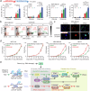
Fig. 7. CDK2/4/6 inhibition induces genomic instability.
a Percentage of cells with >4 N DNA content after 24 h treatment with either palbociclib (1 µM) or PF-06873600 (500 nM), followed by a 24 h drug holiday across 0–3 cycles. Data are shown as mean ± SD (n = 3 biological replicates). Asterisks indicate significant differences in the one-way ANOVA test (*p ≤ 0.05; **p ≤ 0.001; ***p ≤ 0.0001). b Density scatterplot showing DNA content versus EdU staining in MDA-MB-231 cells (n = 2000 cells/condition). c Representative images of Hoechst and EdU staining in MDA-MB-231 cells after 3 cycles of intermittent PF-06873600 (500 nM) treatment. The scale bar represents 20 µm. d Growth curves following 3 cycles of intermittent treatment with either DMSO or PF-06873600 (500 nM). Data are shown as mean ± SD (n = 3 biological replicates). Asterisks indicate significant differences in the two-tailed unpaired t-test (*p ≤ 0.05; **p ≤ 0.001). e The proposed model illustrating the selection between mitosis and WGD in G2-arrested cells.

References

    1. Barnum, K. J. & O’Connell, M. J. Cell cycle regulation by checkpoints. Methods Mol. Biol.1170, 29–40 (2014). - PMC - PubMed
    1. Johnson, A. & Skotheim, J. M. Start and the restriction point. Curr. Opin. Cell Biol.25, 717–723 (2013). - PMC - PubMed
    1. Pennycook B. R. & Barr A. R. Restriction point regulation at the crossroads between quiescence and cell proliferation. FEBS Lett. 594, 2046–2060 (2020). - PubMed
    1. Sherr, C. J. G1 phase progression: cycling on cue. Cell79, 551–555 (1994). - PubMed
    1. Matson, J. P. & Cook, J. G. Cell cycle proliferation decisions: the impact of single cell analyses. FEBS J.284, 362–375 (2017). - PMC - PubMed

MeSH terms

LinkOut - more resources