. 2025 Jul 21;15(1):26435.
doi: 10.1038/s41598-025-10316-3.

Ribosomal protein mutation suppresses gonadal leader cell migration defects in mig-17/ADAMTS mutants in Caenorhabditis elegans

Affiliations

Ribosomal protein mutation suppresses gonadal leader cell migration defects in mig-17/ADAMTS mutants in Caenorhabditis elegans

Hon-Song Kim et al. Sci Rep. .

Abstract

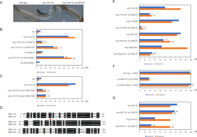
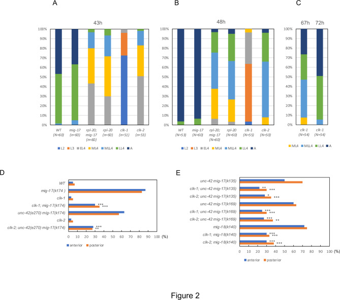
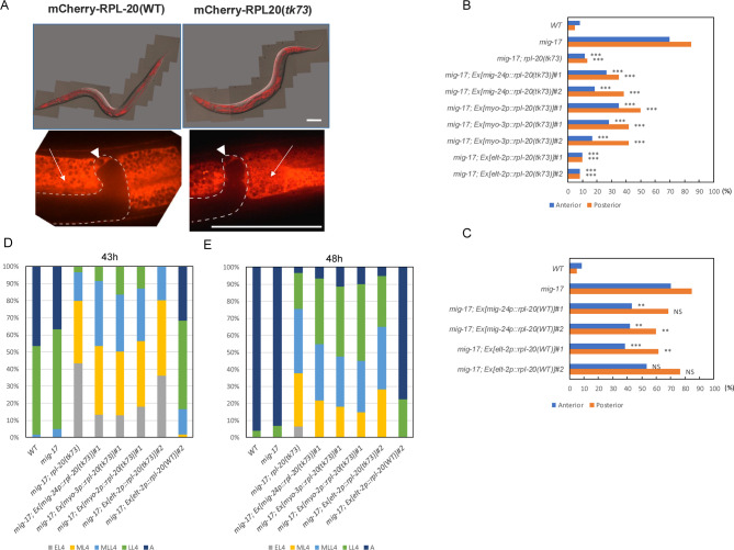
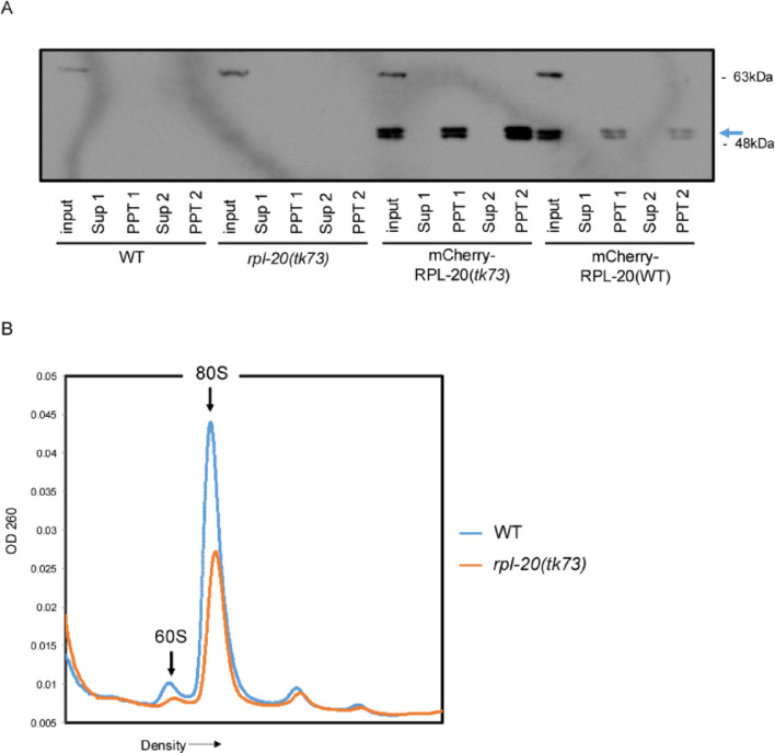
The migration of gonadal distal tip cells (DTCs) in Caenorhabditis elegans serves as an excellent model for studying the migration of epithelial tubes during organogenesis. Mutations in the mig-17/ADAMTS gene cause misdirected DTC migration during gonad formation, resulting in deformed gonad arms. An amino acid substitution in RPL-20, the ortholog of mammalian RPL18a/eL20, a component of the 60 S ribosomal large subunit, exhibited a slow-growth phenotype and strongly suppressed the mig-17 gonadal defects. Slow-growing mutations clk-1 and clk-2 also suppressed mig-17. Intestine-specific overexpression of mutant RPL-20 protein resulted in a slow-growth phenotype and suppressed the mig-17 gonadal defects, but these effects were much weaker when wild-type RPL-20 was overexpressed, suggesting that the mutant RPL-20 protein acquired a novel function. Analysis of ribosome profiles revealed reduced biogenesis of the 60 S subunit, leading to a reduction of 80 S ribosomes in the rpl-20 mutant. These results suggest that DTC migration defects in mig-17/ADAMTS mutants can be partly suppressed by growth retardation caused by the rpl-20 mutation. While defective ribosome biogenesis may contribute to the observed growth retardation, further investigation is needed to clarify the molecular basis of this phenomenon.

Keywords: ADAMTS; Growth retardation; Organogenesis; RPL18a/eL20; Ribosome protein mutation.


Conflict of interest statement

Declarations. Competing interests: The authors declare no competing interests.

Figures

Fig. 1
Fig. 1
Suppression of mig-17 and mig-18 DTC migration defects by rlp-20(tk73). (A) Gonad morphology (arrows) of wild type, mig-17(k174), and mig-17(k174); rpl-20(tk73) young-adult hermaphrodites. Posterior gonads are shown. Anterior to the left, dorsal to the top. Bar, 20 μm. (B) Quantitative analysis of mig-17 gonadal defects combined with rpl-20(tk73). N = 60 for each strain. P-values for Fisher’s exact test against mig-17(k174) for mig-17(k174) carrying strains are indicated: ***, P < 0.001; NS, not significant. (C) Transgenic rescue experiments of mig-17. mig-17 animals introduced with transgenic arrays containing PCR-amplified fragments of the rpl-20(tk73) gene were suppressed for their gonadal defects. #1 and #2 are independently established transgenic lines. N = 60 for each strain. P-values for Fisher’s exact test against mig-17(k174) are indicated: ***, P < 0.001. (D) Amino acid sequence homology between RPL-20 and human RPL18a. Identical amino acids are shown in black boxes. G82 mutated in rpl-20(tk73) is depicted with an arrow. (E-G) rpl-20(tk73) suppresses the DTC migration defects of mig-17 and mig-18 alleles (E), but not gon-1(RNAi) (F) or sqv-5(k175) and mig-22(k141) mutants (G). sqv-5(k175) and mig-22(k141) were marked with unc-13(e1091) and unc-32(e189), respectively. N = 60 for each strain. P-values for Fisher’s exact test against mig-17, mig-18, rpl-20(tk73) control RNAi, sqv-5, and mig-22 for the respective double mutants are indicated: ***, P < 0.001; **, P < 0.01; NS, not significant.
Fig. 2
Fig. 2
Growth rate analysis of mig-17, rpl-20, mig-17; rpl-20, clk-1, and clk-2 mutants. (A-C) Percentages of larval stages were determined at 43 h (A) and 48 h (B) and 67 and 72 h (C) after hatching, as described in Methods. Color codes represent larval stage 2 (L2), larval stage 3 (L3), early larval stage 4 (EL4), mid-larval stage 4 (ML4), mid-late larval stage 4 (MLL4), late larval stage 4 (LL4), and adult stage (A), respectively. (D, E) Suppression of mig-17(k174) (D) and mig-17 and mig-18 alleles (E) by clk-1 and clk-2. N = 60 for each strain. P-values for Fisher’s exact test against mig-17 and unc-42 mig-17 for the respective double mutants are indicated: ***, P < 0.001; **, P < 0.01; *, P < 0.05.
Fig. 3
Fig. 3
Tissue-specific expression of RPL-20. (A) Expression of mCherry-RPL-20(WT) and mCherry-RPL-20(tk73). Upper panels are Nomarski and confocal merged images for young adult animals and lower panels represent close-up images of the gonadal tip regions. Arrows and arrowheads indicate intestine and the distal tip cells, respectively. Bars, 50 μm. (B, C) Rescue experiments of the mig-17 gonadal defect by expressing rpl-20(tk73) (B) or rpl-20(WT) (C) genes under various tissue-specific promoters. N = 60 for each strain. P-values for Fisher’s exact test against mig-17(k174) for mig-17(k174) carrying strains are indicated: ***, P < 0.001; **, P < 0.01; NS, not significant. #1 and #2 are independently established transgenic lines. (D, E) Growth rate analysis of mig-17 animals carrying various plasmid constructs. Percentages of larval stages were determined at 43 h (D) and 48 h (E) after hatching, as described in Methods. #1 and #2 are independently established transgenic lines. Color codes represent larval stages, early L4 (EL4), mid-L4 (ML4), mid-late L4 (MLL4), late L4 (LL4), and adult (A), respectively. N ≥ 60 for each strain.
Fig. 4
Fig. 4
Analysis of ribosomes. (A) Western blot analysis of ribosomes for wild type animals expressing mCherry-RPL-20(tk73) or mCherry-RPL-20(WT). Ribosomes were precipitated by dual sucrose cushion centrifugation and the samples of supernatants and precipitates were analyzed. Sup 1 and 2 and PPT 1 and 2 represent supernatant and precipitate samples from first and second centrifugation, respectively. Arrow indicates the bands corresponding to mCherry-RPL-20. The double bands may be generated due to post-translational modification of RPL-20. Original blot is presented in Supplementary Figure S3. (B) Polysome profiles of wild type and rpl-20(tk73) animals. The amounts of 60 S subunit and 80 S ribosome in rpl-20(tk73) were reduced relative to the wild type.

Similar articles

References

    1. Apte, S. S. A disintegrin-like and metalloprotease (reprolysin-type) with thrombospondin type 1 motif (ADAMTS) superfamily: functions and mechanisms. J. Biol. Chem.284, 31493–31497. 10.1074/jbc.R109.052340 (2009). - PMC - PubMed
    1. Enomoto, H. et al. Cooperation of two ADAMTS metalloproteases in closure of the mouse palate identifies a requirement for versican proteolysis in regulating palatal mesenchyme proliferation. Development137, 4029–4038. 10.1242/dev.050591 (2010). - PMC - PubMed
    1. McCulloch, D. R. et al. ADAMTS metalloproteases generate active versican fragments that regulate interdigital web regression. Dev. Cell.17, 687–698. 10.1016/j.devcel.2009.09.008 (2009). - PMC - PubMed
    1. Russell, D. L., Doyle, K. M., Ochsner, S. A., Sandy, J. D. & Richards, J. S. Processing and localization of ADAMTS-1 and proteolytic cleavage of versican during cumulus matrix expansion and ovulation. J. Biol. Chem.278, 42330–42339. 10.1074/jbc.M300519200 (2003). - PubMed
    1. Shindo, T. et al. ADAMTS-1: a metalloproteinase-disintegrin essential for normal growth, fertility, and organ morphology and function. J. Clin. Invest.105, 1345–1352. 10.1172/JCI8635 (2000). - PMC - PubMed

MeSH terms

LinkOut - more resources