. 2025 Jul 21;15(1):26469.
doi: 10.1038/s41598-025-11600-y.

Synergistic activation of bat SARS-like coronaviruses spike protein by elastase and TMPRSS2

Affiliations

Synergistic activation of bat SARS-like coronaviruses spike protein by elastase and TMPRSS2

Yuichiro Yamamoto et al. Sci Rep. .

Abstract

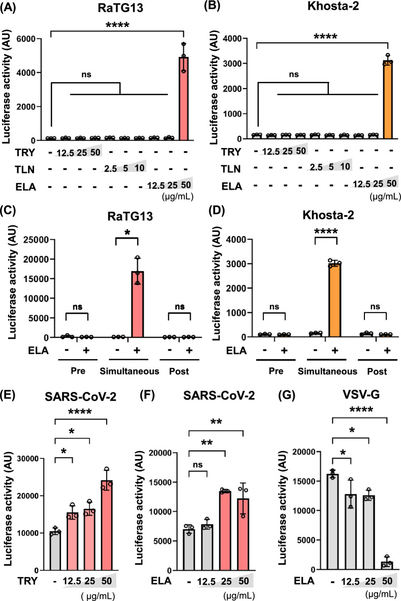
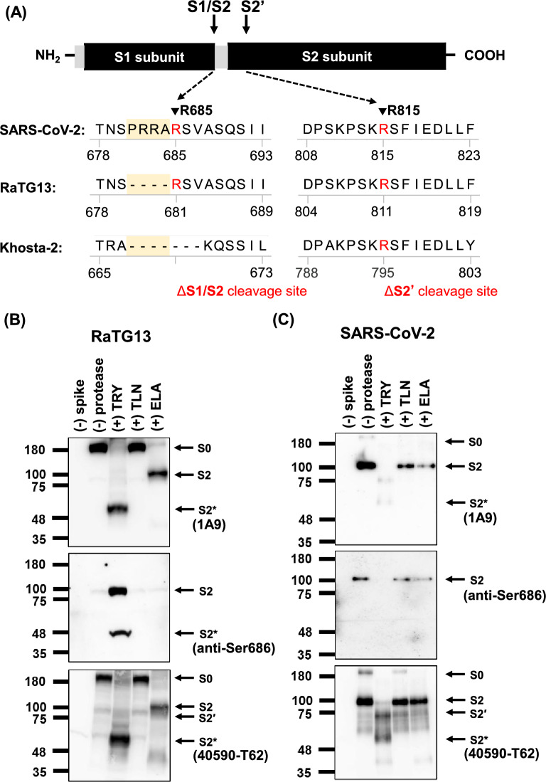
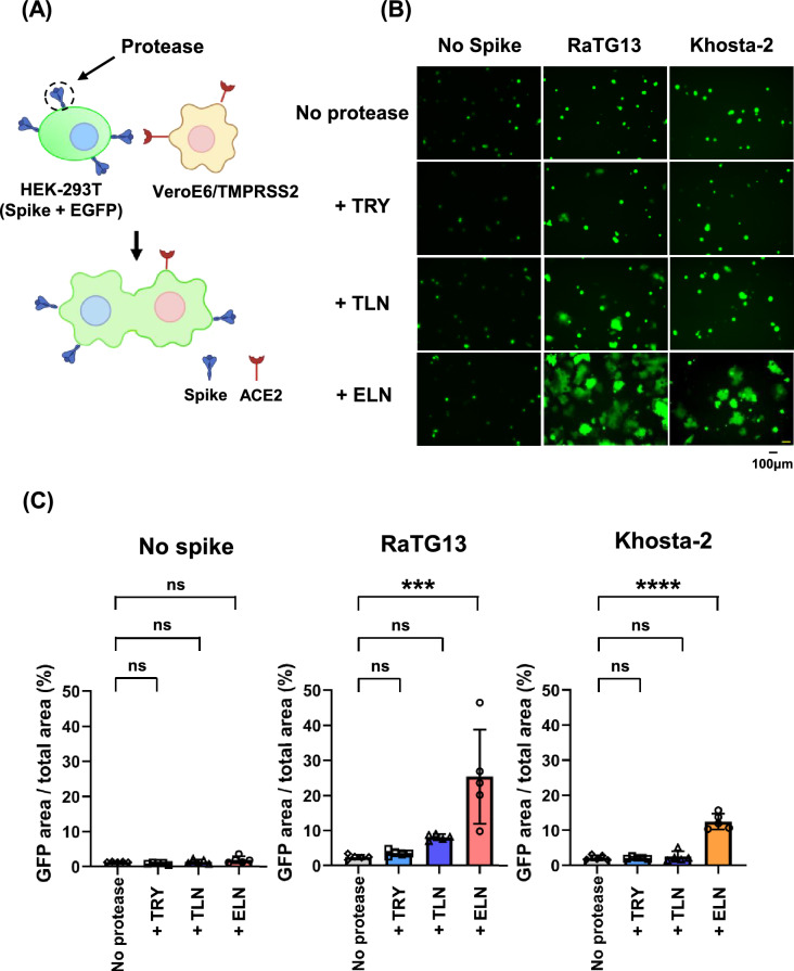
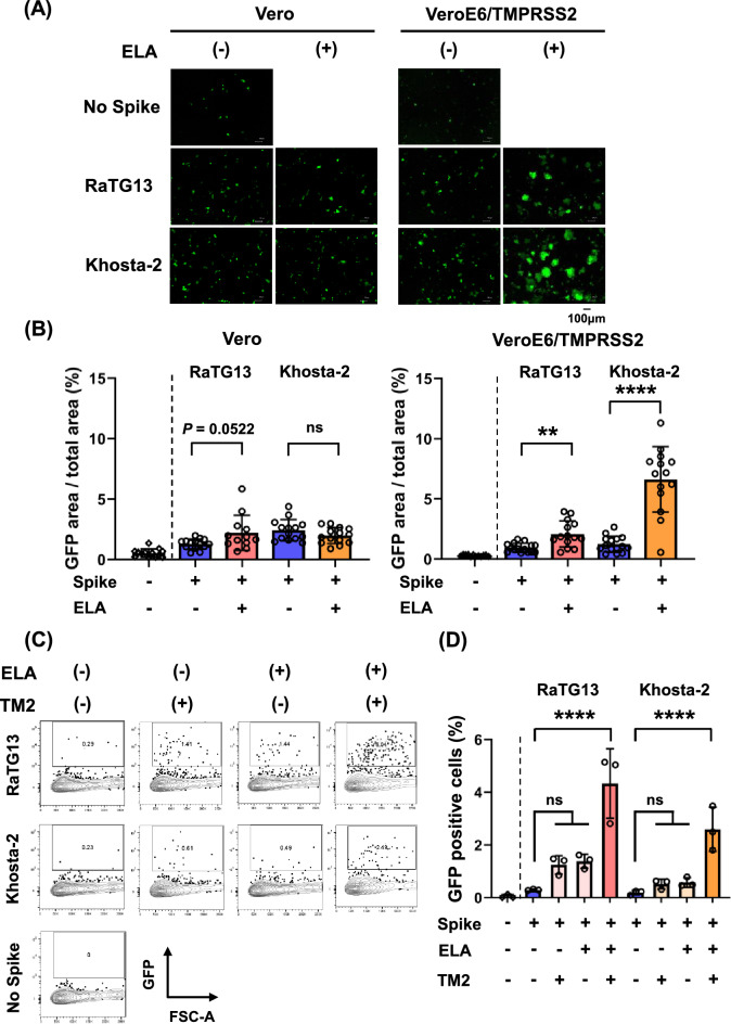
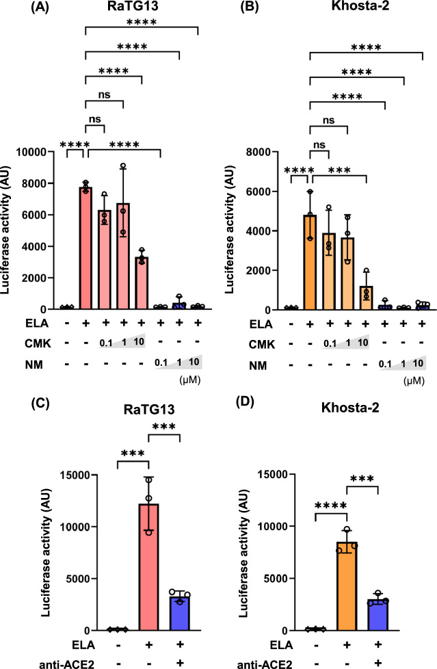
Although numerous sarbecoviruses have been identified in bats, but most lack the ability to infect human cells. Some barriers limit coronavirus zoonosis, including susceptibility to host proteases. Here, we investigated whether exogenous protease treatment can circumvent host restrictions in two severe acute respiratory syndrome (SARS)-related bat coronaviruses. We found that the spike proteins of RaTG13 and Khosta-2, which are sarbecoviruses obtained from horseshoe bats in China and Russia, respectively, facilitated the ACE2-mediated entry of pseudotyped viruses into VeroE6/TMPRSS2 cells following elastase treatment. In contrast, trypsin and thermolysin exhibited no effects. Elastase-enhanced infectivity correlated with increased fusogenicity driven by the cleavage of spike proteins. This process was TMPRSS2-dependent and was inhibited by nafamostat, a TMPRSS2 inhibitor. Additionally, mutation of residue 809 within the S2 subunit of the RaTG13 spike protein (S809D) impaired elastase-induced cleavage and infectivity. Hence, proteolytic processing of the spike protein serves as a restriction to RaTG13 and Khosta-2 infections, which can be overcome by elastase. This suggests that elastase secreted in inflamed tissues during viral infection may increase the zoonotic potential of sarbecoviruses by facilitating human cell entry.

Keywords: Bat SARS-like coronavirus; Elastase; Khosta-2; RaTG13; Spike protein.


Conflict of interest statement

Declarations. Competing interests: The authors declare no competing interests.

Figures

Fig. 1
Fig. 1
Elastase facilitates RaTG13 and Khosta-2 S protein-mediated cell entry Elastase, but not trypsin or thermolysin, facilitates cell entry of pseudotyped RaTG13 (A) and Khosta-2 (B) viruses. Particles bearing S proteins were preincubated for 5 min at room temperature with or without each protease and then added to VeroE6/TMPRSS2 cells. S protein-mediated cell entry was analyzed by measuring the activity of virus-encoded luciferase in the cell lysate at 3 days post inoculation. Experiments were independently repeated three times, and similar results were observed. Data from a representative experiment are shown (mean ± SD). Statistical analysis was performed using one-way ANOVA and subsequent Dunnett’s test. **** indicates p < 0.0001, ns: no significance. (C, D) Elastase treatment of viral particles but not target cells facilitates entry, nor does it facilitate post inoculation. VeroE6/TMPRSS2 cells or pseudotyped particles bearing RaTG13 (panel C) or Khosta-2 (panel D) S proteins were treated with elastase (final concentration: 50 µg/mL). In the “Pre” condition, cells were treated with elastase for 5 min at room temperature before virus inoculation. In the “Simultaneous” condition, pseudotyped virus particles were pretreated with elastase and immediately added to the cells. In the “Post” condition, cells were first inoculated with virus and then treated with elastase 24 h later under the same conditions. S protein-mediated cell entry was analyzed as described in panels (A, B). Experiments were independently repeated three times, and similar results were observed. Data from a representative experiment are shown (mean ± SD). Statistical analysis was performed using the Student’s t-test or Welch’s t-test. * indicates p < 0.05, **** indicates p < 0.0001, ns: no significance. Trypsin and elastase treatments facilitate cell entry of pseudotyped SARS-CoV-2, as shown in panels (E) and (F), respectively. Protease treatments were performed in the same manner as described in panels (A and B). Experiments were independently repeated twice, and similar results were observed. Data from a representative experiment are shown (mean ± SD). Statistical analysis was performed using one-way ANOVA and subsequent Dunnett’s test. * indicates p < 0.05, ** indicates p < 0.01, **** indicates p < 0.0001, ns: no significance. (G) Elastase treatment has no effect on VSV-G-mediated cell entry. Particles bearing the VSV-G were pre-incubated for 5 min with or without elastase and then added to VeroE6/TMPRSS2 cells. The experiments were independently repeated twice, and similar results were observed. Data from a representative experiment are shown (mean ± SD). Statistical analysis was performed using one-way ANOVA and subsequent Dunnett’s test. * indicates p < 0.05, **** indicates p < 0.0001, ns: no significance.
Fig. 2
Fig. 2
Elastase cleaves the RaTG13 S protein at the S2 and S2′ site. (A) Alignment of the S1/S2 loop and the adjacent to the S2′ region sequences of SARS-CoV-2, RaTG13 and Khosta-2 S proteins. (B, C) Cleavage of S proteins by three proteases. Particles pseudotyped with and RaTG13 (panel B) and SARS-CoV-2 (panel C) S protein were incubated with the highest tested concentrations of three proteases for 5 min at 37 ℃ and S protein expression analyzed by immunoblot with SARS-CoV-2 spike antibody. The experiments were independently repeated twice, and similar results were observed. The spike antibodies detected bands corresponding to the uncleaved S protein (S0), the S2 subunit (S2), and S2 subunit cleaved at the S2′ site (S2′) and additional S2 cleavage fragments (S2*). Original blots are presented in Fig. S9.
Fig. 3
Fig. 3
Elastase facilitates RaTG13 and Khosta-2 S protein-mediated cell–cell fusion (A) Schematics of the cell–cell fusion assay are presented in the panel (A). (B) Spike- and EGFP-transfected HEK293 T cells were suspended, pre-incubated for 5 min with each protease (final concentration: 100 µg/mL), and overlaid onto a monolayer of VeroE6/TMPRSS2 cells. After 3 h, the GFP-positive fused cells were photographed (panel B). Scale bar = 100 μm. (C) The green signal area of GFP-positive fused cells, as in (B), was quantified using ImageJ, and the results are shown as a bar graph. The experiments were independently repeated twice, and similar results were observed. Data from a representative experiment are shown (n = 5). Statistical analysis was performed using one-way ANOVA and subsequent Dunnett’s test. *** indicates p < 0.001, **** indicates p < 0.0001, ns: no significance.
Fig. 4
Fig. 4
Elastase does not facilitate RaTG13 and Khosta-2 S protein-mediated cell entry in Vero cells Elastase does not mediate cell entry of pseudotyped RaTG13 (A) and Khosta-2 (B) viruses in Vero cells. Pseudotyped viruses were pre-incubated for 5 min with or without trypsin or elastase and then added to Vero cells. S protein-mediated cell entry was analyzed by measuring the activity of virus-encoded luciferase in the cell lysate at 3 days post inoculation. The experiments were independently repeated three times, and similar results were observed. Data from a representative experiment are shown (mean ± SD). Statistical analysis was performed using one-way ANOVA and subsequent Dunnett’s test. ns: no significance.
Fig. 5
Fig. 5
Elastase and TMPRSS2 synergistically facilitate RaTG13 and Khosta-2 S protein-mediated syncytium formation and cell–cell fusion (A) Syncytium formation by elastase-treated RaTG13 or Khosta-2 S proteins. Each S protein was co-expressed with EGFP in Vero or VeroE6/TMPRSS2 cells. S protein-expressing cells were treated with elastase (final concentration: 100 µg/mL) for 5 min at 37℃, washed, and then observed by a fluorescent microscope 24 h later. Syncytium formation was visualized by the green signal from GFP. Large green cells indicate syncytium formation. Scale bar = 100 μm. (B) The green signal area of GFP-positive fused cells, as in A, were quantified using ImageJ, and the results are shown as a bar graph. The experiments were independently repeated three times, and similar results were observed. Data from a representative experiment are shown (n = 12–15). Statistical analysis was performed using Student’s t-test or Welch’s t-test. ** indicates p < 0.01, **** indicates p < 0.0001, ns: no significance. (C) Cell–cell fusion assay was performed using the split-GFP system. As effector cells, HEK293T cells were co-transfected with RaTG13 or Khosta-2 spike, split GFP 1–10 and DsRed. As target cells, HEK293T cells were co-transfected with ACE2, split GFP11 and DsRed. To investigate the effect of TMPRSS2, target cells were additionally transfected with TMPRSS2. After two days, effector cells were treated with elastase (final concentration: 100 µg/mL) for 30 min at 37 °C, washed and then the effector cell suspensions were mixed with target cells to generate fusion cells, and the reconstituted GFP signals were detected. After 24 h, the GFP-positive fused cells were photographed (panel C). (D) GFP-positive fused cells, as in (C), were quantified by flow cytometry. The experiments were independently repeated twice, and similar results were observed. Data from a representative experiment are shown (mean ± SD). Statistical analysis was performed using one-way ANOVA and subsequent Dunnett’s test. **** indicates p < 0.0001, ns: no significance.
Fig. 6
Fig. 6
Nafamostat and anti-ACE2 inhibits RaTG13 and Khosta-2 S protein-mediated cell entry by elastase NM efficiently inhibits RaTG13 (A) and Khosta-2 (B) S protein-mediated cell entry by elastase and TMPRSS2. Particles bearing S proteins were preincubated with elastase and CMK or NM at the indicated concentrations for 5 min at room temperature and then added to VeroE6/TMPRSS2 cells. Anti-ACE2 efficiently inhibit RaTG13 (C) and Khosta-2 (D) S protein-mediated cell entry by elastase and TMPRSS2. VeroE6/TMPRSS2 cells were preincubated with anti-ACE2 antibody for 1 h at 37 °C, and then the pseudotyped virus was preincubated with elastase (final concentration: 50 µg/mL) for 5 min at room temperature before being added to the VeroE6/TMPRSS2 cells. S protein-mediated cell entry was analyzed by measuring the activity of virus-encoded luciferase in the cell lysate at 3 days post inoculation. The experiments were independently repeated three times, and similar results were observed. Data from a representative experiment are shown (mean ± SD). Statistical analysis was performed using one-way ANOVA and subsequent Dunnett’s test. *** indicates p < 0.001, **** indicates p < 0.0001, ns: no significance.
Fig. 7
Fig. 7
Mutation of Ser809 impairs RaTG13 S protein-mediated cell entry by elastase Elastase-enhanced cell entry is impaired in the S809D-RaTG13 S protein. (A) Alignment of the adjacent to the S2′ region sequences of SARS-CoV, SARS-CoV-2 and RaTG13 S proteins. (B) HEK293T cells were transfected with plasmids encoding the wild-type (WT)-RaTG13 S protein or the S809D mutant RaTG13 S protein. For comparison, HEK293T cells were transfected with WT-SARS-CoV-2 S protein or S813D mutant SARS-CoV-2 S protein. Two days after transfection, the cells were lysed using RIPA buffer, and the lysates were analyzed the expression levels of the RaTG13 S protein by Western blotting. The experiments were independently repeated twice, and similar results were observed. Original blots are presented in Fig. S10 (upper panel). (C) Pseudotyped viruses bearing WT-RaTG13 S protein or S809D mutant RaTG13 S protein were preincubated with elastase (final concentration: 50 µg/mL) for 5 min at room temperature and then added to VeroE6/TMPRSS cells. S protein-mediated cell entry was analyzed by measuring the activity of virus-encoded luciferase in the cell lysate at 3 days post inoculation. The experiments were independently repeated three times, and similar results were observed. Data from a representative experiment are shown (mean ± SD). Statistical analysis was performed using two-way ANOVA and subsequent Tukey’s test. * indicates p < 0.05, **** indicates p < 0.0001, ns: no significance. (D) Particles pseudotyped with WT-RaTG13 S protein or S809D mutant RaTG13 S protein were incubated with elastase (final concentration: 50 µg/mL) for 5 min at 37 ℃ and S protein expression analyzed by immunoblot with SARS-CoV-2 spike antibody. The spike antibody detected bands corresponding to the uncleaved S protein (S0), the S2 subunit (S2), and S2 subunit cleaved at the S2′ and S2* site (S2′, S2*). Original blots are presented in Fig. S10 (lower panel). (E) The S2 + S2′ + S2*/S0 ratio, as in (D), was quantified using ImageJ software. The experiments were independently repeated three times, and similar results were observed. Data from three experiments are shown (mean ± SD). Statistical analysis was performed using two-way ANOVA and subsequent Tukey’s test. ** indicates p < 0.01, *** indicates p < 0.001, ns: no significance.

Similar articles

References

    1. Memish, Z. A. et al. Middle east respiratory syndrome coronavirus in bats, Saudi Arabia. Emerg. Infect. Dis.19, 1819–1823 (2013). - PMC - PubMed
    1. Andersen, K. G., Rambaut, A., Lipkin, W. I., Holmes, E. C. & Garry, R. F. The proximal origin of SARS-CoV-2. Nat. Med.26, 450–452 (2020). - PMC - PubMed
    1. Li, W. et al. Bats are natural reservoirs of SARS-like coronaviruses. Science310, 676–679 (2005). - PubMed
    1. Ruiz-Aravena, M. et al. Ecology, evolution and spillover of coronaviruses from bats. Nat. Rev. Microbiol.20, 299–314 (2022). - PMC - PubMed
    1. Liu, K. et al. Binding and molecular basis of the bat coronavirus RaTG13 virus to ACE2 in humans and other species. Cell184, 3438–3451 (2021). - PMC - PubMed

MeSH terms

LinkOut - more resources