. 2025 Jul 22;15(1):26515.
doi: 10.1038/s41598-025-12017-3.

Sex dependent effects of GPR109A gene deletion in myeloid cells on bone development in mice

Affiliations

Sex dependent effects of GPR109A gene deletion in myeloid cells on bone development in mice

Perry C Caviness et al. Sci Rep. .

Abstract

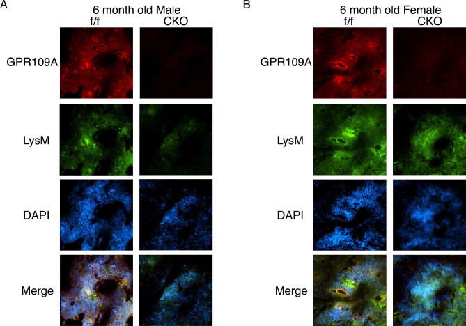
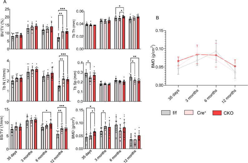
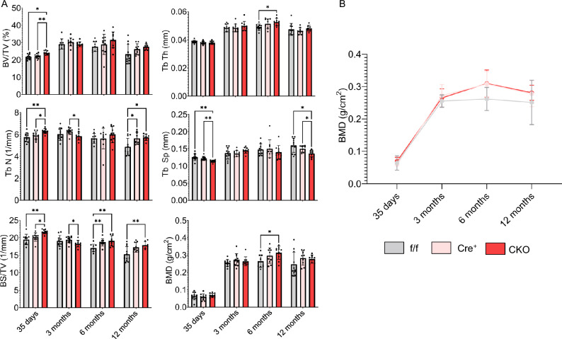
Blueberry metabolite-derived phenolic acids are thought to suppress bone resorption via interactions with the G protein-coupled receptor 109A (GPR109A). Previously, global GPR109A knockout (GPR109A-/-) mice exhibited increased bone mass and a diminished bone-protective response to phenolic acids. While GPR109A is highly expressed in osteoclast precursor macrophages, its role in bone development remains unclear. To address this, we generated a myeloid cell-specific GPR109A knockout (GPR109Aflox/flox/LysM-Cre⁺; CKO) mouse model and assessed bone phenotypes in male and female mice at 35 days, 3 months, 6 months, and 12 months using µCT. At 35 days, CKO males showed significantly increased trabecular bone in both tibia and vertebrae when compared to control genotypes (f/f, Cre⁺). However, at later time points (6 and 12 months), Cre⁺ males exhibited similar trabecular bone phenotypes compared to CKO mice. In contrast, female CKO mice displayed significantly increased trabecular bone at 6 and 12 months. Using three-point bending analysis it was found that only 12-month-old Cre⁺ and CKO male mice exhibited altered bone mechanical properties when compared to f/f mice, while for females no significant changes in bone mechanical properties were observed. These findings suggest that GPR109A regulates bone turnover pathways in a sex-specific manner.

Keywords: Bone; Osteoclast; Sexual dimorphism; µCT.


Conflict of interest statement

Competing interests: The authors declare no competing interests.

Figures

Fig. 1
Fig. 1
Targeting vector and breeding strategy for generation of myeloid cell specific GPR109A CKO (GPR109Aflox/flox/LysM-Cre+) mouse model. (A) GPR109A CDS was cloned into pcDNA3.1 vector whose cloning site is flanked by LoxP regions. Floxed GPR109A region was incorporated into mouse embryotic stem cell allele via electroporation and homologous recombination. Embryotic stem cells carrying floxed GPR109A CDS were injected into C57BL/6 embryos which were carried to term. Adult mice carrying floxed GPR109A CDS were backcrossed to develop a stable mouse line carrying a conditional GPR109A CKO allele. (B) Female GPR109Aflox/flox mice were crossed with male LysM-Cre+ mice to give offspring with either GPR109Aflox/+/LysM-Cre+ or GPR109Aflox/+/LysM-Cre- genotypes. Male GPR109Aflox/+/LysM-Cre+ and female GPR109Aflox/+/LysM-Cre- offspring were crossed to give Cre+, f/f and CKO genotypes as well as others needed for this study. (C) Genotyping was performed on mouse tails using PCR followed by agarose gel electrophoresis (1.5%). Primers were designed to detect the presence (350 bp) or absence (400 bp) of floxed regions flanking GPR109A CDS as well as presence of LysM-Cre (750 bp) or Cre (350 bp) in DNA isolated from mouse tail samples.
Fig. 2
Fig. 2
Best representative images from immunohistochemistry confirm deletion of GPR109A in tibia of both male and female CKO mice. (A, B) Immunohistochemistry was performed on tibia isolated from male and female f/f and CKO mice at 6 months of age. Loss of GPR109A protein expression (in red) was confirmed for CKO mice is present in tibia as well as the presence of LysM-Cre (in green). LysM and DAPI staining (in blue) are unaffected for CKO mice compared to f/f mice confirming myeloid cell deletion of GPR109A did not affect tibia at the cell level.
Fig. 3
Fig. 3
µCT analysis of male tibia reveals early life improvements in trabecular bone for CKO mice compared to f/f and Cre+ controls. (A) µCT parameters for male tibia (f/f, Cre+ and CKO) at different time points (35 days, 3 months, 6 months, 12 months). BMD was significantly increased at 35 days for CKO mice compared to f/f and Cre+ controls. Yet at 12 months, Cre recombinase effect is observed as BV/TV, Tb N and BS/TV are significantly increased while Tb Sp is significantly decreased for both Cre+ and CKO mice compared to f/f controls. (B) Line graph of tibia BMD over all time points shows that early life BMD increase for CKO mice compared to other genotypes is not sustained throughout life. For One-way ANOVA, *P ≤ 0.05, **P ≤ 0.01, ***P ≤ 0.001, **** P ≤ 0.0001.
Fig. 4
Fig. 4
µCT analysis of female tibia reveals later in life improvements in trabecular bone for CKO mice compared to f/f and Cre+ controls. (A) µCT parameters for female tibia (f/f, Cre + and CKO) at different time points (35 days, 3 months, 6 months, 12 months). For female mice, impact of GPR109A myeloid cell deletion is primarily observed at 12 months as evident by significant increases in BV, BV/TV, Tb N, BS/TV and BMD for CKO mice compared to both f/f and Cre+ mice. For CKO mice, BMD is also significantly increased at 6 months when compared to both f/f and Cre+ controls. (B) Line graph of female mice tibia BMD over all time points showing peak BMD occurs at 6 months for CKO mice. For One-way ANOVA, *P ≤ 0.05, **P ≤ 0.01, ***P ≤ 0.001, **** P ≤ 0.0001.
Fig. 5
Fig. 5
µCT analysis of male mice L5 vertebrae reveal early life improvements in CKO trabecular bone compared to f/f and Cre+ controls. (A) µCT parameters for male L5 vertebrae (f/f, Cre+ and CKO) at different time points (35 days, 3 months, 6 months, 12 months). BV/TV, Tb N and BS/TV are significantly increased while Tb Sp is significantly decreased for 35-day-old CKO mice compared to f/f and Cre+ controls. As with tibia, a Cre recombinase effect was observed at latter time points as BS/TV is significantly increased for 6-month-old Cre+ and CKO mice compared to f/f and Tb N is significantly increased for Cre+ and CKO mice compared to f/f at 12 months. In addition, at both 6 and 12 months, BMD for Cre+ and CKO are similar, and both are increased compared to f/f mice but not to a significant extent. (B) Line graph of male L5 vertebrae BMD over all time points shows no change between genotypes at early time points but similar increases for both Cre+ and CKO mice compared to f/f at 6 and 12 months. For One-way ANOVA, *P ≤ 0.05, **P ≤ 0.01, ***P ≤ 0.001, **** P ≤ 0.0001.
Fig. 6
Fig. 6
µCT analysis of female L5 vertebrae reveal later in life improvements in CKO trabecular bone compared to both f/f and Cre+ controls. (A) µCT parameters for L5 vertebrae from female mice (f/f, Cre+ and CKO) at different time points (35 days, 3 months, 6 months, 12 months). Impact of GPR109A myeloid cell deletion is primarily observed at later time points as observed by significant increases in BV, BV/TV and Tb N as well as a significant decrease in Tb Sp at both 6 and 12 months for CKO mice compared to both f/f and Cre+ controls. In addition, BS/TV was significantly increased for CKO mice at 12 months when compared to Cre+ but not f/f mice. Finally, at 6 months BMD for CKO mice was found to be significantly increased compared to f/f and Cre+ controls. (B) Line graph of female L5 vertebrae BMD over all time points showing peak BMD occurs at 6 months for CKO mice. For One-way ANOVA, *P ≤ 0.05, **P ≤ 0.01, ***P ≤ 0.001, **** P ≤ 0.0001.
Fig. 7
Fig. 7
Cre recombinase male specific effect is also observed in 3-point bending analysis results. (A) For males, 3-point bending analysis of femur from 12-month old mice reveals significant increases in Yield Stress, Ultimate Stress and Elastic Modulus for both Cre+ and CKO mice compared to f/f mice. (B) For females, 3-point bending analysis of 12-month old femur shows no significant changes in Yield Stress, Ultimate Stress and Elastic Modulus between f/f, Cre+ or CKO mice. For One-way ANOVA, *P ≤ 0.05, **P ≤ 0.01, ***P ≤ 0.001, **** P ≤ 0.0001.

Update of

Similar articles

References

    1. Chen, J.-R. et al. GPR109A mediates the effects of hippuric acid on regulating osteoclastogenesis and bone resorption in mice. Commun. Biol.10.1038/s42003-020-01564-2 (2021). - PMC - PubMed
    1. Singh, N. et al. Activation of Gpr109a, receptor for niacin and the commensal metabolite butyrate, suppresses colonic inflammation and carcinogenesis. Immunity40, 128–139. 10.1016/j.immuni.2013.12.007 (2014). - PMC - PubMed
    1. Ren, N. et al. Phenolic acids suppress adipocyte lipolysis via activation of the nicotinic acid receptor GPR109A (HM74a/PUMA-G). J. Lipid Res.50, 908–914. 10.1194/jlr.M800625-JLR200 (2009). - PMC - PubMed
    1. Tunaru, S. et al. PUMA-G and HM74 are receptors for nicotinic acid and mediate its anti-lipolytic effect. Nat. Med.9, 352–355. 10.1038/nm824 (2003). - PubMed
    1. Miyamoto, J. et al. Ketone body receptor GPR43 regulates lipid metabolism under ketogenic conditions. Proc. Natl. Acad. Sci. USA116, 23813–23821. 10.1073/pnas.1912573116 (2019). - PMC - PubMed

Substances

LinkOut - more resources