

IL-37 ameliorates acetaminophen-induced acute liver injury by limiting MAPK/NFκB signaling-mediated liver inflammation
- PMID: 40691258
- PMCID: PMC12280191
- DOI: 10.1038/s41598-025-10764-x
IL-37 ameliorates acetaminophen-induced acute liver injury by limiting MAPK/NFκB signaling-mediated liver inflammation
Abstract
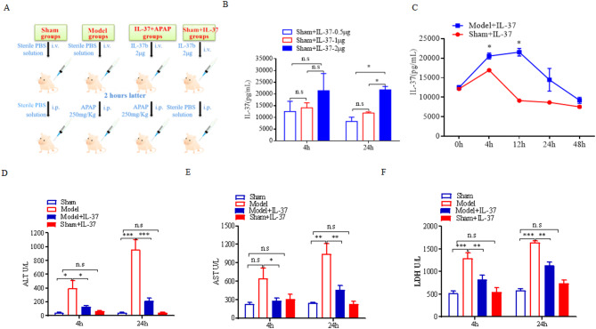
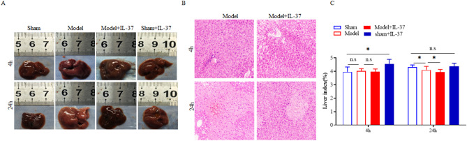
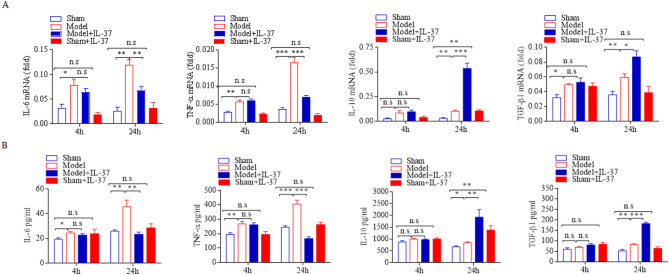
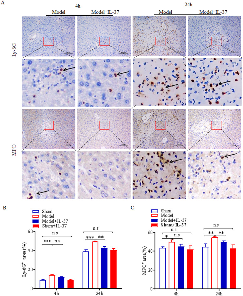
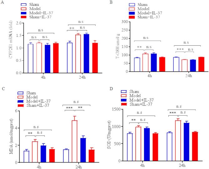
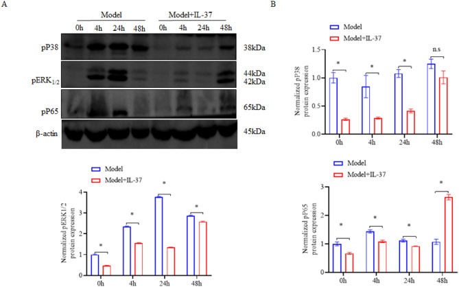
Acetaminophen (APAP) overdose is a common cause of drug-induced liver injury (DILI), which can lead to sterile inflammation and progress to acute liver failure and even death. However, there are currently limited therapeutic options available. Iinterleukin-37 (IL-37) is considered as an anti-inflammatory cytokine. The role and novel mechanism of IL-37 on DILI are still unknown. Male C57BL/6 mice were pretreated with IL-37 for 2 h prior to intraperitoneal injection of acetaminophen (APAP). Hepatic function was assessed by measuring serum levels of alanine aminotransferase (ALT), aspartate aminotransferase (AST), and lactate dehydrogenase (LDH). Liver tissue damage was evaluated via hematoxylin and eosin (H&E) staining. The inflammatory response was characterized by immunohistochemical (IHC) analysis of myeloperoxidase (MPO) and lymphocyte antigen 6 complex locus G (Ly6G), and the levels of interleukin-6 (IL-6), IL-10, transforming growth factor-beta 1 (TGF-β1), and tumor necrosis factor-alpha (TNF-α) in liver tissue. Oxidative stress status was determined by measuring superoxide dismutase (SOD), malondialdehyde (MDA), and glutathione (GSH) levels. CYP2E1 mRNA expression was analyzed using qPCR. Protein expression of phosphorylated p38 (pP38), phosphorylated extracellular signal-regulated kinase 1/2 (pERK1/2), and phosphorylated p65 (pP65) was evaluated by Western blotting. Compared to the model group without recombinant human IL-37 treatment, the model + IL-37 group exhibited significantly attenuated liver injury, characterized by reduced neutrophil infiltration, decreased levels of pro-inflammatory mediators IL-6 and TNF-α, and elevated levels of the anti-inflammatory cytokines IL-10 and TGF-β1. The levels of pp38, pERK1/2, and pp65 in liver tissue were significantly suppressed in the Model + IL-37 group compared to the Model group at 24 h. Furthermore, MDA levels were significantly lower in the IL-37-treated group relative to the model group, while SOD activity showed no significant difference. Our results also indicate that neither CYP2E1 mRNA relative expression nor GSH levels differed significantly between the model group and the IL-37-treated group at either 4 h or 24 h after APAP exposure. IL-37 has a significant protective effect against acetaminophen-induced liver injury (AILI) by suppressing the inflammatory response involved in the MAPK-NF-κB/p65 signalling pathway. Our study suggests IL-37 as a potential therapeutic strategy for DILI.
Keywords: APAP; IL-37; Liver injury; Oxidative stress; Sterile inflammation.
© 2025. The Author(s).
Conflict of interest statement
Declarations. Competing interests: The authors declare no competing interests.
Figures
Similar articles
-
[Vitexin-4 ″-O-glucoside alleviates acetaminophen-induced acute liver injury].Zhejiang Da Xue Xue Bao Yi Xue Ban. 2025 May 25;54(3):307-317. doi: 10.3724/zdxbyxb-2024-0381. Zhejiang Da Xue Xue Bao Yi Xue Ban. 2025. PMID: 40461289 Free PMC article. Chinese.
-
[Mechanism of total saponins of Panax japonicus against liver injury induced by acetaminophen in mice based on ERK/NF-κB/COX-2 signaling pathway].Zhongguo Zhong Yao Za Zhi. 2024 May;49(10):2585-2596. doi: 10.19540/j.cnki.cjcmm.20240205.702. Zhongguo Zhong Yao Za Zhi. 2024. PMID: 38812159 Chinese.
-
Lagerstroemia Speciosa (L.) Pers mitigates acetaminophen-induced acute liver toxicity in rats through modulations of oxidative stress, inflammation, apoptosis and the NF-κB/TNF-α/iNOS, Nrf2/HO-1, signaling pathways.J Mol Histol. 2025 Jul 23;56(4):238. doi: 10.1007/s10735-025-10493-5. J Mol Histol. 2025. PMID: 40699454
-
Risk prediction of hepatotoxicity in paracetamol poisoning.Clin Toxicol (Phila). 2017 Sep;55(8):879-892. doi: 10.1080/15563650.2017.1317349. Epub 2017 Apr 27. Clin Toxicol (Phila). 2017. PMID: 28447858
-
The potential of curcumin in mitigating acetaminophen-induced liver damage.Naunyn Schmiedebergs Arch Pharmacol. 2025 Jul;398(7):8169-8188. doi: 10.1007/s00210-025-03907-4. Epub 2025 Feb 26. Naunyn Schmiedebergs Arch Pharmacol. 2025. PMID: 40009170 Review.
References
-
- Zhongping, D., PengFei, Y., Qiao, W. & Yu, C. Research advances in the mechanism of drug-induced liver injury due to paracetamol. J. Clin. Hepatobiliary Dis.35(9), 2108–2112 (2019).
-
- Larson, A. et al. Acetaminophen-induced acute liver failure: Results of a United States multicenter, prospective study. Hepatology (Baltimore, MD)42(6), 1364–1372. 10.1002/hep.20948 (2005). - PubMed
-
- Lee, W. Acetaminophen-related acute liver failure in the United States. Hepatol. Res. Off. J. Jpn. Soc. Hepatol.38, S3-8. 10.1111/j.1872-034X.2008.00419.x (2008). - PubMed
-
- Kon, K. et al. Mitochondrial permeability transition in acetaminophen-induced necrosis and apoptosis of cultured mouse hepatocytes. Hepatology40(5), 1170–1179. 10.1002/hep.20437 (2004). - PubMed
MeSH terms
Substances
Grants and funding
LinkOut - more resources
Full Text Sources
Medical
Research Materials
Miscellaneous
