. 2025 Jul 21;22(1):170.
doi: 10.1186/s12984-025-01700-1.

Transcutaneous auricular vagus nerve stimulation enhances structural and functional remodeling in sensorimotor networks following intracerebral hemorrhage in rats

Affiliations

Transcutaneous auricular vagus nerve stimulation enhances structural and functional remodeling in sensorimotor networks following intracerebral hemorrhage in rats

Jun Zhang et al. J Neuroeng Rehabil. .

Abstract

Background: Intracerebral hemorrhage (ICH) leads to severe neurological deficits by disrupting brain structure and function, particularly in the sensorimotor cortex. Effective interventions to promote post-ICH brain remodeling remain limited. This study investigated the effects of transcutaneous auricular vagus nerve stimulation (taVNS) on structural and functional remodeling in the sensorimotor networks of rats with ICH, using multi-scale analyses spanning micro-, meso-, and macro-levels.

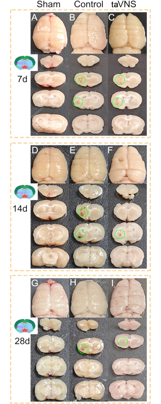
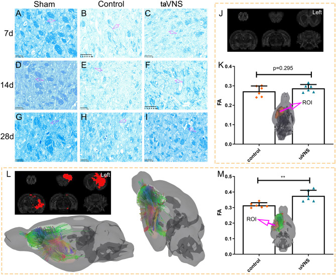
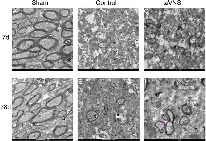
Methods: A rat model of left basal ganglia ICH was established, followed by taVNS intervention. Structural remodeling was assessed through histology, immunofluorescence, and transmission electron microscopy. Functional remodeling was evaluated using fractional amplitude of low-frequency fluctuations (fALFF) and degree centrality (DC) analyses.

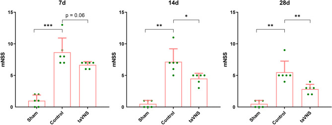
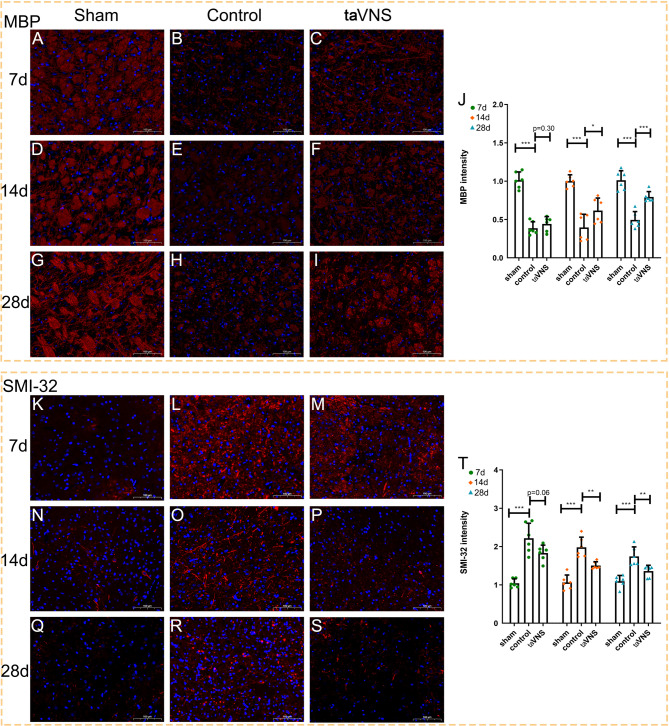
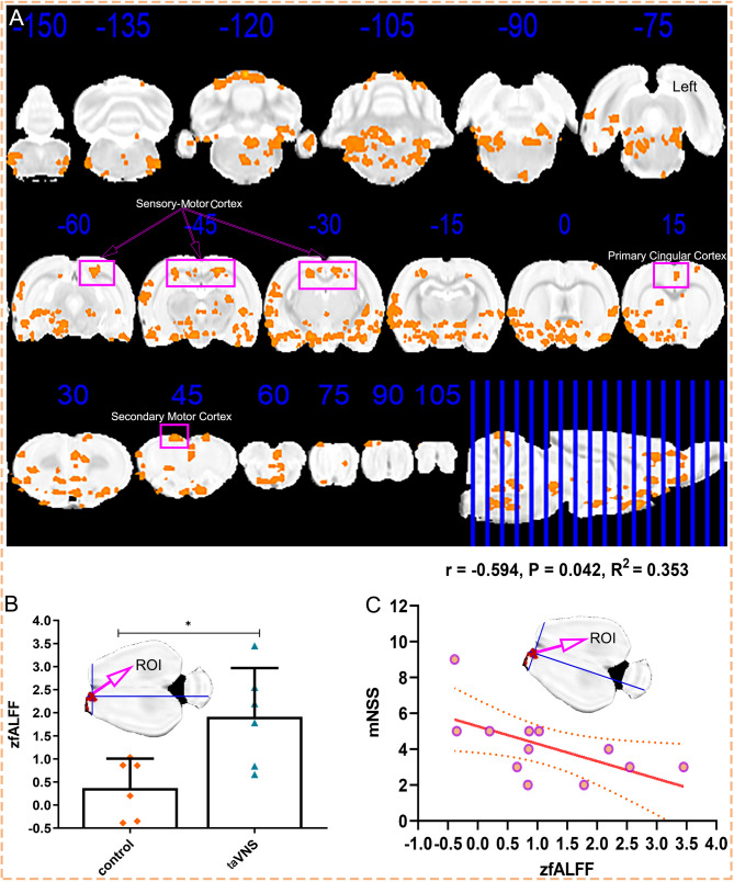
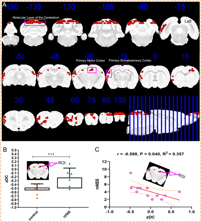
Results: taVNS enhanced myelin repair and axonal remodeling, indicated by increased myelin basic protein (MBP) expression, reduced dephosphorylated neurofilament protein (SMI-32), and partial restoration of synaptic ultrastructure. Functional imaging showed significant longitudinal increases in zfALFF and zDC values in sensorimotor regions, including the primary and secondary motor cortices, which negatively correlated with modified neurological severity scores (mNSS).

Conclusion: taVNS promotes structural and functional remodeling in the sensorimotor cortex after ICH, offering a potential therapeutic strategy for neurological recovery.

Keywords: Brain remodeling; Intracerebral hemorrhage; Neuroplasticity; Sensorimotor networks; TaVNS.


Conflict of interest statement

Declarations. Ethical approval: This study was approved by the Ethics Committee of the Laboratory Animal Science Department, Fudan University (202310008 S). Competing interests: The authors declare no competing interests.

Figures

Fig. 1
Fig. 1
Comparison of mNSS scores across sham, control, and taVNS groups at weeks 1, 2, and 4. mNSS were assessed in the sham, control, and taVNS groups at 1, 2, and 4 weeks post-ICH modeling. At week 1, mNSS scores were significantly elevated in the control and taVNS groups compared to the sham group, with no significant difference between the control and taVNS groups (p > 0.05). By week 2, mNSS scores in the taVNS group were significantly reduced compared to the control group (p < 0.05). At week 4, the taVNS group demonstrated further mNSS reductions compared to the control group (p < 0.01). Statistical significance: *p < 0.05, **p < 0.01, ***p < 0.001. mNSS, modified neurological severity score; taVNS, transcutaneous auricular vagus nerve stimulation
Fig. 2
Fig. 2
Comparative gross anatomy of brain tissue at 1, 2, and 4 weeks post-ICH. Macroscopic brain sections from the sham, control, and taVNS groups were collected at 1, 2, and 4 weeks post-ICH modeling. (A-C) Brain sections from week 1 reveal marked structural damage in the left basal ganglia in the control and taVNS groups compared to the sham group. (D-F) At week 2, residual hematoma was less pronounced in the taVNS group compared to the control group. (G-I) By week 4, residual hematoma and structural damage in both the control and taVNS groups were nearly resolved. ROI, region of interest; taVNS, transcutaneous auricular vagus nerve stimulation
Fig. 3
Fig. 3
Myelin staining and CST reconstruction highlight taVNS-induced recovery. (A-I) LFB staining reveals myelin density in the left basal ganglia at 1, 2, and 4 weeks post-ICH in the sham, control, and taVNS groups. At week 1, myelin density is significantly disrupted in the control and taVNS groups compared to the sham group (A-C). By week 2, the taVNS group shows increased myelin density relative to the control group (D-F). At week 4, the taVNS group demonstrates near-restoration of myelin density compared to the sham group (G-I). (J) CST reconstruction at week 1 shows no significant differences between the control and taVNS groups. (K) FA values of the left basal ganglia at week 1 also show no significant differences between groups. (L) At week 4, CST reconstruction in the taVNS group shows significant restoration compared to the control group. (M) FA values of the left basal ganglia at week 4 are significantly higher in the taVNS group than in the control group (**p < 0.01). taVNS, transcutaneous auricular vagus nerve stimulation; ROI, region of interest; FA, fractional anisotropy. Scale bar = 50 μm
Fig. 4
Fig. 4
Immunofluorescence analysis of myelin and axonal remodeling proteins post-taVNS. (A-I) Immunofluorescence staining of MBP in the left basal ganglia at 1, 2, and 4 weeks post-ICH reveals reduced MBP expression in the control and taVNS groups compared to the sham group at week 1 (A-C). MBP expression significantly increased in the taVNS group at weeks 2 and 4 compared to the control group (D-I, J). (K-S) SMI-32 staining for dephosphorylated neurofilament protein shows decreased expression in the control and taVNS groups compared to the sham group at week 1 (K-M). The taVNS group displayed a significant reduction in SMI-32 expression at weeks 2 and 4 compared to the control group (N-S, T). Statistical significance: *p < 0.05, **p < 0.01, ***p < 0.001. MBP, myelin basic protein; SMI-32, dephosphorylated neurofilament protein; taVNS, transcutaneous auricular vagus nerve stimulation; scale bar = 100 μm
Fig. 5
Fig. 5
Ultrastructural evaluation of basal ganglia recovery post-taVNS. (A-C) TEM images of the left basal ganglia at week 1 show extensive axonal damage and near-complete loss of synaptic connections in both the control and taVNS groups compared to the sham group. (D-F) At week 4, TEM images demonstrate partial recovery of myelin, axons, and synaptic structures, with notable improvement in the taVNS group compared to the control group. taVNS, transcutaneous auricular vagus nerve stimulation; Scale bar = 1 μm
Fig. 6
Fig. 6
Longitudinal remodeling of sensorimotor cortex spontaneous activity and effectiveness of taVNS modulation. (A) Mixed-effects analysis reveals significant longitudinal remodeling of spontaneous brain activity in the sensorimotor cortex, with enhanced zfALFF in the taVNS group compared to the control group. (B) At week 4, zfALFF values in the right secondary motor cortex were significantly higher in the taVNS group compared to the control group, confirming the efficacy of taVNS modulation. (C) A significant negative correlation was observed between zfALFF values in the right secondary motor cortex and mNSS scores (r = -0.594, P = 0.042). zfALFF, z-transformed fractional amplitude of low-frequency fluctuations; ROI, region of interest; taVNS, transcutaneous auricular vagus nerve stimulation
Fig. 7
Fig. 7
Longitudinal remodeling of sensorimotor core nodes and taVNS modulation effectiveness. (A) Mixed-effects analysis reveals significant longitudinal remodeling of core nodes (zDC) in the sensorimotor cortex, with greater increases in zDC values in the taVNS group compared to the control group. (B) At week 4, zDC values in the left primary motor cortex were significantly higher in the taVNS group compared to the control group, highlighting the effectiveness of taVNS modulation. (C) zDC values in the left primary motor cortex were negatively correlated with mNSS scores (r = -0.598, p = 0.040). zDC, z-transformed degree centrality; ROI, region of interest; taVNS, transcutaneous auricular vagus nerve stimulation

Similar articles

References

    1. Kittner SJ, Sekar P, Comeau ME, Anderson CD, Parikh GY, Tavarez T, et al. Ethnic and racial variation in intracerebral hemorrhage risk factors and risk factor burden. JAMA Netw Open. 2021;4:e2121921. - PMC - PubMed
    1. Morotti A, Brouwers HB, Romero JM, Jessel MJ, Vashkevich A, Schwab K, et al. Intensive blood pressure reduction and spot sign in intracerebral hemorrhage: a secondary analysis of a randomized clinical trial. JAMA Neurol. 2017;74:950–60. - PMC - PubMed
    1. Liu F, Chen C, Hong W, Bai Z, Wang S, Lu H, et al. Selectively disrupted sensorimotor circuits in chronic stroke with hand dysfunction. CNS Neurosci Ther. 2022;28:677–89. - PMC - PubMed
    1. Jiang L, Xu H, Yu C. Brain connectivity plasticity in the motor network after ischemic stroke. Neural Plast. 2013;2013:924192. - PMC - PubMed
    1. Griffis JC, Metcalf NV, Corbetta M, Shulman GL. Structural disconnections explain brain network dysfunction after stroke. Cell Rep. 2019;28:2527–e25402529. - PMC - PubMed

MeSH terms

LinkOut - more resources