

The recurrence and multi-organ involvement of giant penile plexiform neurofibroma in an adult with neurofibromatosis type 1: a case report
- PMID: 40691787
- PMCID: PMC12278514
- DOI: 10.1186/s12894-025-01881-w
The recurrence and multi-organ involvement of giant penile plexiform neurofibroma in an adult with neurofibromatosis type 1: a case report
Abstract
Background: Neurofibromatosis type 1 (NF1) is an autosomal dominant genetic disorder with malignant potential, affecting various anatomical regions, predominantly the head and neck. Urogenital involvement is relatively uncommon. In particular, penile neurofibromatosis is exceedingly rare, with fewer than 20 cases documented globally. In China, only four pediatric cases have been reported, with no known occurrences in adults. To date, no recurrent penile plexiform neurofibromas have been reported in adults. Here, we report a rare case of a recurrent giant penile plexiform neurofibroma, accompanied by multiple neurofibromas involving the prostate, bladder, and retroperitoneum, in an adult NF1 patient.
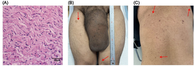
Case report: A 37-year-old male patient initially presented to the Department of Urology, Shandong First Medical University Affiliated Provincial Hospital, on April 17, 1994 (at the age of 8), with a seven-year history of painless penile enlargement. He underwent resection of the penile mass, with histopathological analysis confirming a diagnosis of plexiform neurofibroma. Following surgery, the penile morphology normalized. However, at the age of 20, he experienced recurrent painless penile enlargement but did not pursue further medical evaluation. On October 7, 2023, the patient was readmitted due to right flank pain and hydronephrosis. Physical examination revealed marked penile enlargement with a palpable large mass and multiple café-au-lait macules across the body. Although the patient had no urinary complaints, he was unable to achieve normal erectile function. Laboratory findings indicated an elevated serum creatinine level of 124.20 µmol/L. Imaging modalities, including computed tomography urography (CTU), magnetic resonance imaging (MRI), and positron emission tomography/computed tomography (PET/CT), identified a massive penile tumor with concomitant multiple tumors in the prostate, bladder, and retroperitoneum. Penile tumor biopsy confirmed the diagnosis of plexiform neurofibroma. Immunohistochemical analysis demonstrated S-100 (+), recombinant human SOX10 protein (SOX10) (+), and Ki-67+ (< 1%). Given the histopathological results and clinical findings, the patient was diagnosed with neurofibromatosis type 1. To alleviate right flank pain, percutaneous nephrostomy was performed, resulting in a reduction in serum creatinine to 90.80 µmol/L within three days post-procedure, with subsequent symptom relief. Owing to extensive tumor infiltration and financial limitations, the patient opted against radical surgery or systemic therapy. He was subsequently discharged and has since been monitored with biannual follow-ups, including regular nephrostomy tube replacement. As of the latest follow-up, his clinical status remains stable without evidence of significant disease progression.
Conclusion: This case report describes a rare adult patient with neurofibromatosis type 1, presenting with recurrent giant penile plexiform neurofibroma accompanied by multiple neurofibromas involving the prostate, bladder, and retroperitoneum. To the best of our knowledge, this combination of manifestations represents the first reported case. The patient underwent only percutaneous nephrostomy to alleviate symptoms, without pursuing additional treatment. Follow-up evaluations indicated a stable clinical course, suggesting that certain NF1 patients may achieve long-term stability without aggressive therapeutic intervention. This case underscores the importance of comprehensive evaluation, prolonged surveillance, and multidisciplinary management for NF1 patients, particularly those with extensive and atypical disease manifestations. Treatment decisions should consider a balance between patient preferences and the natural progression of the disease. This report offers valuable insights into the management of similar cases.
Keywords: Bladder; Metastasis; Neurofibroma; Neurofibromatosis Type 1; Penis; Plexiform Neurofibroma; Prostate; Recurrence; Retroperitoneum.
© 2025. The Author(s).
Conflict of interest statement
Declarations. Ethics approval and consent to participate: Written informed consent to participate was obtained from the patient of this case report. The studies involving humans were approved by Biomedical Research Ethic Committee of Shandong Provincial Hospital Affiliated to Shandong First Medical University. The studies were conducted in accordance with the local legislation and institutional requirements. Consent for publication: Written informed consent was obtained from the patient for publication of this case report and any accompanying images. Competing interests: The authors declare no competing interests.
Figures


References
-
- Andres-Sanchez N, Fisher D, Krasinska L. Physiological functions and roles in cancer of the proliferation marker Ki-67. J Cell Sci. 2022;135(11). 10.1242/jcs.260184. - PubMed
-
- Issa B, Mansour E, Jabbour G, Chikhani C, Mansour H, Jabbour M. Plexiform penile neurofibroma: A case report of a rare entity in a Pre-Pubertal child. Urology. 2021;156:e124–6. - PubMed
-
- Wu F, Ji XN, Chen Q. [Research progress on pathogenesis and treatment of neurofibromatosis type 1]. Zhonghua Er Ke Za Zhi. 2023;61(8):757–60. - PubMed
Publication types
MeSH terms
Grants and funding
- ZLKY-HS24007-4/Health Development Promotion Project, China
- ZLKY-HS24007-4/Health Development Promotion Project, China
- ZLKY-HS24007-4/Health Development Promotion Project, China
- ZLKY-HS24007-4/Health Development Promotion Project, China
- ZLKY-HS24007-4/Health Development Promotion Project, China
- ZLKY-HS24007-4/Health Development Promotion Project, China
- ZR2021MH251/Shandong Provincial Natural Science Foundation, China
- ZR2021MH251/Shandong Provincial Natural Science Foundation, China
- ZR2021MH251/Shandong Provincial Natural Science Foundation, China
- ZR2021MH251/Shandong Provincial Natural Science Foundation, China
- ZR2021MH251/Shandong Provincial Natural Science Foundation, China
- ZR2021MH251/Shandong Provincial Natural Science Foundation, China
LinkOut - more resources
Full Text Sources
Research Materials
Miscellaneous
