. 2025 Jul 21;16(1):390.
doi: 10.1186/s13287-025-04502-3.

Programmed cell death regulates hematopoietic cell homeostasis under radiation conditions

Affiliations

Programmed cell death regulates hematopoietic cell homeostasis under radiation conditions

Manling Shu et al. Stem Cell Res Ther. .

Abstract

Background: It is well-known that hematopoietic cells are sensitive to irradiation exposure. Apoptosis, necroptosis, pyroptosis and ferroptosis might contribute to irradiation-induced hematopoietic injury. However, it is uncertain whether different hematopoietic cells apply specific cell death pathways under irradiation exposure.

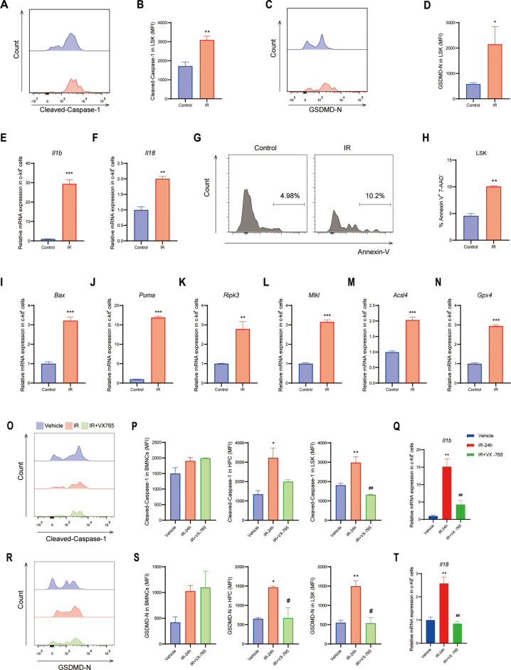
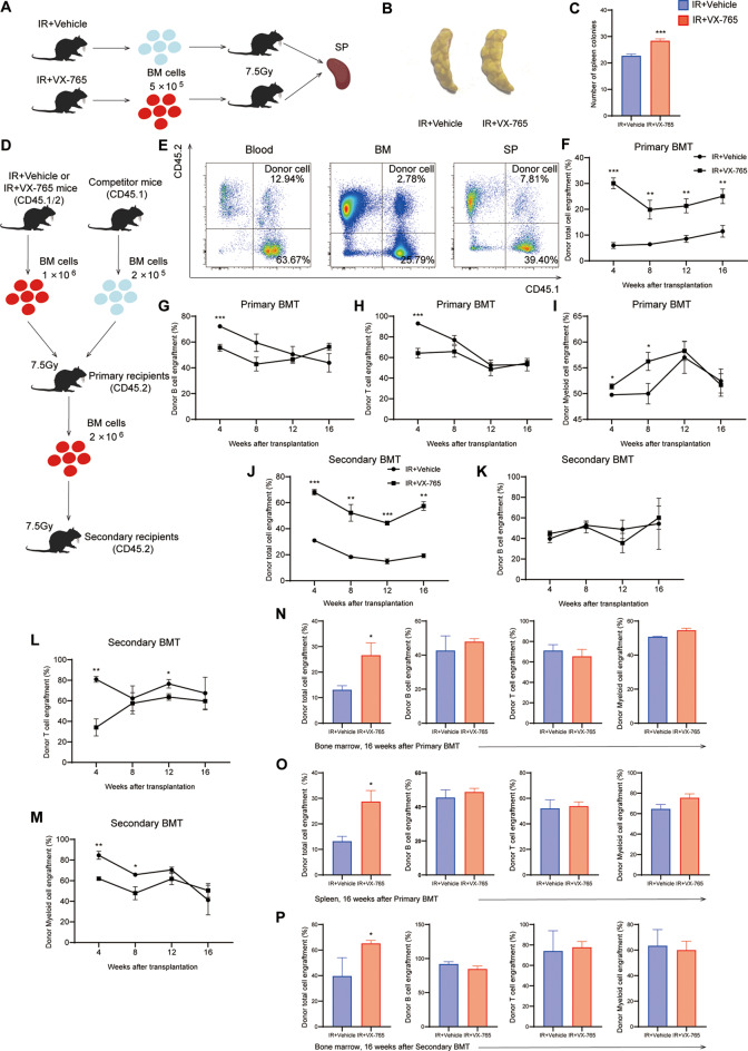
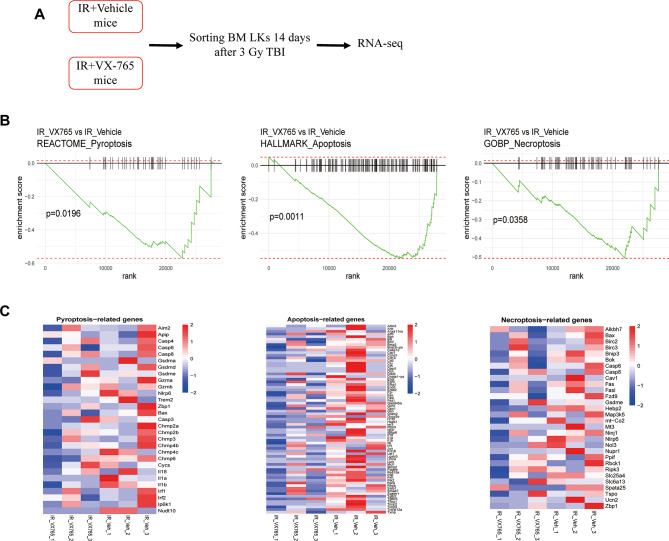
Methods: We investigated the role of different programmed cell death pathways in irradiation-induced hematopoietic cell injury. In order to study the acute and long-term effects of ionizing radiation on hematopoietic system, we established injury models of mice at different time points after irradiation and measured the proportion of hematopoietic stem progenitor cells by flow cytometry. The pattern of programmed cell death involved in radiation-induced hematopoietic cell injury was identified through the analysis of different populations of hematopoietic cells in the bone marrow by immunomagnetic bead sorting combined with qRT-PCR and flow cytometry. The role of pyroptosis in radiation injury of hematopoietic stem cells was further studied by Caspase-1 inhibitor VX-765 application. In vivo spleen colony formation, competitive bone marrow transplantation and secondary transplantation were used to verify the protective effect of inhibiting Caspase-1 on hematopoietic stem cells damaged by radiation. RNA sequencing (RNA-Seq) using Lin-c-Kit+ cell populations revealed the mechanism by which inhibition of Caspase-1 mitigates post-irradiation hematopoietic stem cell damage.

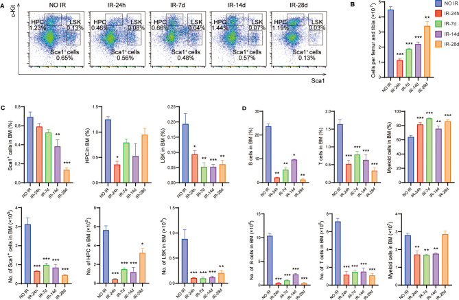
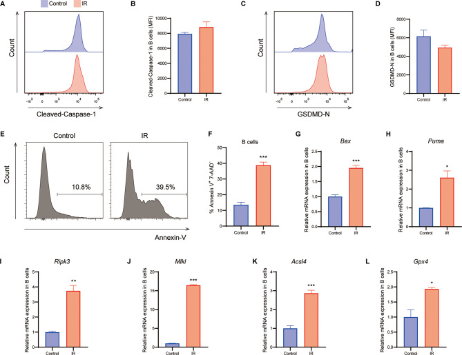
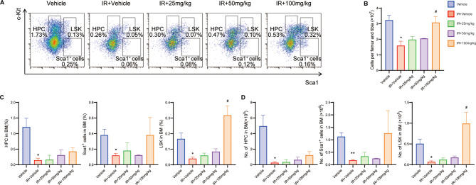
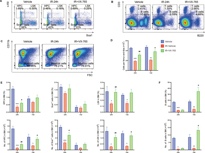
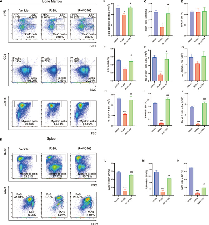
Results: A single exposure to whole-body ionizing radiation of 3 Gy causes acute bone marrow injury and long-term myelosuppression, resulting in hematopoietic imbalances and a bias toward myeloid differentiation. Ionizing radiation induced bone marrow B cell apoptosis and necroptosis, bone marrow T cell apoptosis. Various programmed cell death modes were involved in radiation injury of hematopoietic stem cells. Inhibition of Caspase-1 by VX-765 accelerated the recovery of hematopoietic stem cells after radiation. It is worth noting that inhibition of Caspase-1 promotes the proliferation and differentiation of hematopoietic stem cells after ionizing radiation. VX-765 treatment under ionizing radiation stress increased numbers of spleen colony formation, ability of long-term hematopoietic reconstitution in vivo and self-renewal. VX-765 alleviates post-irradiation hematopoietic stem cell injury by inhibiting pyroptosis, apoptosis and necroptosis.

Conclusions: These data suggest that multiple programmed cell death pathways are involved in radiation-induced damage to hematopoietic cells. Inhibiting Caspase-1 activity can be used as a strategy for protecting against radiation-induced injury to hematopoietic stem cells.

Keywords: Apoptosis; Hematopoietic cells; Ionizing radiation; Necroptosis; Pyroptosis.


Conflict of interest statement

Declarations. Ethics approval and consent to participate: This study did not include clinical trials. All experiments were approved by the Experimental Animal Welfare and Ethics Committee of Nanchang University under the project “The role of activating exogenous apoptosis to remove aging hematopoietic stem cells in bone marrow reconstruction of radiation-induced chronic injury”. The approval number is NCU-CLA-2019-319. The approval date for these animal experiments is 2019-03-16. Our contributions are reported in accordance with the ARRIVE guidelines 2.0. Consent for publication: Not applicable. Competing interests: The authors declare no competing interests.

Figures

Fig. 1
Fig. 1
Ionizing radiation results in acute injuries and the long-term effects of on hematopoietic cells in mice. Mice were adaptively fed for one week before being exposed to a single whole-body irradiation with 3 Gy X-rays. Bone marrow cells were analyzed at 1, 7, 14, and 28 days post-irradiation. Flow cytometry was employed to measure hematopoietic stem/progenitor cells, B cells, T cells, and Myeloid cells in the bone marrow. (A) Representative flow cytometry plots illustrating changes in hematopoietic stem/progenitor cell populations at different time points following irradiation. (B) Total nucleated cell counts in the bone marrow. (C) Proportions and numbers of hematopoietic stem/progenitor and precursor cells in the bone marrow. (D) Proportions and numbers of B cells, T cells, and Myeloid cells. Data are expressed as the mean ± SEM. n = 4 per group. *p < 0.05, **p < 0.01, ***p < 0.001 versus NO IR group
Fig. 2
Fig. 2
Ionizing radiation induces apoptosis and necroptosis in bone marrow B cells. Flow cytometry was used to detect apoptosis and pyroptosis through measuring expression levels of programmed cell death target genes in bone marrow cells with and without irradiation. (A) Flow cytometry plot showing expression of Cleaved-Caspase-1. (B) Mean Fluorescence Intensity (MFI) of Cleaved-Caspase-1. (C) Flow cytometry plot showing expression levels of GSDMD-N. (D) MFI of GSDMD-N. (E) Representative flow cytometry plot for apoptosis. (F) Percentages of early apoptotic cells in B cells. (G-H) Expression of Bax and Puma in B cells by qRT-PCR. (I-J) Expression of Ripk3 and Mlkl in B cells by qRT-PCR. (K-L) Expression of Acsl4 and Gpx4 in B cells by qRT-PCR. Data are reported as the mean ± SEM. n = 3 per group. *p < 0.05, **p < 0.01, ***p < 0.001 versus Control group
Fig. 3
Fig. 3
Ionizing radiation leads to various programmed cell death in bone marrow hematopoietic stem cells. Flow cytometry was used to detect pyroptosis and apoptosis. c-Kit+ cells were magnetic bead sorted to examine the expression levels of target genes related to programmed cell death in bone marrow after irradiation. Mice were administered 100 mg/kg VX-765 24 h before and 6 h after 3 Gy whole-body irradiation, and the protein expression level in bone marrow was measured 24 h after irradiation. (A) Representative flow chart of Cleaved-Caspase-1 expression. (B) MFI of Cleaved-Caspase-1 in LSK cells. (C) Representative flow chart of GSDMD-N expression. (D) MFI of GSDMD-N in LSK cells. (E-F) Expression of Il1b and Il18 by qRT-PCR. (G) Representative flow chart for apoptosis in LSK cells. (H) Ratio of early apoptotic in LSK cells. (I-J) Expression of Bax and Puma by qRT-PCR. (K-L) Expression of Ripk3 and Mlkl by qRT-PCR. (M-N) Expression of Acsl4 and Gpx4 by qRT-PCR. (O) Representative flow chart of Cleaved-Caspase-1 expression in LSKs. (P) MFI of Cleaved-Caspase-1 in HSPCs. (Q) Representative flow chart of GSDMD-N expression in HSCs. (R) MFI of GSDMD-N in HSPCs. (S) Expression of Il1b in c-Kit+ cells by qRT-PCR. (T) Expression of Il18 in c-Kit+ cells by qRT-PCR. Data are reported as the mean ± SEM. n = 3–5 per group. *p < 0.05, **p < 0.01, ***p < 0.001 versus Control group or Vehicle group. #p < 0.05, ##p < 0.01 versus IR-24 h group
Fig. 4
Fig. 4
The protective effects of VX-765 on hematopoietic cell quantity and proportion in acute ionizing radiation-induced injury. Mice were administered varying doses of VX-765 24 h before and 6 h after receiving a total body ionizing radiation dose of 3 Gy. Bone marrow cells were collected and analyzed 24 h after ionizing radiation. (A) Representative flow cytometry plots depicting hematopoietic stem/progenitor cells after irradiation with different drug dosages. (B) Total numbers of nucleated cells in the bone marrow after irradiation. (C-D) Proportion and quantity of hematopoietic stem/progenitor cells and precursor cells. Data are reported as the mean ± SEM. n = 3 per group. *p < 0.05, **p < 0.01 versus Vehicle group. #p < 0.05 versus IR + Vehicle group
Fig. 5
Fig. 5
The protective effects of VX-765 on the abundance and composition of hematopoietic cells in acute ionizing radiation-induced injury. Mice were administered a dose of 100 mg/kg VX-765 24 h before and 6 h after receiving a total body irradiation dose of 3 Gy. Bone marrow cells were collected 24 h and 14 days post-irradiation for subsequent analysis. (A) Representative flow cytometry plot depicting hematopoietic stem/progenitor cells. (B) Representative flow cytometric plots of B and T cells. (C) Representative flow cytometry plot showing Myeloid cells. (D) Total nucleated cell counts in the bone marrow after VX-765 treatment. (E) The numbers and percentages of hematopoietic stem/progenitor and precursor cells were evaluated by flow cytometry. (F) The numbers and percentages of B cells were determined after VX-765 treatment. Data are reported as the mean ± SEM. n = 3–5 per group. *p < 0.05, **p < 0.01, ***p < 0.001 versus Vehicle group. #p < 0.05, ##p < 0.01 versus IR + Vehicle group
Fig. 6
Fig. 6
The protective effects of VX-765 on the long-term effects of ionizing radiation-induced damage to the hematopoietic immune system. A dose of 100 mg/kg of VX-765 was administered 24 h before and 6 h after total-body irradiation at a dose of 3 Gy. After 28 days post-irradiation, bone marrow, spleen, and thymus were collected for analysis. (A) Representative flow cytometry plot of the bone marrow hematopoietic cells. (B) The total numbers of nucleated cells in bone marrow after VX-765 treatment. (C-H) The percentages and numbers of hematopoietic stem cells in the bone marrow after VX-765 treatment. (I-J) Percentages and numbers of bone marrow B cells after VX-765 treatment. (K) Representative flow cytometry of B cells and subpopulations in the spleen. (L-N) Quantification and characterization of B cell subsets in the spleen. Data are reported as the mean ± SEM. n = 4 per group. *p < 0.05, **p < 0.01, ***p < 0.001 versus Vehicle group. #p < 0.05, ##p < 0.01, ###p < 0.001 versus IR-28d group
Fig. 7
Fig. 7
The effects of VX-765 on proliferation, differentiation, in vivo reconstitution and self-renewal capacity of hematopoietic stem cells after ionizing radiation. (A) Experimental design for endogenous splenic colony formation. (B) Representative images depicting splenic nodules in mice 9 days after BMT. (C) Statistical analysis of the number of splenic colonies in mice. (D) The scheme of competitive BMT and secondary BMT. (E) PB, BM, SP chimerism representative flow cytometry. (F) Proportion of donor CD45.1/2+ cells in PB of recipient mice. (G-I) The percentages of donor-derived cells in the PB of recipient mice after BMT. (J) Proportion of donor-derived CD45.1/2+ cells in the PB of secondary BMT recipient mice. (K-M) The percentages of donor-derived cells in the PB of recipient mice after secondary BMT. (N) Implantation of donor cell sources in BM was measured at 16 weeks after BMT. (O) Implantation of donor cell sources in SP was measured at 16 weeks after BMT. (P) Implantation of donor cell sources in BM was measured at 16 weeks after secondary BMT. Data are reported as the mean ± SEM compared to IR + Vehicle. n = 3–5 per group. *p < 0.05, **p < 0.01, ***p < 0.001
Fig. 8
Fig. 8
Inhibition of caspase-1 significantly altered the transcriptional profile associated with programmed cell death in the Linc-Kit+ cell population 14 days after IR. RNA-seq was performed on LKs sorted from bone marrow cells of IR + Vehicle and IR + VX-765 group mice after 3 Gy TBI (n = 3). (A) Schematic flow diagram of flow sorting process. (B) GSEA plot showing the correlated enrichment of pyroptosis, apoptosis and necroptosis in the LKs cell populations of IR + Vehicle and IR + VX-765 groups. (C) Heatmap showing the expression of programmed cell death-related genes in IR + Vehicle and IR + VX-765 groups

Similar articles

References

    1. Shao L, Luo Y, Zhou D. Hematopoietic stem cell injury induced by ionizing radiation. Antioxid Redox Signal. 2014;20(9):1447–62. - PMC - PubMed
    1. Gutschalk CM, Herold-Mende CC, Fusenig NE, Mueller MM. Granulocyte colony-stimulating factor and granulocyte-macrophage colony-stimulating factor promote malignant growth of cells from head and neck squamous cell carcinomas in vivo. Cancer Res. 2006;66(16):8026–36. - PubMed
    1. Cheng H, Zheng Z, Cheng T. New paradigms on hematopoietic stem cell differentiation. Protein Cell. 2020;11(1):34–44. - PMC - PubMed
    1. Milyavsky M, Gan OI, Trottier M, Komosa M, Tabach O, Notta F, Lechman E, Hermans KG, Eppert K, Konovalova Z, et al. A distinctive DNA damage response in human hematopoietic stem cells reveals an apoptosis-independent role for p53 in self-renewal. Cell Stem Cell. 2010;7(2):186–97. - PubMed
    1. Wang J, Sun Q, Morita Y, Jiang H, Groß A, Lechel A, Hildner K, Guachalla LM, Gompf A, Hartmann D, et al. A differentiation checkpoint limits hematopoietic stem cell Self-Renewal in response to DNA damage. Cell. 2014;158(6):1444. - PubMed

MeSH terms

LinkOut - more resources