

A whole-brain voxel-based analysis of structural abnormalities in PTSD: An ENIGMA-PGC study
- PMID: 40692500
- PMCID: PMC12344465
- DOI: 10.1192/j.eurpsy.2025.10062
A whole-brain voxel-based analysis of structural abnormalities in PTSD: An ENIGMA-PGC study
Abstract
Background: Patients with posttraumatic stress disorder (PTSD) exhibit smaller regional brain volumes in commonly reported regions including the amygdala and hippocampus, regions associated with fear and memory processing. In the current study, we have conducted a voxel-based morphometry (VBM) meta-analysis using whole-brain statistical maps with neuroimaging data from the ENIGMA-PGC PTSD working group.
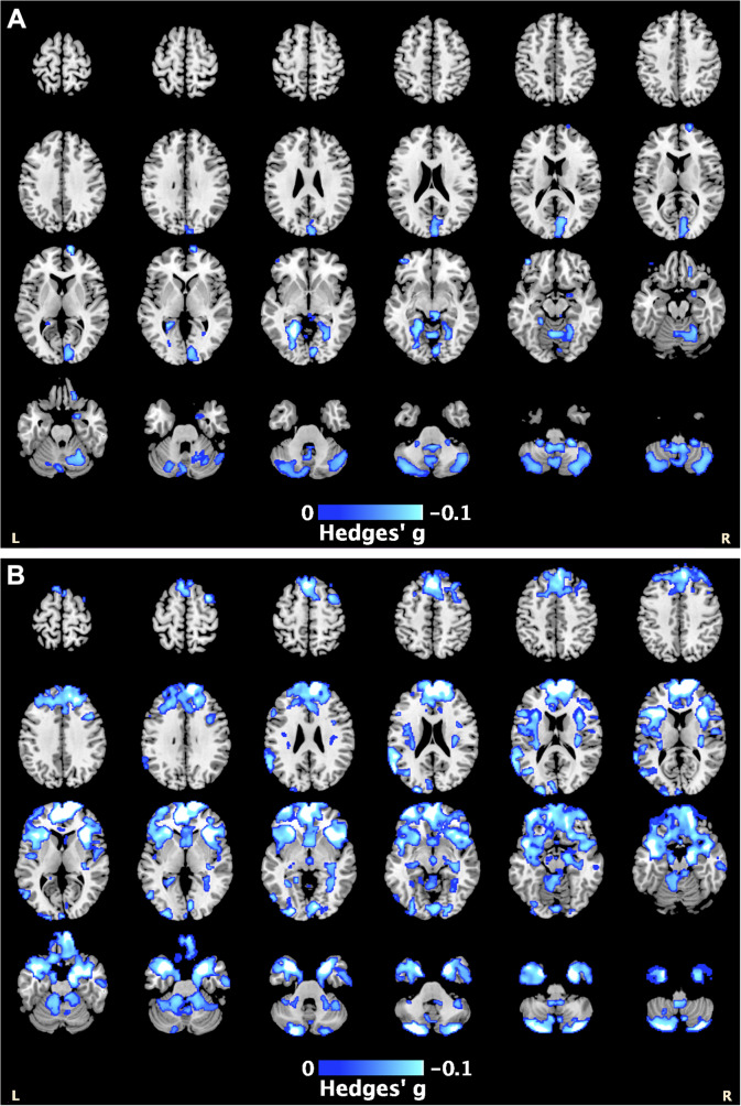
Methods: T1-weighted structural neuroimaging scans from 36 cohorts (PTSD n = 1309; controls n = 2198) were processed using a standardized VBM pipeline (ENIGMA-VBM tool). We meta-analyzed the resulting statistical maps for voxel-wise differences in gray matter (GM) and white matter (WM) volumes between PTSD patients and controls, performed subgroup analyses considering the trauma exposure of the controls, and examined associations between regional brain volumes and clinical variables including PTSD (CAPS-4/5, PCL-5) and depression severity (BDI-II, PHQ-9).
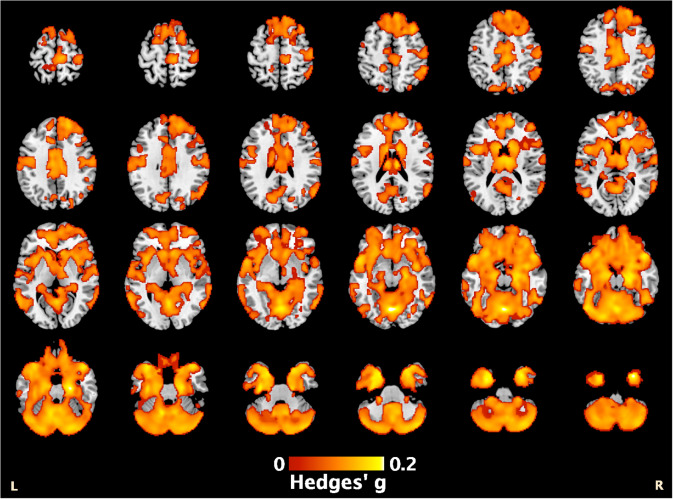
Results: PTSD patients exhibited smaller GM volumes across the frontal and temporal lobes, and cerebellum, with the most significant effect in the left cerebellum (Hedges' g = 0.22, pcorrected = .001), and smaller cerebellar WM volume (peak Hedges' g = 0.14, pcorrected = .008). We observed similar regional differences when comparing patients to trauma-exposed controls, suggesting these structural abnormalities may be specific to PTSD. Regression analyses revealed PTSD severity was negatively associated with GM volumes within the cerebellum (p corrected = .003), while depression severity was negatively associated with GM volumes within the cerebellum and superior frontal gyrus in patients (p corrected = .001).
Conclusions: PTSD patients exhibited widespread, regional differences in brain volumes where greater regional deficits appeared to reflect more severe symptoms. Our findings add to the growing literature implicating the cerebellum in PTSD psychopathology.
Keywords: PTSD; brain structure; gray matter volume; neuroimaging; trauma; voxel-based morphometry.
Conflict of interest statement
N Jahanshad received partial research support from Biogen, Inc. (Boston, USA) for research unrelated to the content of this article. P Thompson received partial research support from Biogen, Inc. (Boston, USA) for research unrelated to the topic of this manuscript. R Davidson is the founder and president of, and serves on the board of directors for, the non-profit organization Healthy Minds Innovations, Inc. L Lebois reports unpaid membership on the Scientific Committee for the International Society for the Study of Trauma and Dissociation (ISSTD), grant support from the National Institute of Mental Health, K01 MH118467, and spousal IP payments from Vanderbilt University for technology licensed to Acadia Pharmaceuticals unrelated to the present work. ISSTD and NIMH were not involved in the analysis or preparation of the manuscript. C Abdullah has served as a consultant, speaker and/or on advisory boards for Douglas Pharmaceuticals, Freedom Biosciences, FSV7, Lundbeck, Psilocybin Labs, Genentech, Janssen and Aptinyx; served as editor of Chronic Stress for Sage Publications, Inc; and filed a patent for using mTOR inhibitors to augment the effects of antidepressants (filed on August 20, 2018). J Krystal is a consultant for AbbVie, Inc., Amgen, Astellas Pharma Global Development, Inc., AstraZeneca Pharmaceuticals, Biomedisyn Corporation, Bristol-Myers Squibb, Eli Lilly and Company, Euthymics Bioscience, Inc., Neurovance, Inc., FORUM Pharmaceuticals, Janssen Research & Development, Lundbeck Research USA, Novartis Pharma AG, Otsuka America Pharmaceutical, Inc., Sage Therapeutics, Inc., Sunovion Pharmaceuticals, Inc., and Takeda Industries; is on the Scientific Advisory Board for Lohocla Research Corporation, Mnemosyne Pharmaceuticals, Inc., Naurex, Inc., and Pfizer; is a stockholder in Biohaven Pharmaceuticals; holds stock options in Mnemosyne Pharmaceuticals, Inc.; holds patents for Dopamine and Noradrenergic Reuptake Inhibitors in Treatment of Schizophrenia, US Patent No. 5,447,948 (issued September 5, 1995), and Glutamate Modulating Agents in the Treatment of Mental Disorders, U.S. Patent No. 8,778,979 (issued July 15, 2014); and filed a patent for Intranasal Administration of Ketamine to Treat Depression. U.S. Application No. 14/197,767 (filed on March 5, 2014); US application or Patent Cooperation Treaty international application No. 14/306,382 (filed on June 17, 2014). Filed a patent for using mTOR inhibitors to augment the effects of antidepressants (filed on August 20, 2018). S Nelson consults for Turing Medical, which commercializes FIRMM. This interest has been reviewed and managed by the University of Minnesota in accordance with its Conflict of Interest policies. E Olson is employed by the nonprofit organization Crisis Text Line, for work unrelated to the content of this manuscript. All other authors report no potential competing interests or disclosures.
Figures
References
-
- Association AP. Diagnostic and statistical manual of mental disorders. 5th ed. American Psychiatric Association; 2013.
Publication types
MeSH terms
Grants and funding
- I01 RX002172/RX/RRD VA/United States
- R21 MH098198/MH/NIMH NIH HHS/United States
- T32 MH018931/MH/NIMH NIH HHS/United States
- I01 RX002170/RX/RRD VA/United States
- HD071982, HD085850/Eunice Kennedy Shriver National Institute of Child Health and Human Development
- R61 NS120249/NS/NINDS NIH HHS/United States
- W81XWH-10-1-0925/U.S. Department of Defense
- P41 EB015922/EB/NIBIB NIH HHS/United States
- MRC- RFA-IFSP-01-2013/SHARED ROOTS/South African Medical Research Council
- R01MH105535/MH/NIMH NIH HHS/United States
- I01 RX002174/RX/RRD VA/United States
- 1IK2RX000709/Rehabilitation Research and Development Service
- R56 MH071537/MH/NIMH NIH HHS/United States
- K01 MH122774/MH/NIMH NIH HHS/United States
- I01 RX003444/RX/RRD VA/United States
- IK2 RX002922/RX/RRD VA/United States
- MH101380/MH/NIMH NIH HHS/United States
- via its support of (Clinical and Translational Science Awards) Yale Center for Clinical Investigation/TR/NCATS NIH HHS/United States
- R21 MH098212/MH/NIMH NIH HHS/United States
- K12 HD085850/HD/NICHD NIH HHS/United States
- R01 MH071537/MH/NIMH NIH HHS/United States
- R21 MH106998/MH/NIMH NIH HHS/United States
- I01 RX003442/RX/RRD VA/United States
- IK2 CX001680/CX/CSRD VA/United States
- I01 CX002097, I01 CX002096, I01 CX001820, I01 HX003155, I01 RX003444, I01 RX003443, I01 RX003442, I01 CX001135, I01 CX001246, I01RX001774, I01 RX001135, I01 RX002076, I01 RX001880, I01 RX002172, I01 RX002173, I01RX 002171, I01 RX002174, and I01 RX002170/U.S. Department of Veterans Affairs
- I01 CX001135/CX/CSRD VA/United States
- R21 MH112956/MH/NIMH NIH HHS/United States
- 40-00812-98-10041/ZONMW_/ZonMw/Netherlands
- K01 MH118428/MH/NIMH NIH HHS/United States
- UL1TR000454/TR/NCATS NIH HHS/United States
- R01 MH105355/MH/NIMH NIH HHS/United States
- MH098212, MH071537/MH/NIMH NIH HHS/United States
- NIMH K01 MH118428/MH/NIMH NIH HHS/United States
- R01 MH105535/MH/NIMH NIH HHS/United States
- M01 RR000039/RR/NCRR NIH HHS/United States
- I01 RX003443/RX/RRD VA/United States
- R01 MH111671/MH/NIMH NIH HHS/United States
- R01 MH106574/MH/NIMH NIH HHS/United States
- R01MH111671, R01MH117601, R01AG059874, MJFF 14848/U.S. Department of Defense
- W81XWH-13-2-0025/Medical Research and Materiel Command
- R61NS120249/NS/NINDS NIH HHS/United States
- R01 MH116147/MH/NIMH NIH HHS/United States
- HFP90-020/Health Services Research and Development
- I01 RX001880/RX/RRD VA/United States
- I01 RX000622/RX/RRD VA/United States
- MR/N013700/1/MRC_/Medical Research Council/United Kingdom
- I01 RX002171/RX/RRD VA/United States
- via its support for (P50) Center for the Translational Neuroscience of Alcohol/AA/NIAAA NIH HHS/United States
- Pilot Grant/Brain and Behavior Research Foundation
- W81XWH08-2-0159/U.S. Department of Defense
- R01-MH043454 and T32-MH018931/MH/NIMH NIH HHS/United States
- I01 HX003155/HX/HSRD VA/United States
- NARSAD Young Investigator Award/National Alliance for Research on Schizophrenia and Depression
- W81XWH- 08-2-0038/Congressionally Directed Medical Research Programs
- R21MH106998/MH/NIMH NIH HHS/United States
- U54 EB020403/EB/NIBIB NIH HHS/United States
- R01 MH117601/MH/NIMH NIH HHS/United States
- I01 RX001774/RX/RRD VA/United States
- I01 CX002097/CX/CSRD VA/United States
- R01MH107382-02/MH/NIMH NIH HHS/United States
- R01MH105355-01A/MH/NIMH NIH HHS/United States
- U21A20364, No. 31971020/National Natural Science Foundation of China
- I01 RX002076/RX/RRD VA/United States
- R01 MH119227/MH/NIMH NIH HHS/United States
- K01MH118467/MH/NIMH NIH HHS/United States
- 1K2RX002922/Rehabilitation Research and Development Service
- R21MH112956 and R01MH119227/MH/NIMH NIH HHS/United States
- 1R01MH110483 and 1R21 MH098198/MH/NIMH NIH HHS/United States
- 110614/Academisch Medisch Centrum
- R01 MH110483/MH/NIMH NIH HHS/United States
- UL1 TR000454/TR/NCATS NIH HHS/United States
- R01 MH107382/MH/NIMH NIH HHS/United States
- I01 CX001246/CX/CSRD VA/United States
- R56 AG058854/AG/NIA NIH HHS/United States
- A core grant to the Waisman Center/National Institute of Child Health and Human Development
- K23 MH101380/MH/NIMH NIH HHS/United States
- K01MH122774/MH/NIMH NIH HHS/United States
- R01 MH043454/MH/NIMH NIH HHS/United States
- R21 MH102634/MH/NIMH NIH HHS/United States
- R01 HD071982/HD/NICHD NIH HHS/United States
- I01 RX002173/RX/RRD VA/United States
- 1R21MH102634/MH/NIMH NIH HHS/United States
- K01 MH118467/MH/NIMH NIH HHS/United States
- I01 CX002096/CX/CSRD VA/United States
- I01 CX001820/CX/CSRD VA/United States
- SFB/TRR 58: C06, C07/Deutsche Forschungsgemeinschaft
- I01RX000622/Rehabilitation Research and Development Service
- IK2 RX000709/RX/RRD VA/United States
- I01 RX001135/RX/RRD VA/United States
- R01 MH096987/MH/NIMH NIH HHS/United States
- 27040/National Alliance for Research on Schizophrenia and Depression
- W81XWH-12-2-0012/U.S. Department of Defense
- No. 20ZDA079/Major Basic Research Project of the Natural Science Foundation of the Jiangsu Higher Education Institutions
- R01 AG059874/AG/NIA NIH HHS/United States
LinkOut - more resources
Full Text Sources
Medical
Miscellaneous
