

3D visualization of uterus and ovary: tissue clearing techniques and biomedical applications
- PMID: 40692613
- PMCID: PMC12277315
- DOI: 10.3389/fbioe.2025.1610539
3D visualization of uterus and ovary: tissue clearing techniques and biomedical applications
Abstract
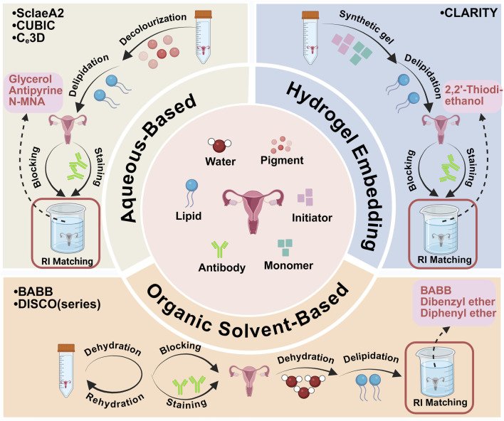
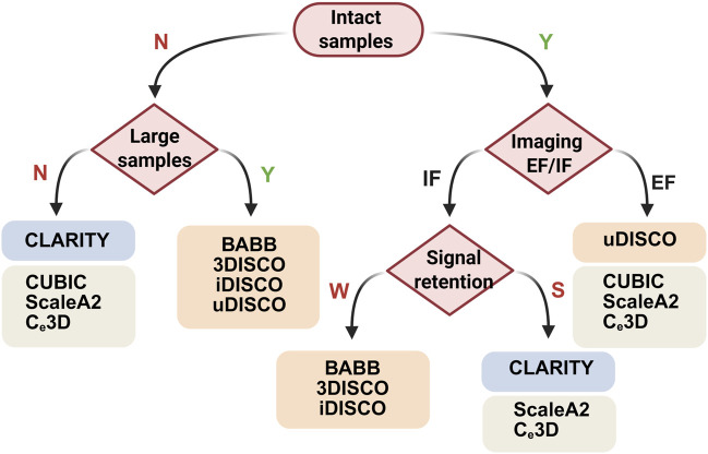
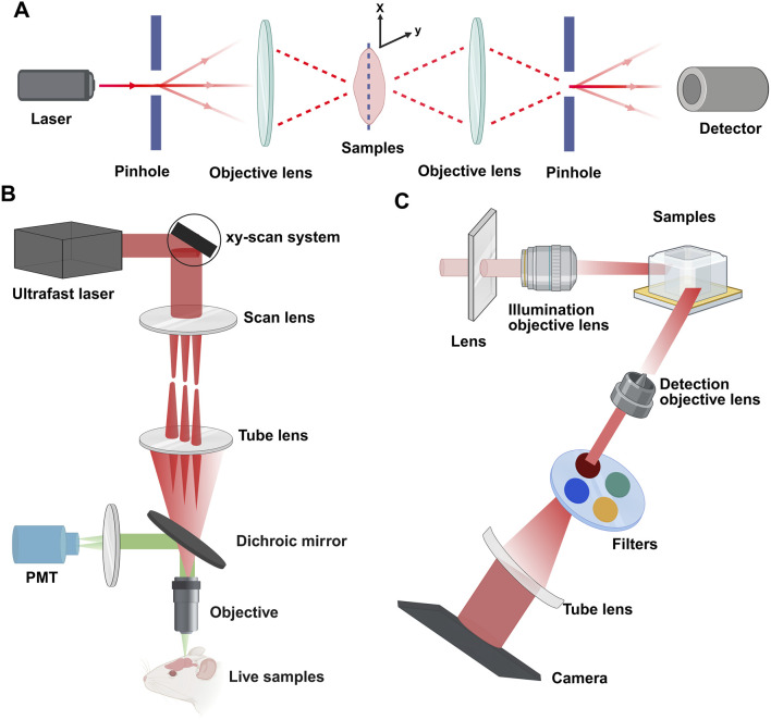
Recent advancements in tissue clearing and three-dimensional (3D) visualization technologies have enabled subcellular-level examination of entire organs, particularly in complex structures such as the ovary and uterus. Traditional histological approaches are limited by two-dimensional views, which restrict our understanding of female reproductive system functions. In this review, we highlight the innovations in 3D tissue clearing techniques applied to uterine and ovarian tissues, which, combined with analytical tools, facilitate comprehensive 3D visualization and image analysis. We evaluate the advantages and disadvantages of three primary categories of tissue clearing techniques: organic solvent-based, hydrogel-based, and hydrogel-embedded methods, specifically regarding the uterus and ovary. Light-sheet and multiphoton microscopy complement these techniques, providing unprecedented capabilities for high-resolution imaging of large tissue volumes. Tissue clearing technologies provide a robust strategy for early diagnosis of uterine and ovarian pathologies. Additionally, we explore the integration of tissue clearing technologies with spatial transcriptomics and AI-driven analytical tools to achieve comprehensive 3D molecular mapping. We hope this review contributes to a better understanding of tissue clearing techniques and can help researchers in navigating methodological choices for uterine and ovarian investigations.
Keywords: 3D visualization; AI; ovary; spatial omics; tissue clearing; uterus.
Copyright © 2025 Liu, Song, Liu, Dong, Zheng, Leung and Chen.
Conflict of interest statement
The authors declare that the research was conducted in the absence of any commercial or financial relationships that could be construed as a potential conflict of interest.
Figures
Similar articles
-
Automatic Segmentation and Alignment of Uterine Shapes from 3D Ultrasound Data.Comput Biol Med. 2024 Aug;178:108794. doi: 10.1016/j.compbiomed.2024.108794. Epub 2024 Jun 27. Comput Biol Med. 2024. PMID: 38941903
-
Baseline anatomical assessment of the uterus and ovaries in infertile women: a systematic review of the evidence on which assessment methods are the safest and most effective in terms of improving fertility outcomes.Hum Reprod Update. 2017 Sep 1;23(5):533-547. doi: 10.1093/humupd/dmx019. Hum Reprod Update. 2017. PMID: 28903473
-
Generative interpolation and restoration of images using deep learning for improved 3D tissue mapping.bioRxiv [Preprint]. 2024 Mar 28:2024.03.07.583909. doi: 10.1101/2024.03.07.583909. bioRxiv. 2024. Update in: Nat Methods. 2025 Jul;22(7):1556-1567. doi: 10.1038/s41592-025-02712-4. PMID: 38496512 Free PMC article. Updated. Preprint.
-
Fast Tissue Clearing and Volume Imaging Techniques for Anatomy.Microsc Res Tech. 2025 Jun 27. doi: 10.1002/jemt.70013. Online ahead of print. Microsc Res Tech. 2025. PMID: 40579818 Review.
-
Optimization of Tissue Clearing Methods and Imaging Conditions for 3D Visualization of the Vasculature of the Adult Murine Knee.bioRxiv [Preprint]. 2025 Jun 27:2025.06.26.661802. doi: 10.1101/2025.06.26.661802. bioRxiv. 2025. PMID: 40642113 Free PMC article. Preprint.
References
Publication types
LinkOut - more resources
Full Text Sources