

Diagnostic challenges of rarely well-differentiated adenocarcinoma of the stomach
- PMID: 40692725
- PMCID: PMC12277191
- DOI: 10.3389/pore.2025.1612163
Diagnostic challenges of rarely well-differentiated adenocarcinoma of the stomach
Abstract
Background: Fundic gland tumors are a rare subtype of gastric tumors with fundic gland differentiation. This group of tumors has a low incidence rate and shows indistinctive cellular atypia, obvious structural atypia, special tissue morphology, and clinical prognosis, thus leading to diagnostic challenges.
Aim: We aimed to investigate the clinical and endoscopic characteristics and pathological features of gastric adenocarcinoma of the fundic gland (GA-FG) to provide a better understanding of this disease.
Methods: We collected data from patients diagnosed as having GA-FG at Guangdong Provincial People's Hospital between January 2019 and April 2024. The analysis focused on their clinical data, endoscopic characteristics, pathological morphological characteristics, immunohistochemistry results, treatment, and prognosis.
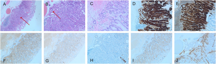
Results: Among the four patients were two men and two women (age range, 52-65 years). The tumors were mainly located in the gastric fundus and gastric body, and the lesions commonly had a superficial bulge. Three patients had an initial diagnosis of oxyntic gland adenoma, which was diagnosed as GA-FG after complete resection. These tumors were negative for MUC5AC, but showed diffuse strong positivity for MUC6 and pepsinogen I, and synaptophysin expression.
Conclusion: GA-FG is a rare gastric tumor with unique morphological features. As it is difficult to diagnose with a biopsy, immunohistochemistry plays an important role in the differential diagnosis. Oxyntic gland adenoma can be regarded as the intramucosal stage of GA-FG. Although all patients were negative for MUC5AC expression, MUC6 and pepsinogen I can help the diagnosis of GA-FG.
Keywords: MUC6; diagnosis; gastric adenocarcinoma of the fundic gland; stomach neoplasms; welldifferentiated.
Copyright © 2025 Qin, Wang, Xiao, Ma, Fan, Zhu, Luo, Zhang and Liu.
Conflict of interest statement
The authors declare that the research was conducted in the absence of any commercial or financial relationships that could be construed as a potential conflict of interest.
Figures

Similar articles
-
Pathological reevaluation of gastric neoplasms with differentiation into the fundic gland.Scand J Gastroenterol. 2025 Jul;60(7):664-674. doi: 10.1080/00365521.2025.2515428. Epub 2025 Jun 6. Scand J Gastroenterol. 2025. PMID: 40478615
-
Gastric epithelial neoplasm of fundic-gland mucosa lineage: representative of the low atypia differentiated gastric tumor and Ki67 may help in their identification.Pathol Oncol Res. 2024 May 30;30:1611734. doi: 10.3389/pore.2024.1611734. eCollection 2024. Pathol Oncol Res. 2024. PMID: 38873175 Free PMC article.
-
Systemic pharmacological treatments for chronic plaque psoriasis: a network meta-analysis.Cochrane Database Syst Rev. 2021 Apr 19;4(4):CD011535. doi: 10.1002/14651858.CD011535.pub4. Cochrane Database Syst Rev. 2021. Update in: Cochrane Database Syst Rev. 2022 May 23;5:CD011535. doi: 10.1002/14651858.CD011535.pub5. PMID: 33871055 Free PMC article. Updated.
-
Impact of residual disease as a prognostic factor for survival in women with advanced epithelial ovarian cancer after primary surgery.Cochrane Database Syst Rev. 2022 Sep 26;9(9):CD015048. doi: 10.1002/14651858.CD015048.pub2. Cochrane Database Syst Rev. 2022. PMID: 36161421 Free PMC article.
-
[Characteristics of gastric hepatoid adenocarcinoma: a clinicopathological and molecular analysis].Zhonghua Bing Li Xue Za Zhi. 2025 Jul 8;54(7):748-754. doi: 10.3760/cma.j.cn112151-20250106-00012. Zhonghua Bing Li Xue Za Zhi. 2025. PMID: 40619264 Chinese.
References
-
- Ueyama H, Yao T, Akazawa Y, Hayashi T, Kurahara K, Oshiro Y, et al. Gastric epithelial neoplasm of fundic-gland mucosa lineage: proposal for a new classification in association with gastric adenocarcinoma of fundic-gland type. J Gastroenterol (2021) 56:814–28. 10.1007/s00535-021-01813-z - DOI - PMC - PubMed
MeSH terms
Substances
LinkOut - more resources
Full Text Sources
Medical
Research Materials
Miscellaneous