. 2025 Jul 7:13:1571770.
doi: 10.3389/fcell.2025.1571770. eCollection 2025.

Wnt-dependent mechanism of the apical constriction of roof plate cells in developing mouse spinal cord

Affiliations

Wnt-dependent mechanism of the apical constriction of roof plate cells in developing mouse spinal cord

Takuma Shinozuka et al. Front Cell Dev Biol. .

Abstract

Apical constriction of epithelial cells usually occurs in a local portion of epithelial sheet, which results in bending of epithelial tissues. However, it is uncertain whether diffusible signal molecules, like Wnt, regulate such locally restricted events. Here, we show that Wnt ligands are required for apical constriction of Wnt1-expressing roof plate (RP) cells during development of the neural tube. Analysis of Wntless conditional knock-out (cKO) embryos, in which Wnt secretion from Wnt1-expressing roof plate cells is impaired, revealed that RP-derived Wnt ligands are required for phosphorylation of myosin light chain (MLC) and apical constriction of RP cells. Loss- or gain-of-function analysis of β-catenin reveals that this apical constriction is regulated in a β-catenin-dependent manner. Consistent with the timing of apical constriction, Wnt ligands accumulate on the apical side of RP cells. In embryos with Wnt1-expressing RP-specific defects in synthesis of heparan sulfate proteoglycan, apical accumulation of Wnt ligands and apical constriction are impaired. Therefore, we propose that specific accumulation of Wnt ligands on RP cells drives apical constriction of these cells.

Keywords: Wnt; apical constriction; cytoskeleton; morphogenesis; neural tube; roof plate.


Conflict of interest statement

The authors declare that the research was conducted in the absence of any commercial or financial relationships that could be construed as a potential conflict of interest. The author(s) declared that they were an editorial board member of Frontiers, at the time of submission. This had no impact on the peer review process and the final decision.

Figures
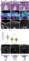
FIGURE 1
FIGURE 1
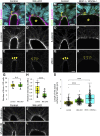
Apical constriction occurs in roof plate. (A–C) Hematoxylin and eosin (H&E) staining of the transverse sections at E9.5 (A), E10.5 (B) and E11.5 (C). (D–M) Transverse sections at the forelimb level showing apical constriction of RP cells at E9.5 (D,G,K), E10.5 (E,H,L) and E11.5 (F,I,M) using anti-β-catenin (D–I) and anti-pMLC (K–M) antibodies. A statistical summary of the length of the apical surface of RP cells is shown in (J). The length of the apical surface of RP cells (the 10 most dorsal cells per section) was quantified based on localization of beta-catenin at the apical membrane. For each developmental stage, a total of 90 cells from three embryos were examined. ***P < 0.001 (Student’s t-test). Nuclei were counterstained with DAPI. Brackets indicate the region from the apical surface to the nucleus of each cell. Three embryos were examined in each experiment. (N) Schematic of RP cell morphology and distribution of pMLC based on K-M. Scale bars: 10 µm.
FIGURE 2
FIGURE 2
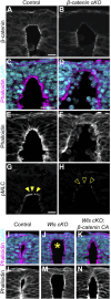
Wnt secretion from roof plate cells is required for apical constriction. (A–L) Transverse sections at the forelimb level in Wls cKO embryos (B,D,F,J,L) and littermate controls (A,C,E,I,K) at E11.5. To visualize morphology of RP cells, we stained sections with anti-Ezrin (A,B) and anti-β-catenin antibodies (A–D). To visualize the actomyosin cytoskeleton, we stained sections with anti-pMLC (E,F) antibody, phalloidin (I,J) and anti-Myosin IIB (K,L) antibody. A statistical summary indicating the number of Ezrin-positive RP cells is shown in (G). A total of 14 sections from four control and 13 sections from four Wls cKO embryos were examined. n.s., not significant. A statistical summary of the relative intensity of pMLC normalized to β-catenin signal intensity is shown in (H). For each genotype, a total of 60 cells from three embryos were examined. ***P < 0.001 (Student’s t-test). (M–S) Transverse sections at the forelimb level in Wnt1; Wnt3a double KO embryos (N,P,R) and littermate controls (M,O,Q) at E11.5 stained with anti-Ezrin (M,N), anti-β-catenin (M–P) and anti-pMLC (Q,R) antibodies. Nuclei were counterstained with DAPI. An asterisk indicates expansion of the apical surface. Arrowheads indicate pMLC signals in RP cells in control, while open arrowheads indicate reduced signals in Wls cKO or Wnt1; Wnt3a double KO embryos. Three embryos were examined in each experiment. A statistical summary indicating the length of the apical surface of RP cells is shown in (S). The length of the apical surface of Ezrin-positive roof plate cells was quantified based on the localization of beta-catenin at the apical membrane. A total of 192 cells prepared from three control, 218 cells from four Wls cKO and 204 cells from three Wnt1; Wnt3a double KO embryos were examined. ***P < 0.001 (Student’s t-test). Scale bars: 10 µm.
FIGURE 3
FIGURE 3
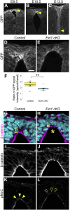
Apical constriction in roof plate cells occurs in a β-catenin-dependent manner. (A–H) Transverse sections at the forelimb level in β-catenin cKO embryos (B,D,F,H) and littermate controls (A,C,E,G) at E10.5 stained with anti-β-catenin (A,B) antibody, phalloidin (C–F) and anti-pMLC (G,H) antibody. (I–N) Transverse sections at the forelimb level in control (I,L), Wls cKO (J,M) and Wls cKO; β-catenin CA (K,N) embryos at E10.5 stained with phalloidin. Nuclei were counterstained with DAPI. Arrowheads indicate pMLC signals in RP cells, while open arrowheads indicate reduced signals. An asterisk indicates expansion at the apical surface. Three embryos were examined in each experiment. Scale bars: 10 µm.
FIGURE 4
FIGURE 4
HSPGs are required for apical constriction. (A–C) Transverse sections of EGFP-Wnt3a knock-in embryos at the forelimb level in the developing spinal cord at E9.5 (A), E10.5 (B) and E13.5 (C) were stained with anti-GFP antibody. Arrowheads indicate the apical tips of RP cells. (D–L) Transverse sections at the forelimb level in Ext1-cKO embryos (E,H,J,L) and littermate controls (D,G,I,K) at E10.5 using anti-GFP (D,E), anti-β-catenin (G–J) and anti-pMLC (K,L) antibodies. Nuclei were counterstained with DAPI. Brackets indicate the region from the apical surface to the nucleus of each cell. An asterisk indicates expansion of the apical surface. Arrowheads indicate pMLC signals in RP cells, while open arrowheads indicate reduced signals. A statistical summary indicating the ratio of the GFP signal intensity from apical to basal is shown in (F). A total of 9 sections from two Ext1 cKO and 10 sections from two littermate control embryos were examined. **P < 0.01 (Student’s t-test). Scale bar: 10 µm.

Similar articles

References

    1. Alexandre C., Baena-Lopez A., Vincent J. P. (2014). Patterning and growth control by membrane-tethered wingless. Nature 505, 180–185. 10.1038/nature12879 - DOI - PMC - PubMed
    1. Baeg G. H., Lin X., Khare N., Baumgartner S., Perrimon N. (2001). Heparan sulfate proteoglycans are critical for the organization of the extracellular distribution of wingless. Development 128, 87–94. 10.1242/dev.128.1.87 - DOI - PubMed
    1. Banziger C., Soldini D., Schutt C., Zipperlen P., Hausmann G., Basler K. (2006). Wntless, a conserved membrane protein dedicated to the secretion of wnt proteins from signaling cells. Cell 125, 509–522. 10.1016/j.cell.2006.02.049 - DOI - PubMed
    1. Bartscherer K., Pelte N., Ingelfinger D., Boutros M. (2006). Secretion of wnt ligands requires evi, a conserved transmembrane protein. Cell 125, 523–533. 10.1016/j.cell.2006.04.009 - DOI - PubMed
    1. Brault V., Moore R., Kutsch S., Ishibashi M., Rowitch D. H., McMahon A. P., et al. (2001). Inactivation of the beta-catenin gene by Wnt1-Cre-mediated deletion results in dramatic brain malformation and failure of craniofacial development. Development 128, 1253–1264. 10.1242/dev.128.8.1253 - DOI - PubMed

LinkOut - more resources