. 2025 Dec;14(1):2534912.
doi: 10.1080/2162402X.2025.2534912. Epub 2025 Jul 22.

The human STING agonist E7766 induces immunogenic tumor clearance, independent of tumor-intrinsic STING expression in the KRASG12D/+ Trp53-/- murine model of sarcoma

Affiliations

The human STING agonist E7766 induces immunogenic tumor clearance, independent of tumor-intrinsic STING expression in the KRASG12D/+ Trp53-/- murine model of sarcoma

Karys M Hildebrand et al. Oncoimmunology. 2025 Dec.

Abstract

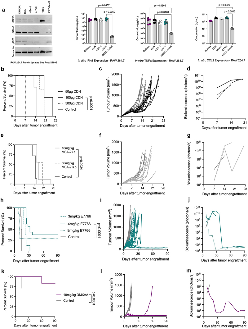
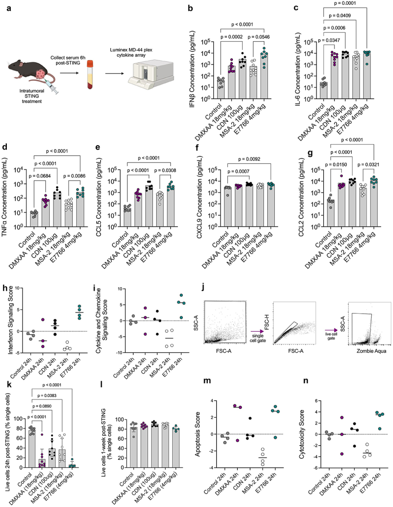
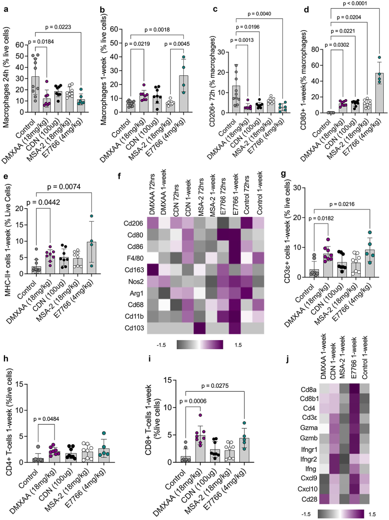
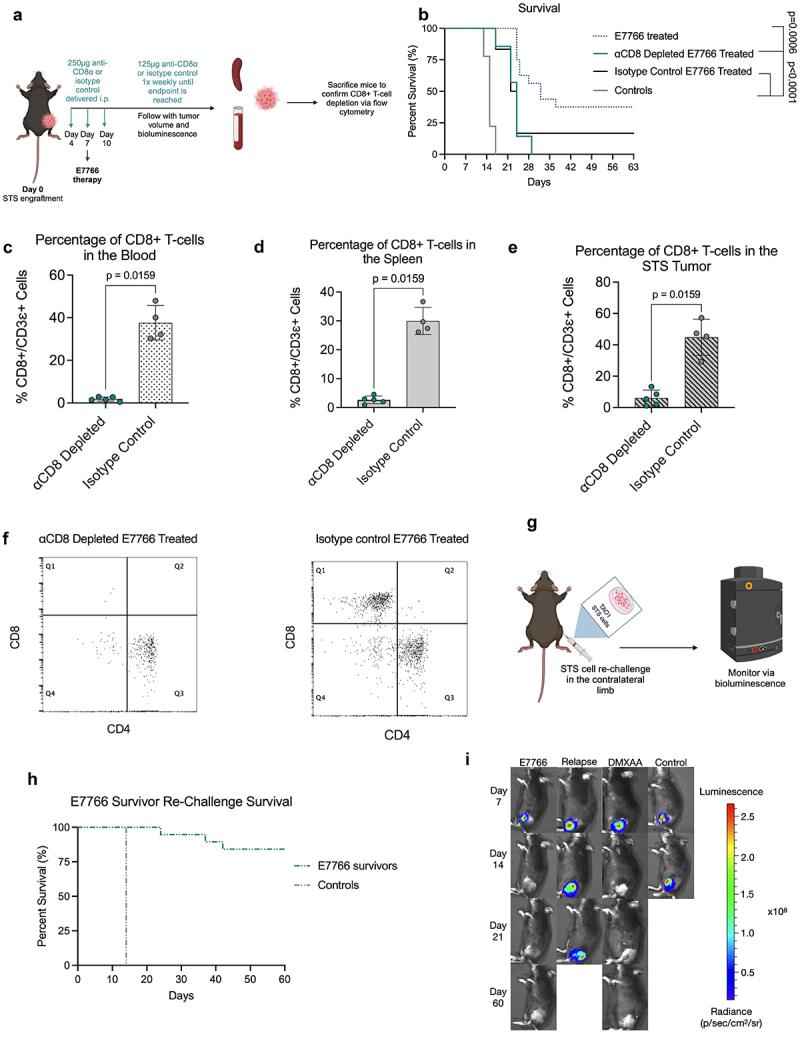
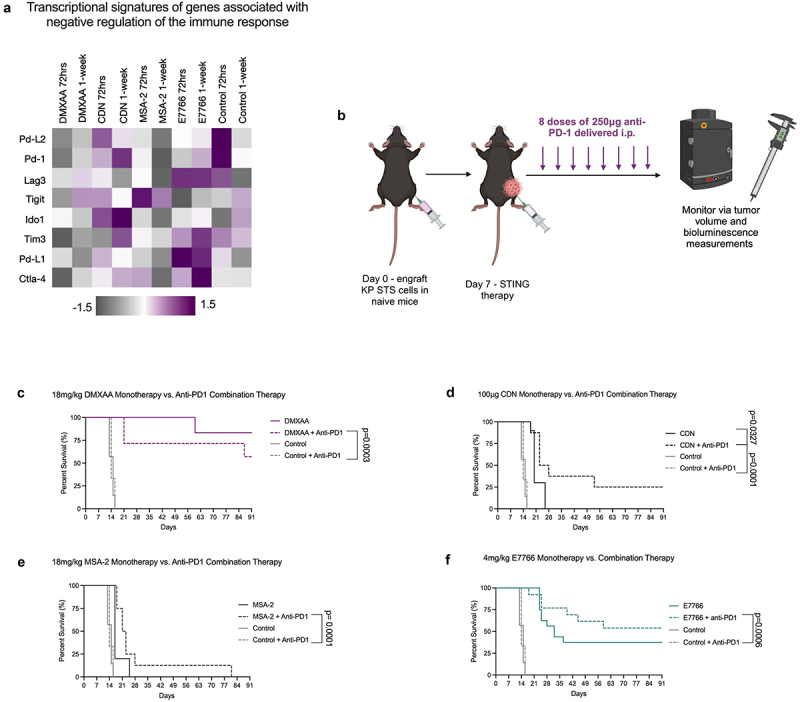
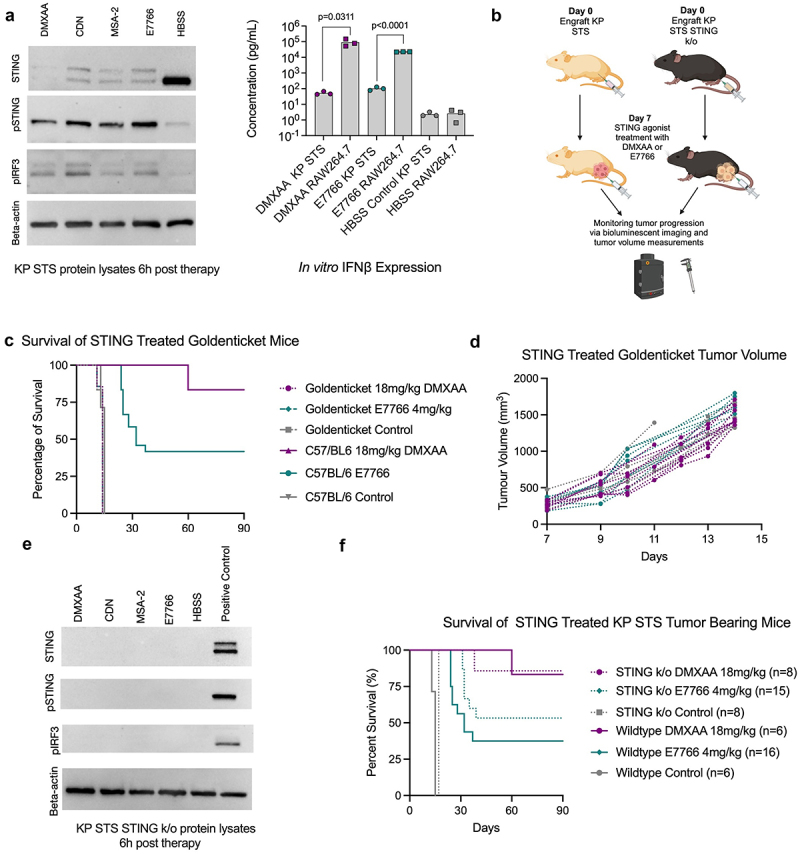
Soft tissue sarcomas (STS) are aggressive high-fatality cancers that affect children and adults. Most STS subtypes harbor an immunosuppressive tumor microenvironment (TME) and respond poorly to immunotherapy. Therapies capable of dismantling the immunosuppressive TME are needed to improve sensitivity to emerging immunotherapies. Activation of the Stimulator of INterferon Genes (STING) pathway has shown promising anti-tumor effects in preclinical models of carcinoma, but evaluations in sarcoma are lacking. Herein, we sought to examine the immune modulation and therapeutic efficacy of three translational small molecule STING agonists in an immunologically cold model of STS. Three classes of STING agonists, ML RR-S2 CDA, MSA-2, and E7766 were evaluated in an orthotopic KrasG12D/+ Trp53-/- model of STS. Dose titration survival studies, cytokine serology, and tumor immune phenotyping were used to examine STING agonist efficacy following intra-tumoral treatment. All STING agonists significantly increased survival time, however, only E7766 resulted in durable tumor clearance, inducing CD8+ T-cell infiltration and activated lymphocyte transcriptomic signatures in the TME. Antibody depletion was used to assess the dependency of treatment responses on CD8+ T-cells, showing that in their absence, tumor clearance did not occur following E7766 therapy. Using STING deficient mice, and CRISPR/Cas9 gene editing, we demonstrated that STS clearance following STING therapy was dependent on host STING and not tumor-intrinsic STING pathway functionality. E7766 represents a promising candidate able to remodel the TME of murine STS tumors toward an inflamed phenotype independent of tumor-intrinsic STING functionality, and should be considered for potential translation in STS treatment.

Keywords: Immunotherapy; STING; Solid tumor; sarcoma.


Conflict of interest statement

The authors of this manuscript do not have any relevant competing interests or conflicts of interest to disclose.

Figures

Figure 1.
Figure 1.
E7766 induces tumor clearance in KP sarcomas. a. Immunoblot and cytokine concentrations from RAW264.7 cells following equimolar treatment with STING agonists. cGAMP treatment was used as a positive control condition. b. Overall survival of STS bearing mice treated i.T. With 50 µg (n=6), 100 µg (n=10), or 500 µg of CDN (n=10) and control (n=7) (log-rank Mantel-Cox test). c & d. Tumor volume (c) and bioluminescence (d) of CDN treated mice over time. e. Overall survival of STS bearing mice treated with 18 mg/kg of MSA-2 i.T. (n=9) or 50 mg/kg of MSA-2 subcutaneously (n=11) and control (n=7) (log rank Mantel-Cox test). f & g Tumor volume (f) and bioluminescence (g) of MSA-2 treated mice over time. h. Overall survival of STS bearing mice treated i.T. With 3 mg/kg (n=8), 4 mg/kg (n=16), or 9 mg/kg (n=7) of E7766 and control (n=10) (log rank Mantel-Cox test). i & j Tumor volume (i) and bioluminescence (j) measurements of E7766 treated mice over time. k. Overall survival of STS bearing mice treated i.t. With 18 mg/kg (n=6) and control (n=10) (log rank Mantel-Cox test). l & m. Tumor volume (i) and bioluminescence (j) measurements of E7766 treated mice over time. All bioluminescence figures represent the average BLI signal for each condition.
Figure 2.
Figure 2.
E7766 and CDN lead to similar systemic cytokine production phenotypes 6h following therapy, and all STING agonists induced significant amounts of cell death within the tumor microenvironment at 24h post-therapy. a. Schematic of experimental design created with BioRender.com. Mice were engrafted with 100,000 KP STS cells and one week later were treated with either 18 mg/kg DMXAA, 100 µg CDN, 18 mg/kg MSA-2, 4 mg/kg E7766, or control. Six hours follow treatment, serum was collected for cytokine analyses. b-g. Serum concentrations of IFNβ, IL-6, TNFα, CCL5, CXCL9, CCL2, respectively (Kruskal-Wallis test with Dunn’s test for multiple comparisons, n= 8–10 per condition). H&I. interferon signaling (h) and cytokine and chemokine signaling (i) scores generated from STING treated tumors using the Nanostring® advanced analysis tool 24h following STING therapy. j. Flow cytometry gating strategy for identifying dead cells. k&l Percentage of live cells at 24h (k) and 1-week (l) following STING therapy. m&n apoptosis (m) and cytotoxicity (n) scores generated from STING treated tumors using the Nanostring® advanced analysis tool 24h following STING therapy.
Figure 3.
Figure 3.
E7766 therapy appears to polarize the myeloid TME compartment of the KP STS towards an immunogenic phenotype.a&b. Proportion of macrophages (F480+/CD11b+) cells at 24h and 1-week following STING agonist administration, respectively, as a percentage of live cells. c. Proportion of CD206+ macrophages as a percentage of macrophages 72hrs following STING therapy. d. Proportion of CD80+ macrophages as a percentage of macrophages 1-week following STING therapy. e. Proportion of MHC-II+ myeloid cells as a percentage of live cells 1-week following STING therapy. f. Heatmap of transcriptomic expression of genes associated with markers of myeloid lineage cells. Proportion of CD3ε+ (g), CD4+ (h), and CD8+ (i) as a percentage of live cells 1-week after STING agonist administration. j. Heatmap of gene expression signatures associated with markers of lymphocyte activation 1-week following STING therapy (Kruskal-Wallis test with Dunn’s test for multiple comparisons, n=5–8 per condition).
Figure 4.
Figure 4.
E7766 induced KP tumor clearance is CD8 T lymphocyte dependent and results in systemic protection against tumor rechallenge.a. Schematic of CD8+ T-cell depletion protocol. b. Survival of E7766 treated mice under CD8+ T-cell depleted or non-depleted states (log rank Mantel-Cox test). c-e. Proportion of CD8+ T-cells in the blood (c), spleen (d), and tumor (e), following STING agonist treatment as a percentage of CD3ε+ cells. f. Dot plots of flow cytometry samples in the depleted or undepleted condition depicting CD4+ and CD8+ T-cell populations. g. Schematic of KP STS re-challenge in STING agonist induced survivors created with BioRender.com. h. Kaplan-Meier survival plots of survivor mice re-challenged with KP STS (n=3–24 mice per condition. I. Tumor bioluminescence Images of survivor mice following STS re-challenge from days 7–60. Each mouse image is a representative animal from each group (Mann-Whitney U test, n=5–8 mice per condition). The relapse group is specific to mice from the E7766 treatment condition that have relapsed tumors.
Figure 5.
Figure 5.
E7766 survivors are protected against KP STS re-challenge and STING therapy coupled with anti-PD-1 immune checkpoint blockade therapy is additive. a. Transcriptomic profiling of genes associated with negative regulation anti-tumor immunity. b. Schematic of anti-PD-1 + STING combination therapy dosing schedule created with BioRender.com. c. Overall survival of mice treated with DMXAA, anti-PD-1, or in combination. d. Overall survival of mice treated with CDN, anti-PD-1, or CDN + anti-PD-1. e. Overall survival of mice treated with MSA-2, MSA-2 + anti-PD-1, or anti-PD-1. f. Overall survival of mice treated with E7766, E7766 + anti-PD-1, or anti-PD-1. (log-rank Mantel-Cox tests n= 5–16 mice per group). Control and anti-PD-1 monotherapy conditions are repeated in figures c-f.
Figure 6.
Figure 6.
STING expression in host cells, but not tumor cells, is required for STS clearance following STING agonist therapy. a. Immunoblot showing expression of STING (33-35kDa), phosphorylated STING (pSTING) (41kDa), and phosphorylated IRF3 (pIRF3) (45kDa), from the protein lysates of STING agonist treated RAW 264.7 cells and supernatant cytokine concentrations of KP cells and RAW 264.7 for IFNβ (multiple Mann-Whitney U tests comparing IFNβ expression of DMXAA, E7766, and HBSS control treated KP and RAW 264.7 cells, n=3 per condition). b. Schematic of KP STS cell line engraftment on day 0, i.T. STING treatment on day 7, and subsequent monitoring of goldenticket mice and C57BL/6 mice over time via bioluminescence imaging and tumor volume measurements created with Biorender.com. c. Overall survival of KP STS bearing mice treated i.T. With 4 mg/kg E7766, 18 mg/kg DMXAA, or control (n=6–16 per group). d. Tumor volume measurements of STING treated goldenticket mice over time. e. Western blot showing expression of STING, phosphorylated STING (pSTING), and phosphorylated IRF3 (pIRF3) from the protein lysates of STING treated KP STS cells. f. Overall survival of KP STING−/− bearing mice and KP wildtype tumor bearing mice following STING treatment and tumor volume, respectively.

Similar articles

References

    1. Fletcher C, Bridge JA, Hogendoorn PCW, Mertens F.. WHO classification of tumours of soft tissue and bone: WHO classification of tumours. Vol. 5. IARC, World Health Organization; 2013.
    1. Blank A, Fice MP. Challenges in the management of complex soft-tissue sarcoma clinical scenarios. jaaos-J Am Acad Orthopaedic Surgeons. 2024;32(3):e115–19. doi: 10.5435/JAAOS-D-22-00865. - DOI - PubMed
    1. Casali P, Jost L, Sleijfer S, Verweij J, Blay J-Y, Group EGW. Soft tissue sarcomas: ESMO clinical recommendations for diagnosis, treatment and follow-up. Ann Oncol. 2008;19:ii89–ii93. doi: 10.1093/annonc/mdn101. - DOI - PubMed
    1. Bergsma EJ, Elgawly M, Mancuso D, Orr R, Vuskovich T, Seligson ND. Atezolizumab as the first systemic therapy approved for alveolar soft part sarcoma. Ann Pharmacother. 2024;58(4):407–415. doi: 10.1177/10600280231187421. - DOI - PubMed
    1. D’Angelo SP, Mahoney MR, Van Tine BA, Atkins J, Milhem MM, Jahagirdar BN, Antonescu CR, Horvath E, Tap WD, Schwartz GK. Nivolumab with or without ipilimumab treatment for metastatic sarcoma (alliance A091401): two open-label, non-comparative, randomised, phase 2 trials. Lancet Oncol. 2018;19(3):416–426. doi: 10.1016/S1470-2045(18)30006-8. - DOI - PMC - PubMed