. 2025 Dec;16(1):2535470.
doi: 10.1080/21505594.2025.2535470. Epub 2025 Jul 22.

Two strains of Toscana virus show different virulence and replication capacity in mice and cell culture models

Affiliations

Two strains of Toscana virus show different virulence and replication capacity in mice and cell culture models

Marlène Roy et al. Virulence. 2025 Dec.

Abstract

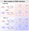
Toscana virus (TOSV), belonging to Phenuiviridae family, is circulating in most Mediterranean countries and is transmitted to humans by infected female sand flies. While most infections are asymptomatic, TOSV is considered as a leading cause of meningitis and encephalitis in humans during summer. Three TOSV genotypes (named A, B, and C) have been identified, although no virus strain belonging to lineage C has been isolated so far. To date, the relationship between TOSV genetic diversity and viral pathogenicity or replication capacity remains unknown. This study aimed to compare two TOSV strains from either lineage A (TOSV-A) or B (TOSV-B) in several cell culture and two mouse models. We showed that TOSV-A replicated more efficiently in BSR and A549 cells, while TOSV-B had a replication advantage in human induced pluripotent stem cells differentiated in neural cells and LL-5 sand fly cells. In vivo, we were unable to detect any virus in the brains of immunocompetent C57BL/6JRj mice infected with either strain of TOSV. On the contrary, we showed that TOSV-A disseminated to the central nervous system of 129/Sv ifnar -/- mice unlike TOSV-B, despite higher viremia of TOSV-B and a greater dissemination of this strain in other organs. The reasons for these differences are not yet known, although we showed that the presence of TOSV neutralizing antibodies in serum was slightly delayed in TOSV-A-infected mice. Altogether, the data presented in this study provide new avenues to study TOSV-induced pathogenesis and ultimately unveil molecular viral determinants modulating TOSV replication capacity.

Keywords: TOSV; Toscana virus; genetic lineage; mouse model; pathogenesis.


Conflict of interest statement

No potential conflict of interest was reported by the author(s).

Figures
None
Graphical abstract
Figure 1.
Figure 1.
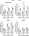
Growth properties of TOSV-A and TOSV-B. Viral growth curves in hIPSc-derived neural cells (MOI ≈ 0.1; 2x10⁴ PFU), LL-5, BSR, and A549 cells (MOI: 0.01) infected by either TOSV-A or TOSV-B. Supernatants were harvested at 24, 48, and 72 hpi, titrated by endpoint dilution and viral titers expressed as TCID50/ml. Comparisons of viral titers at each time point between TOSV-A and TOSV-B were carried out using the Mann–Whitney test; p > 0.05 (ns), p < 0.01 (**), p < 0.001 (***), and p < 0.0001 (****).
Figure 2.
Figure 2.
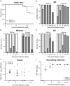
Experimental infection of C57BL/6JRj mice with TOSV-A and TOSV-B. (a) Kaplan-Meier survival curves. 6–8 weeks old female C57BL/6JRj mice (n=12) were infected subcutaneously with 103 PFU of either TOSV-A or TOSV-B. Survival curves were analysed using log-rank (Mantel-Cox) test. (b) Detection of TOSV RNA in the sera (RNAemia). Sera were collected at 3, 5, 7, and 10 dpi. Levels of viral RNA in sera of infected mice were measured by RT-qPCR targeting segment S. Positive mice sera are represented in gray and negative mice sera are represented in white. Note that only 8 mice were analysed at 10 dpi for TOSV-A group because the sera of three mice have not been collected and one mouse died suddenly at 7 dpi. Kinetics of IgM (c) and IgG (d) antibody response against TOSV-A and TOSV-B. Sera were collected at 3, 5, 7, 10, and 13 dpi. Anti-TOSV antibodies within mice sera were detected using in-house IgM and IgG ELISAs. At the indicated day, grey bars represent seroconverted mice and white bars mice with non-detectable anti-TOSV IgM or IgG antibodies. Note that only 10 mice were analysed at 13 dpi for TOSV-A group because the sera of one mouse was not collected and one mouse died suddenly at 7 dpi. (e) Virus neutralisation test. Sera (from one independent experiment only, n=6) containing anti-TOSV IgG and/or IgM were tested for the presence of nAbs against TOSV. The neutralizing titre, expressed as log2, was determined as the highest dilution of serum that caused complete cytopathic effect.
Figure 3.
Figure 3.
Experimental infection of 129/Sv ifnar−/− mice with TOSV-A and TOSV-B. (a) Kaplan-Meier survival curves. 6–8 weeks old female 129/Sv ifnar−/− mice (n=18) were infected subcutaneously with 103 PFU of either TOSV-A or TOSV-B. Survival curves were analysed using log-rank (Mantel-Cox) test. (b) Detection of TOSV RNA in the sera (RNAemia). Sera were collected at 3, 5, 7, and 10 dpi. Levels of viral RNA in sera of infected mice were measured by RT-qPCR targeting segment S. Positive mice sera are represented in gray and negative mice sera are represented in white. Note that only 17 mice were analysed at 8–10 dpi for TOSV-B group because one mouse died suddenly at 8 dpi. Euthanised mice at 8 dpi (one in both TOSV-A and TOSV-B groups) and 9 dpi (one in TOSV-B group) were added to 10 dpi condition (8–10 dpi). (c) TOSV titer in sera (viremia). Viral infectious titers were determined by plaque-forming assay only for RT-qPCR positive sera. Kinetics of IgM (d) and IgG (e) Antibody response against TOSV-A and TOSV-B. Sera were collected at 3, 5, 7, 10 and 13 dpi. Anti-TOSV antibodies within mice sera were detected using in-house IgM and IgG ELISAs. At the indicated day, grey bars represent seroconverted mice and white bars mice with non-detectable anti-TOSV IgM or IgG antibodies. Euthanised mice at 8 dpi (one in both TOSV-A and TOSV-B groups) and 9 dpi (one in TOSV-B group) were added to 10 dpi condition (8–10 dpi), when one mouse in TOSV-A group euthanised at 11 dpi was added to 13 dpi condition (11–13 dpi). (f) Virus neutralisation test. Sera (from one independent experiment only, n=6) containing anti-TOSV IgG and/or IgM were tested for the presence of nAbs against TOSV. The neutralizing titre, expressed as log2, was determined as the highest dilution of serum that caused complete cytopathic effect.
Figure 4.
Figure 4.
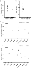
TOSV organ tropism in 129/Sv ifnar−/− mice and viral load in organs/sera. (a) Detection of TOSV RNA in brains of mice euthanised during the experiment (black triangles and circles) or at the end of the experiment (white triangles and circles). Levels of viral RNA were measured by RT-qPCR targeting segment S. (b) Viral loads of TOSV in mouse brains. Viral infectious titers were determined by plaque-forming assay for RT-qPCR positive brains of mice euthanised during the experiment (black triangles) or at the end of the experiment (white triangles and circle). (C) Viral loads of TOSV in mouse organs and serum at 3 (top panel) and 5 (bottom panel) dpi. Mice, subcutaneously infected with 103 PFU of either TOSV-A or TOSV-B, were euthanized at either 3 (white triangles and circles) or 5 dpi (grey triangles and circles). Organs and sera were collected and the presence of TOSV RNA was assessed using RT-qPCR and, if positive, the infectious viral titre (expressed as PFU/g or PFU/ml, respectively) was determined using plaque-forming assay.
Figure 5.
Figure 5.
Growth properties of TOSV-A and TOSV-B in mice OBC. Each OBC, obtained from either C57BL/6JRj or C57BL/6JRj ifnar −/− mice, was infected with 103 PFU of either TOSV-A (n=9 and n=10, respectively) or TOSV-B (n=10, 24 hpi - n=9, 72 hpi, and n=13, respectively). Amount of infectious viral particles were determined by endpoint dilution at 24 and 72 hpi. Mean values are represented and comparisons of viral titers at each time point between TOSV-A and TOSV-B were carried out using the Mann–Whitney test; p > 0.05 (ns), p < 0.05 (*), and p < 0.001 (***).

References

    1. Verani P, Ciufolini MG, Nicoletti L, et al. Ecological and epidemiological studies of toscana virus, an arbovirus isolated from phlebotomus. Annal Dell Supc Sanita. 1982;18(3):397–15. - PubMed
    1. Ayhan N, Prudhomme J, Laroche L, et al. Broader geographical distribution of toscana virus in the Mediterranean region suggests the existence of larger varieties of sand fly vectors. Microorganisms. 2020;8(1):114. doi: 10.3390/microorganisms8010114 - DOI - PMC - PubMed
    1. Charrel RN, Izri A, Temmam S, et al. Toscana virus RNA in Sergentomyia minuta flies. Emerg Infect Dis. 2006;12(8):1299–1300. doi: 10.3201/eid1708.060345 - DOI - PMC - PubMed
    1. Ergunay K, Kasap OE, Orsten S, et al. Phlebovirus and Leishmania detection in sandflies from Eastern Thrace and Northern Cyprus. Parasit Vectors. 2014;7(1):575. doi: 10.1186/s13071-014-0575-6 - DOI - PMC - PubMed
    1. Es-Sette N, Ajaoud M, Anga L, et al. Toscana virus isolated from sandflies, Morocco. Parasit Vectors. 2015;8(1):205. doi: 10.1186/s13071-015-0826-1 - DOI - PMC - PubMed

Publication types

LinkOut - more resources