

Surgical treatment of first branchial cleft anomalies using retrograde facial nerve dissection technique
- PMID: 40694114
- PMCID: PMC12283764
- DOI: 10.1007/s00383-025-06122-7
Surgical treatment of first branchial cleft anomalies using retrograde facial nerve dissection technique
Abstract
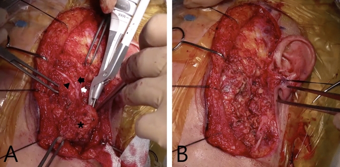
Purpose: First branchial cleft anomalies (FBCAs) are infrequent congenital malformations. In FBCAs removal surgery, due to the previous infection history and the anatomical proximity of the FBCAs tract to the facial nerve, postoperative recurrence and facial paralysis are not uncommon. This study aimed to assess the clinical feasibility and outcomes of FBCAs resection using the retrograde facial nerve dissection technique.
Methods: This retrospective study included 19 patients (mean age, 6.3 ± 4.4 years) who underwent FBCAs excision via retrograde facial nerve dissection between 2017 and 2023. Data on demographics, operative details, histopathology, postoperative complications, and follow-up survey were reviewed.
Results: Preoperative infection history was present in 94.7% of patients; 42.1% had prior incision and drainage and 15.8% had previous excision attempts. Complete resection was achieved in all cases without facial nerve palsy. No recurrence was observed during the follow-up periods (median, 23.9 ± 9.8 months). Postoperative pain and paresthesia showed clinical improvement, while cosmetic satisfaction was relatively limited.
Conclusion: In FBCAs patients, the close proximity of the facial nerve and the adhesion between the tract and facial nerve pose significant challenges. Using retrograde facial nerve dissection is believed to enable complete removal and reduce postoperative facial nerve paralysis.
Keywords: Children; Facial nerve; First branchial cleft anomalies; Retrograde dissection; Surgical treatment.
© 2025. The Author(s).
Conflict of interest statement
Declarations. Conflicts of interest: No potential conflict of interest relevant to this article was reported.
Figures


Similar articles
-
Differences in management outcome for first branchial cleft anomalies: A comparison of infants and toddlers to older children.Int J Pediatr Otorhinolaryngol. 2019 Jul;122:161-164. doi: 10.1016/j.ijporl.2019.04.009. Epub 2019 Apr 11. Int J Pediatr Otorhinolaryngol. 2019. PMID: 31029951
-
Management of a First Branchial Cleft Fistula in an Adult Patient: A Case Report and Literature Review.Cureus. 2025 Jul 11;17(7):e87756. doi: 10.7759/cureus.87756. eCollection 2025 Jul. Cureus. 2025. PMID: 40792327 Free PMC article.
-
The treatment for the first branchial cleft anomalies in children.Eur Arch Otorhinolaryngol. 2017 Sep;274(9):3465-3470. doi: 10.1007/s00405-017-4648-y. Epub 2017 Jun 20. Eur Arch Otorhinolaryngol. 2017. PMID: 28634783
-
Interventions for treating supracondylar elbow fractures in children.Cochrane Database Syst Rev. 2022 Jun 9;6(6):CD013609. doi: 10.1002/14651858.CD013609.pub2. Cochrane Database Syst Rev. 2022. PMID: 35678077 Free PMC article.
-
Congenital first branchial cleft anomalies in children: a study of 100 surgical cases and a review of the literature.Eur Arch Otorhinolaryngol. 2023 Jan;280(1):425-433. doi: 10.1007/s00405-022-07607-0. Epub 2022 Aug 30. Eur Arch Otorhinolaryngol. 2023. PMID: 36040517 Review.
References
-
- Liu W, Chen M, Hao J, Yang Y, Zhang J, Ni X (2017) The treatment for the first branchial cleft anomalies in children. Eur Arch Otorhinolaryngol 274(9):3465–3470. 10.1007/s00405-017-4648-y - PubMed
-
- D’Souza ARUH, Ranit De, Zeitoun H (2002) Updating concepts of first branchial cleft defects: a literature review. Int J Pediatr Otorhinolaryngol 62:103–109 - PubMed
-
- Liston SL (1982) The relationship of the facial nerve and first branchial cleft anomalies–embryologic considerations. Laryngoscope 92(11):1308–1310. 10.1288/00005537-198211000-00017 - PubMed
-
- Yang R, Dong C, Chen Y, Liu H, Zhang C, Lai Y et al (2022) Analysis of the clinical features and surgical outcomes of first branchial cleft anomalies. Laryngoscope 132(5):1008–1014. 10.1002/lary.29896 - PubMed
MeSH terms
Supplementary concepts
Grants and funding
LinkOut - more resources
Full Text Sources
Medical
