. 2025 Jul;106(7):002123.
doi: 10.1099/jgv.0.002123.

Marek's disease virus replication in chicken skin reconstructed in vitro: evidence for viral particles in corneocytes

Affiliations

Marek's disease virus replication in chicken skin reconstructed in vitro: evidence for viral particles in corneocytes

Laurent Souci et al. J Gen Virol. 2025 Jul.

Abstract

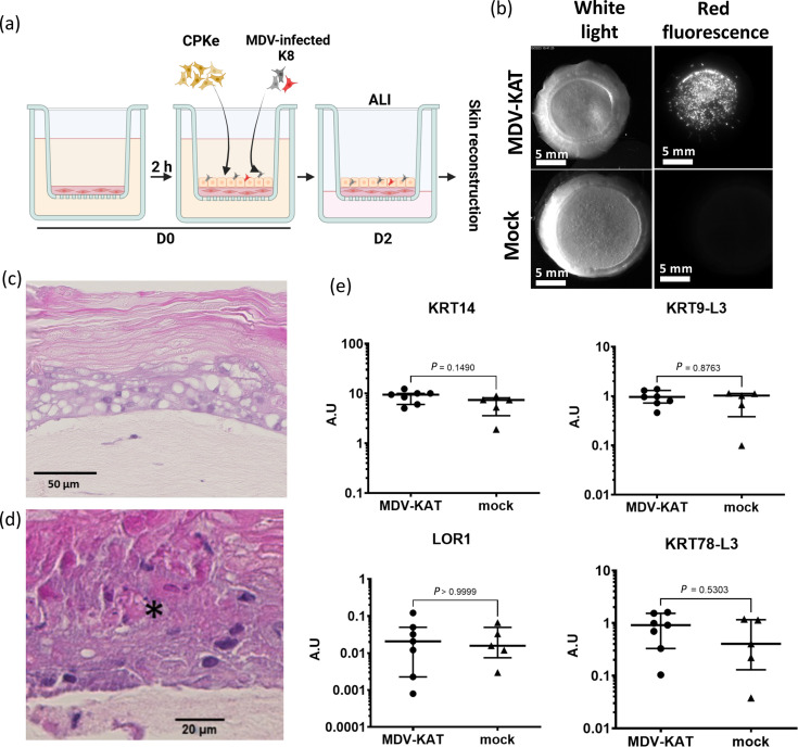
Marek's disease (MD) is a lethal lymphoma of chickens, which is caused by MD virus (MDV), an alphaherpesvirus. MDV infects epithelial cells of the skin appendages, notably feather follicles, replicates in these cells and is shed into the environment exclusively from these tissues. Here, we tested whether chicken skin equivalents (SEs) can be used to model MDV infection. Primary chicken keratinocytes were seeded on a suspension of fibroblasts in collagen and induced to terminally differentiate at the air-liquid interface. A recombinant MDV expressing the Katushka fluorescent protein (MDV-KAT) was introduced into SEs by seeding primary keratinocytes together with MDV-KAT-infected keratinocytes of the K8 cell line. KAT-mediated fluorescence increased during the culture of infected SEs, indicating virus infection and replication, while the expression of keratinocyte differentiation markers was not significantly altered by MDV infection. MDV did not spread to the dermal compartment of SEs but localized to the upper layers of the epidermis. Viral particles were readily observed by electron microscopy in living keratinocytes and for the first time in cornified keratinocytes of the outermost layer of infected SEs, suggesting that viral elements can be released into the environment. Finally, we demonstrated that two fluorescent vaccine strains of MDV, Rispens and herpesvirus of turkey, can infect and replicate in SEs. Taken together, this study establishes chicken SEs as an in vitro model for essential steps of MDV infection.

Keywords: 3D in vitro models; Marek’s disease virus; chicken; herpesvirus; keratinocyte; viral particles.


Conflict of interest statement

The authors declare that there are no conflicts of interest.

Figures

Fig. 1.
Fig. 1.. Validation of reconstructed chicken skin, named SEs, using chicken embryonic cells. (a) Schematic overview of the SE reconstruction. (A) CPKe and dermal fibroblasts were isolated, respectively, from the epidermis and the dermis of leg skin harvested from 19-day-old WL embryos. (B) SEs were produced in an insert, by using a fibroblast (Fb)-populated collagen matrix (salmon colour) on the top of which CPKe were seeded. Cells were cultured for 2 days immerged before being lifted at ALI. ALI facilitates keratinocyte differentiation and stratification into multiple layers, ultimately forming a cornified SE. The SE reconstruction was ended at 14 days post-seeding (D14). SE reconstruction was also performed by mixing K8 cells (a chicken keratinocyte cell line derived from chicken embryonic stem cells) with CPKe at the seeding step (D0). (b) Histological analysis. Images of haematoxylin-phloxin-safranin-stained sections of SE, performed with CPKe alone (left panel) or CPE supplemented with K8 (right panel). Small black arrowheads indicate vacuoles. (c) The transcript expression of skin differentiation markers in the SE was analysed by quantitative PCR, with chicken leg skin (noted skin on graphs), serving as control. In the graphs, each symbol represents independent samples (n=3 to 5). mRNA levels for each gene were normalized to the housekeeping gene RPS17, were expressed in arbitrary unit (A.U.) and were presented in a dot plot showing median values±interquartile range. The Kruskal–Wallis test with a Dunn correction was performed, using the chicken skin as a reference. Exact P-values are indicated on graphs. (d) Cryosections of SE with K8 were stained with an anti-KRT type I antibody (11E10) plus a secondary antibody labelled with Alexa Fluor 555. Nuclei (DNA) were stained with Hoechst 33342. Samples were observed by fluorescence microscopy. The red fluorescent signal indicated that KRT14 was localized in the lower layers of the epidermis (mostly basal layer). The nuclei are visible in the basal and suprabasal layers and very rarely not in the cornified layer.
Fig. 2.
Fig. 2.. Generation and characterization of the MDV-KAT virus in vitro. (a) Overview of the MDV RB-1B genome with a focus on the mini-F cassette with the Katushka fluorescent reporter gene under the TK promotor (MDV-KAT), shown as a red arrow. Image of MDV-KAT-infected CESCs (b) and MDV-KAT-K8 cells (c) showing bright red fluorescent plaques. Scales bars, 500 µm. (d) Plaque size assays of the indicated viruses 4 days post-infection in CESCs. The plaque diameter is shown as bars with mean and sd. P>0.5 (ns, non-significant), Mann–Whitney test (n=50).
Fig. 3.
Fig. 3.. MDV efficiently infects and replicates in SE. (a) Schematic overview of the SE infection with MDV. (b) Morphological macroscopic aspect under white light of SE infected with the very virulent red fluorescent MDV-KAT or mock-infected SE (with K8) at D14 post-seeding and infection (scale bar, 6 mm; 250-ms exposure time). Red fluorescent imaging of MDV-KAT and mock-infected SE using a fluorescence stereomicroscope to evaluate MDV infection (scale bar, 6 mm; 250-ms exposure time). (c, d) Histological analysis included H- and E-stained sections of MDV-KAT-infected SE at two magnifications. In (d), a lesion is highlighted by an asterisk. (e) The mRNA expression levels of skin differentiation markers in MDV-KAT and mock-infected SE were quantified. Each symbol corresponds to one SE, performed on four independent experiments (MDV-KAT, n=7; mock, n=3) presented here (for the mock, same data shown in Fig. 1). mRNA levels for each gene were normalized to the housekeeping gene RPS17, were expressed in A.U. and were presented in a dot plot showing median values±interquartile range.
Fig. 4.
Fig. 4.. MDV infection in SE followed by fluorescence imaging and viral genome loads at different timepoints. SEs were infected at D0 with MDV-KAT, and viral infection was assessed into SEs throughout time, during reconstruction (D2, D6, D9 and D14 post-seeding time). (a) SEs were imaged using a fluorescent stereomicroscope. (A) The entire SEs were imaged using the same exposure time. (B) Portions of SE were imaged at higher magnification (×80). (C) Quantification of the red fluorescent signal on independent SEs (n=1 to 3) from a single kinetic experiment. (b) Histological structure of MDV-infected SEs at indicated timepoints. Formalin-fixed SEs embedded in paraffin were sectioned and stained in HPS. The number of layers in the epidermis increased over time as expected and in consequence its thickness (indicated on the side of enlarged panels). The corneum layer started to appear at D9 and was highly developed at D14. (c) Viral genome loads of infected SE were determined at different timepoints. DNA was extracted from SE and viral genomes were quantified by real-time qPCR, and their numbers were indicated per million nucleated cells. The cell number determination is based on the detection of iNos cellular DNA sequence. Results are presented in a dot plot showing median values±interquartile range. To show inter- and intra-experiment variations between SEs, each independent experiment was represented in a different colour (three independent assays are shown) and each SE with a symbol. Two similar symbols of the same colour mean quantification from two parts of the same SE.
Fig. 5.
Fig. 5.. MDV is located in the upper layers of the reconstructed epidermis, notably in corneocytes. (a) MDV-KAT-infected SE (at D14) was treated with thermolysin, and the reconstructed epidermis was separated from the dermal equivalent. Both the dermis and epidermis were imaged using a fluorescent stereomicroscope under white light (upper panel) and fluorescence (lower panel). The epidermis showed numerous fluorescent infection plaques, but no fluorescence signal was observed in the dermis. (b) Cryosections of MDV-KAT-infected SEs were stained with Hoechst and directly observed by confocal microscopy. The red fluorescent signal indicates that the infection was localized in the suprabasal layers of the epidermis. The white dashed line indicates the boundary between the epidermis and dermis. The white arrows pointed to red fluorescent cells with a typical morphology of corneocytes. (c) Corneocytes were purified from the entire reconstructed epidermis infected with MDV-KAT, stained with Hoechst 33342 dye and subsequently examined by light and fluorescence microscopy. The cells had the shape of corneocytes with no nuclei as expected. (d) Viral genome loads in corneocytes. DNA was extracted from purified corneocytes, and viral loads per 100 ng of total DNA were quantified by RT-qPCR using the ICP4 gene. No normalization relative to iNos copy number was performed due to cellular DNA degradation during final keratinocyte differentiation into corneocytes.
Fig. 6.
Fig. 6.. Viral particles are observed in the inner layers of the epidermis by TEM. MDV-KAT-infected SEs at D14 were collected and subsequently analysed at ultrastructural level (a). Image of the epidermis. The four layers with keratinocytes at different stages of differentiation are visible. Note that the cornified layer is highly electron dense. (b) Image of a zone with three keratinocytes (in framed), inside which viral particles were observed. All viral particles were nuclear capsids, of the three types (a, b, c). An enlargement of the cell framed in yellow is shown on the right panel. Yellow triangles indicate capsids. (c). Image of a zone where complete viral particles, indicated by orange arrows, are visible inside vesicles. An enlargement of a complete particle is shown on the right panel.
Fig. 7.
Fig. 7.. Examination of the cornified layer by fluorescence stereomicroscopy and TEM. The cornified layer was harvested on fresh MDV-KAT-infected SE at D14 and subsequently analysed. (a) Image of the freshly harvested material under a fluorescent microscope on the red channel. Numerous small MDV plaques are visible. (b) Examination of the harvested material at ultrastructural level by TEM. The material has the aspect of several layers of cornified cells as expected for the cornified layer. The cells were highly electron dense as observed earlier in the cornified layer on entire SEs. (c, d) Images of corneocytes at ultrastructural level, in which viral particles were observed. Herpesvirus capsids (A, B, C) are visibly surrounded by cellular fibres. No nucleus was visible. The yellow triangles indicated particles.
Fig. 8.
Fig. 8.. SEs are infectable by vaccinal MD strains, Rispens and HVT. SEs were produced by mixing CPKe with K8 cells infected with recombinant fluorescent vaccinal strains, Rispens mCherry or HVT FarRed, as performed for MDV. Analyses were performed on reconstructed SEs at day 14. (a, b) Red fluorescence images of infected SEs were performed using a fluorescent stereomicroscope to detect infection, with Rispens mCherry (a) or HVT FarRed (b). (c, d) HPS stained sections for both infected SEs, with Rispens mCherry (c) or HVT FarRed SE (d). (e, f) Viral load measurement (viral genome copies/106 cells) in SEs, with Rispens mCherry (e) or HVT FarRed (f).

Similar articles

References

    1. Souci L, Denesvre C. Interactions between avian viruses and skin in farm birds. Vet Res. 2024;55:54. doi: 10.1186/s13567-024-01310-0. - DOI - PMC - PubMed
    1. Souci L, Denesvre C. 3D skin models in domestic animals. Vet Res. 2021;52:21. doi: 10.1186/s13567-020-00888-5. - DOI - PMC - PubMed
    1. Randall MJ, Jüngel A, Rimann M, Wuertz-Kozak K. Advances in the biofabrication of 3D skin in vitro: healthy and pathological models. Front Bioeng Biotechnol. 2018;6:154. doi: 10.3389/fbioe.2018.00154. - DOI - PMC - PubMed
    1. Niehues H, Bouwstra JA, El Ghalbzouri A, Brandner JM, Zeeuwen PLJM, et al. 3D skin models for 3R research: the potential of 3D reconstructed skin models to study skin barrier function. Exp Dermatol. 2018;27:501–511. doi: 10.1111/exd.13531. - DOI - PubMed
    1. Lachner J, Derdak S, Mlitz V, Wagner T, Holthaus KB, et al. An in vitro model of avian skin reveals evolutionarily conserved transcriptional regulation of epidermal barrier formation. J Invest Dermatol. 2021;141:2829–2837. doi: 10.1016/j.jid.2021.04.029. - DOI - PubMed

LinkOut - more resources