. 2025 Jul 22;19(7):e0013317.
doi: 10.1371/journal.pntd.0013317. eCollection 2025 Jul.

STAT1-mediated interferon signaling in the hematopoietic system is essential for restricting Usutu virus infection in vivo

Affiliations

STAT1-mediated interferon signaling in the hematopoietic system is essential for restricting Usutu virus infection in vivo

Amy N Nelson et al. PLoS Negl Trop Dis. .

Abstract

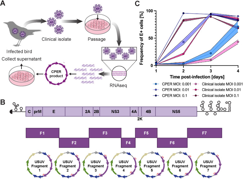
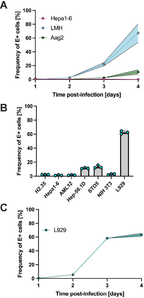
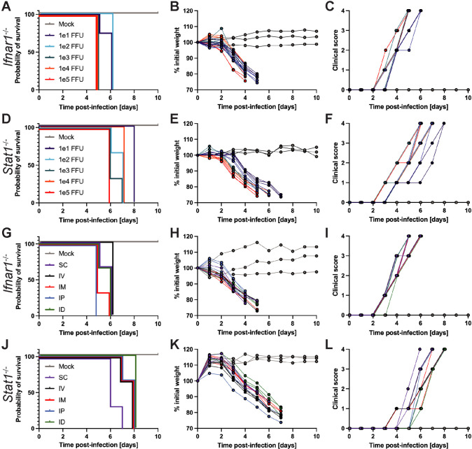
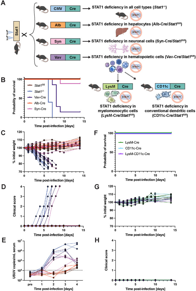
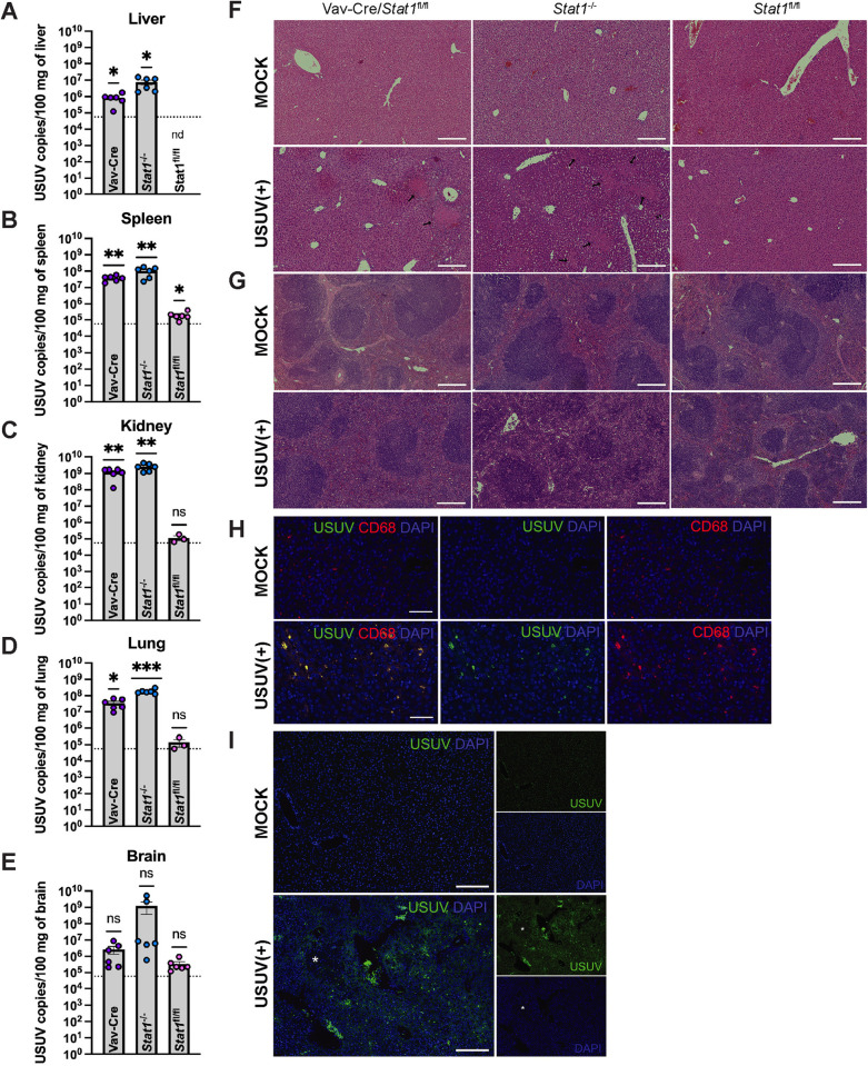
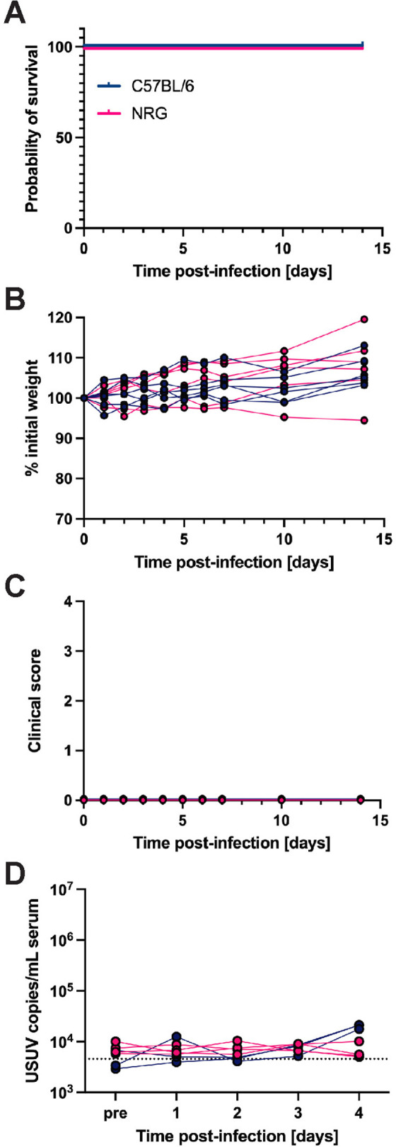
Usutu virus (USUV) is an emerging mosquito-borne flavivirus known to induce neuroinvasive disease in birds, mice, and humans in European and African countries. The mechanisms of infection and dissemination remain poorly understood. Thus, elucidating how USUV spreads in a susceptible host is crucial for identifying therapeutic targets. To investigate host defenses against USUV, we generated an infectious clone of the TC508 isolate. After characterizing its replication dynamics in cultured cells from multiple species, we investigated its pathogenesis in an array of mice with genetic perturbations. Previous studies demonstrated that whole-body deletion of type I interferon (IFN) signaling led to widespread USUV infection and fatality in mice. Here, we observed the same lethal phenotype in STAT1-deficient mice and identified hematopoietic cells specifically as central to USUV pathogenesis in a mammalian host. Deletion of STAT1 in all hematopoietic subsets, but not hepatocytes, neurons, macrophages or conventional dendritic cells, was sufficient for systemic viral dissemination and ultimate fatality. Conversely, mice lacking functional B, T, and natural killer (NK) cells but with intact myeloid cells were resistant to USUV. Our findings provide new insights into the tissue-specific barriers that regulate USUV infection and underscore the importance of innate immunity in host defense for this important emerging flavivirus.


Conflict of interest statement

The authors of this manuscript have read the journal’s policy and declare the following competing interests. R.E.S. is on the scientific advisory boards of Miromatrix Inc. and Lime Therapeutics and is a speaker and consultant for Alnylam Inc.

Figures

Fig 1
Fig 1. A genetically-defined infectious clone of USUV TC508 was generated using the circular polymerase extension reaction (CPER) and remains infectious in cell culture.
(A) Schematic representation of the workflow for generating an USUV infectious clone from an avian clinical isolate. Some figure elements (bird [64], cells [65], and DNA fragments [66]) were sourced from the public domain and are listed as references. (B) Schematic representation of the USUV genome and its division into seven fragments used for CPER. Each fragment was inserted into pCR-Blunt II-TOPO vectors. The purple arrows represent the USUV genes. Other vector components include lacZa (blue), ccdB (blue), antibiotic resistance markers (green), ori (sand), and the lac promoter (gray). The red half arrows indicate the PCR primer binding sites. (C) Replication kinetics of the USUV TC508 infectious clone in human hepatoma (Huh7) cells 1–4 days post-infection (dpi) using a multiplicity of infection (MOI) of 0.001, 0.01, or 0.1. The percentage of infected cells was quantified based on envelope (E) antigen staining detected by flow cytometry. Shading indicates standard error of the mean.
Fig 2
Fig 2. USUV TC508 has variable Infection kinetics in murine, avian, and mosquito cell lines.
(A) Percent of E antigen-positive cells in mouse hepatoma (Hepa1-6), chicken hepatoma (LMH), and embryonic mosquito cells (Aag2) 1–4 days post-infection (dpi) using a multiplicity of infection (MOI) of 0.01. Shading indicates standard error of the mean. (B) Percent of E antigen-positive cells in various murine cell lines at 4 dpi using an MOI of 0.01. (C) Percent of E antigen-positive cells in mouse fibroblasts (L929) 1–4 dpi using an MOI of 0.01. Shading indicates standard error of the mean.
Fig 3
Fig 3. USUV infection is lethal in mice with compromised type I interferon or STAT1-mediated antiviral defenses regardless of inoculation dose or route.
The inoculation doses tested ranged from 101 to 105 focus-forming units (FFU), all of which were administered via subcutaneous (SC) route. The inoculation routes tested were SC, intravenous (IV), intramuscular (IM), intraperitoneal (IP), and intradermal (ID) using a consistent inoculation dose of 103 FFU. (A) Survival, (B) weight changes, and (C) clinical scoring of Ifnar1-/- mice following inoculation with varying doses of USUV via SC injection (101 FFU, n = 4; 102 FFU, n = 4; 103 FFU, n = 7; 104 FFU, n = 3; 105 FFU, n = 3). (D) Survival, (E) weight changes, and (F) clinical scoring of Stat1-/- mice following inoculation with varying doses of USUV via SC injection (n = 3/group). (G) Survival, (H) weight changes, and (I) clinical scoring of Ifnar1-/- mice following inoculation with 103 FFU of USUV by various routes (n = 3/group). (J) Survival, (K) weight changes, and (L) clinical scoring of Stat1-/- mice following inoculation with 103 FFU of USUV by various routes (n = 3/group).
Fig 4
Fig 4. USUV infection in tissue-specific STAT1-deficient mouse strains demonstrates a necessity for functional STAT1-mediated signaling in the hematopoietic compartment.
(A) Schematic representation of the breeding scheme used to generate whole-body, hepatocyte-specific, neuronal cell-specific, hematopoietic cell-specific, macrophage-specific, or dendritic cell-specific STAT1 KO mice. Wild-type mice harboring loxP sites within the Stat1 gene (Stat1fl/fl) were crossed with mice containing the Cre recombinase downstream of a ubiquitous (CMV) or tissue-specific (Alb, Syn, Vav, LysM, CD11c) promoter. Some figure elements (mouse [69], liver [70], neuron [71], and immune cells [–77]) were sourced from the public domain and are listed as references. (B) Survival, (C) weight changes, and (D) clinical scoring of Stat1fl/fl (n = 11), Stat1-/- (n = 10), Vav-Cre/Stat1fl/fl (n = 7) Alb-Cre/Stat1fl/fl (n = 6), and Syn-Cre/Stat1fl/fl (n = 9) mice following inoculation with 103 focus-forming units (FFU) of USUV TC508 via SC injection. (E) Changes in viremia 1–4 days post-infection compared to a pre-bleed sample (“pre”) collected prior to infection (n = 3/mouse genotype). (F) Survival, (G) weight changes, and (H) clinical scoring of LysM-Cre/Stat1fl/fl (n = 8), CD11c-Cre/Stat1fl/fl (n = 8), and LysM-CD11c-Cre/Stat1fl/fl (n = 4) following inoculation with 103 FFU of USUV TC508 via SC injection.
Fig 5
Fig 5. USUV replicates in STAT1-sufficient peripheral tissues leading to spleen and liver pathology.
Vav-Cre/Stat1fl/fl, Stat1-/-, and Stat1fl/fl mice (n = 3/group) were infected with 103 focus-forming units (FFU) of USUV via subcutaneous (SC) injection and euthanized at 4 days post-infection (dpi). Tissues were collected and subjected to molecular and histological analysis. For each mouse genotype, one representative image was chosen. USUV RNA was quantified in the (A) liver, (B) spleen, (C) kidney, (D) lung, and (E) brain of Vav-Cre/Stat1fl/fl, Stat1-/-, and Stat1fl/fl mice by qPCR. Significant levels of USUV RNA were detected in the liver, spleen, kidney, and lung of Vav-Cre/Stat1fl/fl and Stat1-/- mice. In some samples collected from Stat1fl/fl mice (6 out of 6 liver samples, 5 out of 6 spleen samples, and 3 out of 6 kidney samples), the amount of USUV RNA was below the limit of detection and was not plotted (nd indicates “not detected”). For each organ harvested from infected mice, two cuts were processed and used as separate sample inputs for qPCR. The dotted line indicates the limit of detection. Statistical analysis was performed using one sample t tests comparing USUV RNA detected in samples to the limit of detection. (F) H&E staining of liver tissue collected from mock or USUV-infected mice with a 200 µm scale bar. Liver necrosis was observed in Vav-Cre/Stat1fl/fl and Stat1-/- mice and is indicated by black arrows. (G) H&E staining of spleen tissue collected from mock or USUV-infected mice with a 200 µm scale bar. Decreased red pulp due to massive immune cell infiltration was observed in Vav-Cre/Stat1fl/fl and Stat1-/- mice. H&E staining of kidney, lung, and brain tissues can be found in S1 Fig. (H) Immunostaining of liver tissue harvested from a Stat1-/- mouse using the histiocyte marker CD68, anti-JEV NS3 to mark USUV infection, and DAPI with a 50 µm scale bar. (I) Liver tissue harvested from a Stat1-/- mice and stained with anti-JEV NS3 and DAPI with a 200 µm scale bar. The white asterisk indicates an inflammatory lesion.
Fig 6
Fig 6. B, T, and natural killer cells are dispensable for restricting USUV infection.
(A) Survival, (B) weight changes, and (C) clinical scoring of NRG (n = 6) and wild-type C57BL/6 (n = 6) mice following inoculation with 103 focus-forming units (FFU) of USUV TC508 via subcutaneous (SC) injection. (D) Changes in viremia 1–4 days post-infection (dpi) compared to a pre-bleed sample (“pre”) collected prior to infection in NRG (n = 4) and C57BL/6 (n = 3) mice.
Fig 7
Fig 7. Administration of IFN-beta (IFN-β) partially protects Vav-Cre/Stat1fl/fl mice from lethal USUV infection.
(A) Survival, (B) weight changes, and (C) clinical scoring of Vav-Cre/Stat1fl/fl mice that received 104 IU IFN-β via SC injection at two timepoints before or after infection with 103 FFU of USUV TC508. The pre-treatment group (n = 6) received IFN-β injections at 2 hours prior to USUV infection and 24 hours post-infection (hpi). One post-treatment group received IFN-β injections 6 and 24 hpi (n = 8) and the second post-treatment group received IFN-β injections at 24 and 48 hpi (n = 7).

Similar articles

References

    1. McIntosh BM. Usutu (SA Ar 1776), nouvel arbovirus du groupe B. Int Catalogue Arboviruses. 1985;3:1059–60.
    1. Diagne MM, Ndione MHD, Di Paola N, Fall G, Bedekelabou AP, Sembène PM, et al. Usutu Virus Isolated from Rodents in Senegal. Viruses. 2019;11(2):181. doi: 10.3390/v11020181 - DOI - PMC - PubMed
    1. Nikolay B, Diallo M, Boye CSB, Sall AA. Usutu virus in Africa. Vector Borne Zoonotic Dis. 2011;11(11):1417–23. doi: 10.1089/vbz.2011.0631 - DOI - PubMed
    1. Williams MC, Simpson DI, Haddow AJ, Knight EM. The isolation of West Nile virus from man and of Usutu virus from the bird-biting mosquito Mansonia aurites (Theobald) in the Entebbe area of Uganda. Ann Trop Med Parasitol. 1964;58:367–74. - PubMed
    1. Fros JJ, Miesen P, Vogels CB, Gaibani P, Sambri V, Martina BE, et al. Comparative Usutu and West Nile virus transmission potential by local Culex pipiens mosquitoes in north-western Europe. One Health. 2015;1:31–6. doi: 10.1016/j.onehlt.2015.08.002 - DOI - PMC - PubMed

Supplementary concepts