. 2025 Jul 22;21(7):e1013275.
doi: 10.1371/journal.ppat.1013275. eCollection 2025 Jul.

A chaperonin complex regulates organelle proteostasis in malaria parasites

Affiliations

A chaperonin complex regulates organelle proteostasis in malaria parasites

Amanda Tissawak et al. PLoS Pathog. .

Abstract

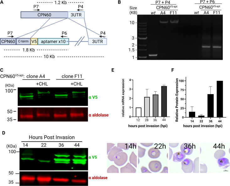
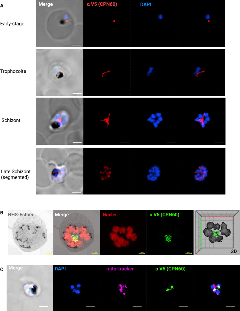
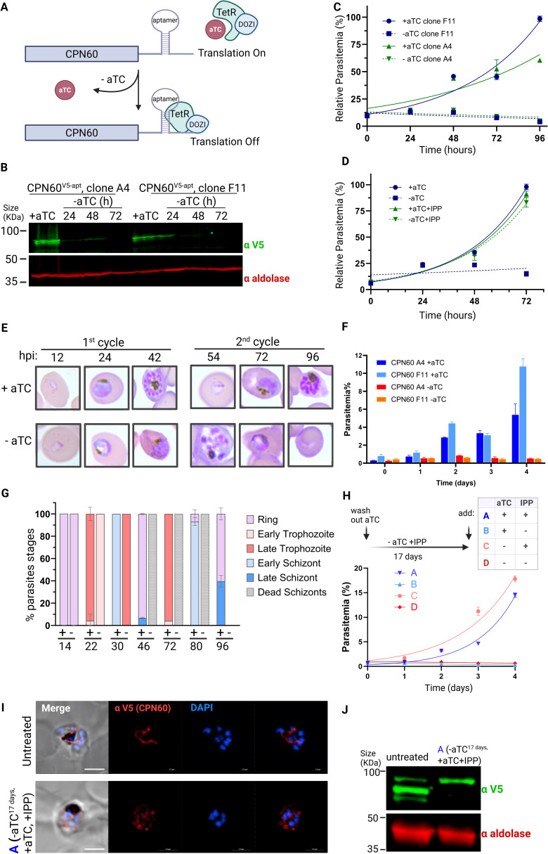
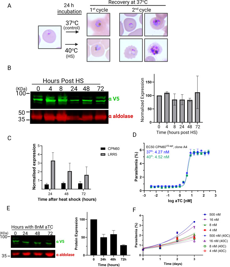
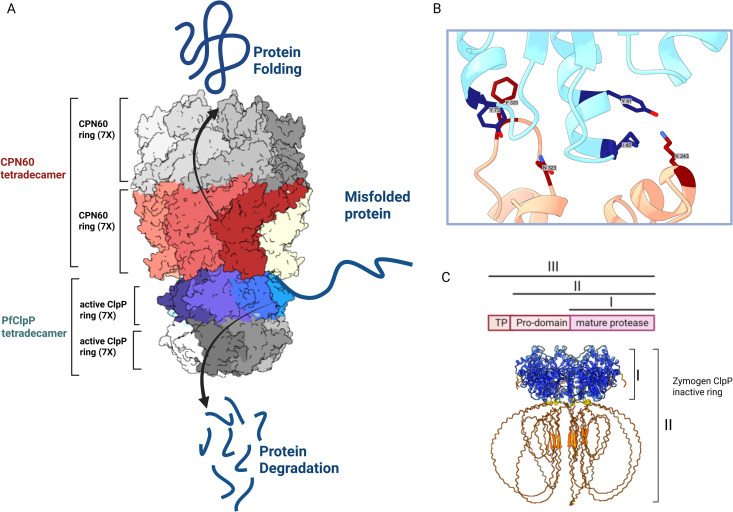
The apicoplast of Plasmodium parasites serves as a metabolic hub that synthesize essential biomolecules. Like other endosymbiotic organelles, 90% of the apicoplast proteome is encoded by the cell nucleus and transported to the organelle. Evidence suggests that the apicoplast has minimal control over the synthesis of its proteome and therefore it is unclear how organelle proteostasis is regulated. Here, we identified and investigated a large and conserved chaperonin (CPN) complex with a previously unknown function. Using genetic tools, we demonstrated that ablation of the apicoplast CPN60 subunit leads to parasite death due to organellar damage, immediately within its first replication cycle, deviating from the delayed death phenotype commonly observed for apicoplast translation inhibitors. Unlike its close orthologues in other prokaryotic and eukaryotic cells, CPN60 is not upregulated during heat shock (HS) and does not affect HS response in the parasite. Instead, we found that it is directly involved in proteostasis through interaction with the Clp (caseinolytic protease) proteolytic complex. We showed that CPN60 physically binds both the active and inactive forms of the Clp complex, and manipulates its stability. A computational structural model of a possible interaction between these two large complexes suggests a stable interface. Finally, we screened a panel of inhibitors for the bacterial CPN60 orthologue GroEL, to test the potential of chaperonin inhibition as antimalarial. These inhibitors demonstrated an anti-Plasmodium activity that was not restricted to apicoplast function, with additional targets outside of this organelle. Taken together, this work reveals how balanced activities of proteolysis and refolding safeguard the apicoplast proteome, and are essential for organelle biogenesis.


Conflict of interest statement

I have read the journal's policy and an author of this manuscript have the following competing interests: S.M.J. is a founder of BioEL Inc, which was formed to commercialize GroEL inhibitors for antibacterial applications.

Figures

Fig 1
Fig 1. Generation and characterization of transgenic CPN60V5-apt parasites.
A. Schematic representation of CRISPR-mediated homologous recombination through CPN60 homology regions enabling the integration of the V5 tag and the tetR-Dozi system. B. PCR amplification of genomic DNA isolated from CPN60V5-apt transfected parasites. Primers used are depicted in panel A and in Table 1. Primers are integration specific and thus can amplify 1.6 KB (P7 + P6) and 11 KB (P7 + P4) products only if integration occurs at the desired genomic locus. C. Western blot depicting two clones of CPN60V5-apt parasites (A4 and F11) following 96-hour incubation with or without 30 μM chloramphenicol (CHL) using an anti-V5 antibody and anti-aldolase as a loading control. D. CPN60V5-apt parasites were synchronized by incubation with 5% sorbitol followed by Percoll-mediated schizont-enrichment the following day. Western blot depicting expression profile of tagged CPN60 throughout parasite life cycle using an anti-V5 antibody. Right: Blood smears demonstrate parasites’ developmental stage at the time of protein extraction. E. CPN60V5-apt parasite clones (A4 and F11) were synchronized using 5% sorbitol, and 8ml of culture were collected at the following time points: early ring stage (~12 h post-invasion), early trophozoite stage (~28 hpi), late trophozoite stage (~36 hpi), and schizont stage (~44 hpi). Parasites were liberated from host red blood cells using 0.1% saponin, and total mRNA was extracted and reverse-transcribed to cDNA. Quantitative real-time PCR (qRT-PCR) was performed to quantify transcript levels of the apicoplast-encoded chaperonin CPN60. Primers P14 + P15 were used to amplify CPN60 and two housekeeping genes (aldolase P16 + P17 and arg-tRNA synthetase P18 + P19) for normalization. Gene expression was normalized to the mean ΔCt of both housekeeping genes (aldolase and arg-tRNA synthetase), and expression levels were calculated relative to the early ring stage using the ΔΔCt method. Data are presented as fold change (2^–ΔΔCt). Values represent the mean expression from two parasite lines, and error bars indicate the standard deviation. Statistical analyses were performed using GraphPad Prism. F. Densitometric analysis of protein bands from Western blot in 1D. V5 signal was normalized to aldolase. Figure created by Biorender.
Fig 2
Fig 2. Organellar Localization and expression of CPN60.
A. Immunofluorescence microscopy of CPN60V5-apt parasites in different developmental stages. Z stack images processed as Maximum Intensity show from left to right: merged DIC contrast, anti-V5 (CPN60, red), DAPI (parasite nucleus, blue), and merge of fluorescent channels. The imaging was performed using a Nikon Spinning Disk confocal fluorescence microscope equipped with a 1005 x/1.4NA objective. Scale bar is 2.5 μM. B. A representative image of an expanded parasite demonstrates uniform apicoplast localization of CPN60 in a well-developed organelle during mid-schizogony (eight nuclei). Images from left: NHS-Esther (total protein, grey), merge, DNA (red), anti-V5 antibody (CPN60, green), and a 3D visualization. Images were captured as Z-stacks (20 slices of 0.1 μm each) at 63x objective using Airyscan microscopy. The images are displayed as maximum intensity projections. Expansion factor is 5X. Scale bar is 5 μm. C. Immunofluorescence microscopy of CPN60V5-apt parasites processed as Maximum Intensity show from left to right: merged DIC contrast, DAPI (parasite nucleus, blue), MitoTracker Deep Red FM (mitochondrion, magenta), anti-V5 antibody (CPN60, green), and merge of fluorescentt channels. The imaging was performed using a Nikon Spinning Disk confocal fluorescence microscope equipped with a 100x/1.4NA objective. Scale bar is 2.5 μM.
Fig 3
Fig 3. CPN60 is essential for parasite’s viability due to its apicoplast function.
A. Schematic representation of the tetR-Dozi system, which enables protein knockdown upon removal of the tetracycline analogue aTC. B. Western blots depicting the expression of the tagged CPN60 using an anti-V5 antibody following knockdown induction. A significant reduction in protein levels is already observed after 24 hours, and falls below detection 72 hours after aTC removal. C. Growth curve of CPN60V5-apt parasites. Independent CPN60V5-apt clones A4 and F11 were grown with or without 0.5 mM aTC, and parasitemia was monitored every 24 h over two cycles via flow cytometry. The 100% of growth represents the highest value of calculated parasitemia (final parasitemia in the presence of aTC). Normalized data are represented as mean ± SEM for three technical replicates. D. Growth curve of CPN60V5-apt parasites, clone A4, supplemented with 200 μM IPP. The 100% of growth represents the highest value of calculated parasitemia (final parasitemia in the presence of aTC). Normalized data are represented as mean ± SEM for three technical replicates. E. CPN60V5-apt parasites were synchronized by incubation with 5% sorbitol followed by Percoll-mediated schizont-enrichment the following day. Synched parasites were washed 8 times and then incubated with or without aTC. Giemsa-stained blood smears were imaged using an upright Eclipse E200 Microscope. F. Growth rates of synchronous CPN60V5-apt parasites. Clones A4 and F11 were grown with or without 0.5 mM aTC, and parasitemia was monitored every 24 h over two cycles via flow cytometry. Data are represented in columns as mean ± SEM for three technical replicates. G. Blood smears of synched CPN60V5-apt parasites with or without aTC were analyzed for specific parasites developmental stages. Minus aTC parasites demonstrate a delayed trophozoite stage already on the first replication cycle, fail to egress, and die during schizogony. Dead schizonts are defined as morphologically aberrant mutant cells with more than one nucleus that linger for more than 48 hours but did not complete their first replication cycle. H. Parasites were cultured without aTC and supplemented with IPP for 17 days. Parasites culture was then divided into four groups treated as following: (A) +aTC, + IPP; (B) +aTC, -IPP; (C) -aTC, + IPP; (D) -aTC, -IPP. Parasitemia of these four groups was monitored every 24 hours over two cycles via flow cytometry. Normalized data are represented as mean ± SEM for three technical replicates. I. Immunofluorescence microscopy of untreated and group (A) CPN60V5-apt parasites. Z stack images processed as Maximum Intensity show from left to right: merged DIC contrast, anti-V5 (CPN60, red), DAPI (parasite nucleus, blue), and merge of fluorescentt channels. The imaging was performed using a Nikon Spinning Disk confocal fluorescence microscope equipped with a 100x/1.4NA objective. Scale bar is 2.5 μM. J. Western blot depicting expression of untreated and group (A) CPN60V5-apt parasites using an anti-V5 antibody and anti-aldolase as a loading control.
Fig 4
Fig 4. CPN60 is not involved in Heat Shock response.
A. Workflow of HS experiments and representative blood smears. Parasites are subjected to HS at 40oC for 24 hours and then allowed to recover at normal temperature (37oC). B. Parasites were subjected to HS and recovery as depicted in (A). Left: Cellular lysates were extracted every 24 hours over a span of four days and subjected to Western blot analysis using an anti-V5 antibody to follow tagged CPN60 levels. Right: Quantification of CPN60 levels post-heat shock demonstrate no significant increase in protein levels. C. P. falciparum culture was subjected to heat shock at 40°C for 24 hours, followed by return to standard conditions (37°C). Twelve milliliters of parasite culture were collected at 24, 48, and 72 hours post-heat shock for RNA extraction and cDNA synthesis. Quantitative real-time PCR (qRT-PCR) was performed to assess the expression levels of CPN60 (P14 + P15), and the heat shock-responsive gene LRR5 (P20 + P21). Gene expression was normalized to the mean ΔCt of two housekeeping genes; aldolase (P16 + P17) and arg-tRNA synthetase (P18 + P19), and values were calculated relative to the non-heat-shocked control using the ΔΔCt method. The graph displays –ΔΔCt values. Error bars represent the standard deviation from three biological replicates. While LRR5 expression increased post-heat shock, CPN60 levels remained unchanged, suggesting that CPN60 is not transcriptionally regulated in response to heat stress. D. To calculate EC50 of aTC, CPN60V5-apt parasites were thoroughly washed to remove aTC, and then incubated in serial aTC dilutions in a 96-well plate. Parasitemia was measured after 4 days using flow cytometry, showing an EC50 of 4.3 nM at 37oc or 4.5 nM following 24 h incubation at 40oc. Data are fit to a dose-response equation and are represented as mean ± SEM. One representative experiment out of three is shown. E. Parasites were thoroughly washed and then incubated with 8 nM aTC (2X EC50). Left: Parasites lysates were obtained every 24 hours and subjected to western blot analyses by being probed with antibodies against V5 (green) and aldolase (loading control, red). The protein marker sizes that co-migrated with the probed protein are shown on the left. Right: Densitometric analysis of protein bands on the left indicating 50% drop in CPN60 expression. F. CPN60V5-apt parasites were washed and incubated with different aTC concentrations (8 nM, 4 nM, 16 nM and 500 nM), subjected to HS and then allowed to grow at 37oC for three days, while being measured daily by flow cytometry. Data are fit to an exponential growth curve and are represented as mean ± SEM for three technical replicates.
Fig 5
Fig 5. CPN60 co-localizes, binds and stabilizes PfClpP.
A. A diagram depicting integration of a transgenic inactive PfClpP with a point mutation in the proteolytic active site (S264A) and a C-terminal Ty tag. CRISPR-mediated integration into the parasite’s genome under the endogenous hsp110 promoter enables constant and moderate expression. B. Western blot analysis of lysates from CPN60V5-apt; ClpPDEAD-Ty parasite line, probed with antibodies against V5 (red) and Ty (green) showing the co-expression of the two proteins. Bottom: Schematic representation of the PfClpP processing: Full length cytoplasmic PfClpP with the transit peptide (TP, III), apicoplast-localized fraction after TP removal (II) and mature PfClpP after proteolytic cleavage of the pro-domain (I). C. EC50 of aTC in CPN60V5-apt; ClpPDEAD-Ty parasite in 37oC (blue) and following 24 hours incubation at 40oC (green). Parasites were washed to remove aTC, and then incubated in serial aTC dilutions in a 96-well plate. Parasitemia was measured after 4 days using flow cytometry, showing comparable EC50s of 4.5 nM and 3.9 nM. Data are fit to a dose-response equation and are represented as mean ± SEM. One representative experiment out of three is shown. D. Immunofluorescence microscopy of CPN60V5-apt; ClpPDEAD-Ty in different developmental stages. Z stack images processed as Maximum Intensity show from left to right: merged fluorescentt channels and DIC contrast, anti-Ty (ClpPDEAD-Ty, green), anti-V5 (CPN60, red), and DAPI (parasite nucleus, blue), along with merged fluorescent channels. The imaging was performed using a Nikon Spinning Disk confocal fluorescence microscope equipped with a 100x/1.4NA objective. Scale bar is 2.5 μM. Right: Cytofluorograms depicting the fluorescence of each pixel in green channel (x-axis) against the red channel (y-axis) of maximum projection confocal images on the left. Pearson’s coefficients (r) above 0.5 indicate significant co-localization between two fluorescent channels. E. Co-IP of CPN60V5-apt and ClpPDEAD-Ty. Parasites were isolated and sonicated, and extracts were incubated with either anti-V5 antibody-conjugated beads (for CPN60 pulldown, left) or anti-Ty antibody-conjugated beads (for PfClpP pulldown, right). Input and IP samples were loaded on SDS-page and blotted with anti-Ty and anti-V5 antibodies. F. CPN60V5-apt; ClpPDEAD-Ty parasites were incubated without aTC for 24 h, and lysates were extracted and subjected to Western blot analysis using antibodies against V5 (CPN60, red), Ty (ClpPDEAD-Ty parasite, green) and aldolase (red). CPN60 knockdown results in the disappearance of the processed form of PfClpP (I) and accumulation of the inactive zymogen (II). Gel image is representative of three biological replicates. G. Densitometric analysis of PfClpP protein bands from Western blot in (F). Each PfClpP processed form (I, II, III) was calculated as a fraction of total PfClpP expression. Results indicate a significant reduction in PfClpP processing upon CPN60 knockdown, suggesting that the interaction with CPN60 is essential for PfClp complex activity.
Fig 6
Fig 6. PBZ compounds used in this study and their EC50 values.
Fig 7
Fig 7. Testing phenylbenzoxazole (PBZ) GroEL inhibitors for anti-Plasmodium activity.
A. Wildtype NF54 parasites were seeded at 0.5% parasitemia and incubated in serial dilutions of PBZ compounds, ranging from 50 μM to 50 nM. Parasitemia was measured after 72 or 96 hours to calculate the drugs’ half-maximal effective concentrations (EC50). Data are fit to a dose-response equation and are represented as mean ± SEM. B. CPN60V5-apt parasites were synchronized and treated with 12.5 μM of different PBZ inhibitors. Up: Giemsa-stained blood smears were imaged using an upright Eclipse E200 Microscope. Bottom: Blood smears of CPN60V5-apt parasites with or without PBZ treatment were analyzed for specific parasites developmental stages. PBZ-treated parasites demonstrate a delayed trophozoite stage already on the first replication cycle, fail to egress, and die during schizogony. C. Immunofluorescence microscopy of CPN60V5-apt parasites following 24-hour incubation with 12.5 μM of different PBZ inhibitors. Z stack images processed as Maximum Intensity show from left to right: merged DIC contrast, anti-V5 antibody (CPN60, red), DAPI (parasite nucleus, blue), and merge of fluorescent channels. The imaging was performed using a Nikon Spinning Disk confocal fluorescence microscope equipped with a 100x/1.4NA objective. Scale bar is 2.5 μM.
Fig 8
Fig 8. Testing specificity of PBZ compounds against apicoplast activity.
A. Wildtype NF54 parasites were incubated with either DMSO (blue, control), 30μM Chloramphenicol (green, CHL) or 12.5μM of different PBZ compounds (shades of red), with (dashed) or without (solid) 200μM IPP. Parasitemia was monitored every 24 h over at least two cycles via flow cytometry. 100% of growth represents the highest value of calculated parasitemia (final parasitemia with DMSO). Normalized data are represented as mean ± SEM for three technical replicates. B. Wildtype NF54 parasites were incubated with DMSO (blue, control) or 12.5μM of PBZ1214 (red), with (dashed) or without (solid) 200μM IPP. Parasitemia was monitored every 24 h over two cycles via flow cytometry. Data are represented as mean ± SEM for three technical replicates. C. Wildtype NF54 parasites were seeded at 0.5% parasitemia and incubated in serial dilutions of PBZ1214 with (red) or without (blue) 200μM IPP. Parasitemia was measured after 72 hours to calculate the drugs’ half-maximal effective concentrations (EC50). Data are fit to a dose-response equation and are represented as mean ± SEM.
Fig 9
Fig 9. A model for regulation of proteostasis in the apicoplast by CPN and Clp complexes.
A. Two opposing mechanisms interact and balance proteostasis in the apicoplast; The CPN complex consists of the ATPase chaperonin CPN60 which is arranged in two heptameric rings assisting in refolding organellar proteins. The Clp proteolytic complex in its core contains the PfClpP protease which is, likewise, arranged in two heptameric rings that degrade organellar proteins. The model shows a complete representation of the interaction between the two tetradecamers of CPN60 and PfClpP as predicted by AF3. PfClpP is depicted in blue and teal shades, while CPN60 is colored in red, brown, and beige. A hypothetical model of the full tetradecamers, constructed based on the solved structures, is shown in grey tones. PfClpP rings can be found in two forms; one as an inactive zymogen which contains an inhibitory pro-domain (shown in C) and a processed form representing the proteolytically active ring (shown in this model). Both forms bind the CPN complex, and the switch between the two forms enable a balance between degradation and folding and determine organellar proteostasis state. B. Alanine scanning predicting hotspot residues in the interaction between PfClpP and CPN60. CPN60 is shown in pink, and PfClpP in light blue. Key interaction residues (“hotspots”) are highlighted in dark blue for PfClpP and dark red for CPN60. C. Structure prediction of the oligomerized PfClpP zymogen coloured by confidence levels according to pLDDT scores. High-confidence regions are shown in blue, while low-confidence regions are depicted in red, indicating the varying reliability of the predicted structure. The upper panel shows schematic representation of PfClpP posttranslational processing.

Similar articles

References

    1. Venkatesan P. The 2023 WHO World malaria report. Lancet Microbe. 2024;5(3):e214. doi: 10.1016/S2666-5247(24)00016-8 - DOI - PubMed
    1. McFadden GI, Reith ME, Munholland J, Lang-Unnasch N. Plastid in human parasites. Nature. 1996;381(6582):482. doi: 10.1038/381482a0 - DOI - PubMed
    1. Köhler S, Delwiche CF, Denny PW, Tilney LG, Webster P, Wilson RJ, et al. A plastid of probable green algal origin in Apicomplexan parasites. Science. 1997;275(5305):1485–9. doi: 10.1126/science.275.5305.1485 - DOI - PubMed
    1. van Dooren GG, Striepen B. The algal past and parasite present of the apicoplast. Annu Rev Microbiol. 2013;67:271–89. doi: 10.1146/annurev-micro-092412-155741 - DOI - PubMed
    1. Ralph SA, van Dooren GG, Waller RF, Crawford MJ, Fraunholz MJ, Foth BJ, et al. Tropical infectious diseases: metabolic maps and functions of the Plasmodium falciparum apicoplast. Nat Rev Microbiol. 2004;2(3):203–16. doi: 10.1038/nrmicro843 - DOI - PubMed