. 2025 Jul 22;20(7):e0322721.
doi: 10.1371/journal.pone.0322721. eCollection 2025.

Busulfan damages spermatogenic function by inducing orchitis

Affiliations

Busulfan damages spermatogenic function by inducing orchitis

Lingjun Zhao et al. PLoS One. .

Abstract

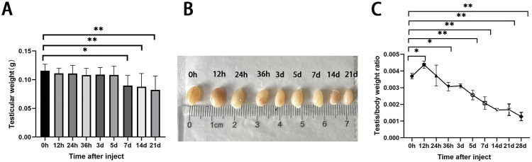
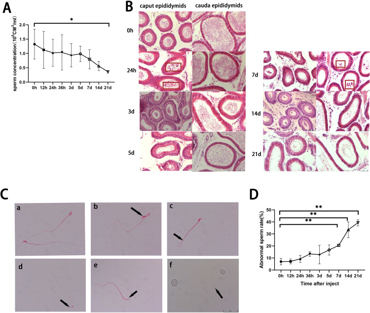
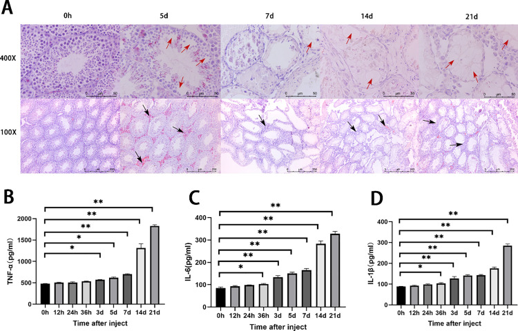
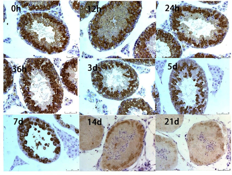
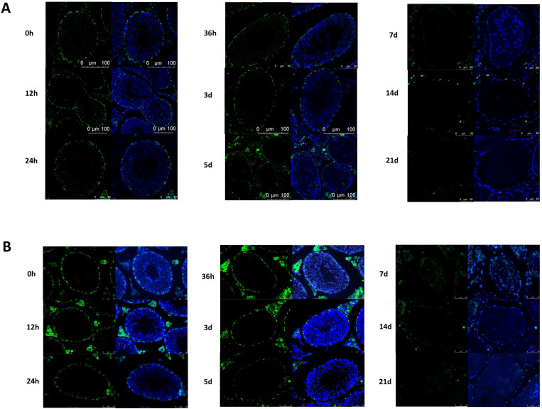
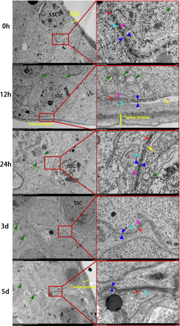
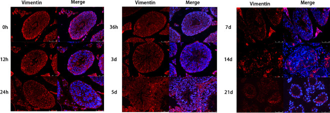
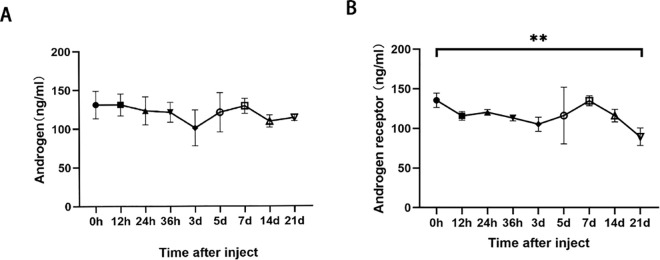
In order to clarify injure mechanism of busulfan to spermatogenic function, we treated mice with busulfan, the testicular and epididymal weights and sperm concentration significantly decreased and the sperm malformation rate increased over time. Moreover, testicular interstitial cell infiltration, a smaller seminiferous tubule, and disorganized and shed spermatogenic cells were also observed by immunohistochemical, immunofluorescence detection after the busulfan treatment. Furthermore, the enzyme-linked absorbance assays showed serum interleukin (IL)-6, IL-1β, and tumor necrosis factor-apha levels (inflammatory factors) were significantly upregulated; blood-testis barrier (BTB)-related protein levels (e.g., N-Cadherin, occludin, and connexin 43) and vimentine gradually decreased. So we infer busulfan treatment induced orchitis, further disrupted the BTB and disrupted the spermatogenic microenvironment, then decreased vimentine and gradually damaged the cytoskeleton, which cause spermatogenic cells losing their supporting from sertoli cells, androgen regulation was also affected, which was detrimental to spermatogenesis. The study result will improve the efficiency and safety in spermatogonial stem cell transplant recipients.


Conflict of interest statement

The authors have declared that no competing interests exist.

Figures

Fig 1
Fig 1. Effects of busulfan treatment (6 mg/kg) on the body and testes weights of mice over three weeks.
(A) testicular volume, and (B) testicular weights decrease over time. (C) The testes-to-body ratio (testicular weight/body weight) significantly decreases with time.
Fig 2
Fig 2. Effects of busulfan treatment (6 mg/kg) on the epididymis and sperm of mice over a three-week period.
(A) The epididymal sperm concentrations decrease over time and significantly differ between hour 0 (control) and day 21 (n = 3 per group). (B) H&E staining of the epididymis (caput and cauda) over time (n = 3). Shed germ cells are identified by red rectangles after 24 hours and 7 days in the caput and cauda epididymis, respectively. Scale bar = 50 μm. (C) Representative images of sperm malformations (eosin stain; 1000 × magnification; n = 3): a) normal sperm; b) head-bent sperm; c) tail-bent sperm; d) hookless sperm; e) headless sperm; f) head-bent sperm; scale bar = 25 μm. (D) The sperm malformation rate increases over time (n = 3 per group).
Fig 3
Fig 3. Busulfan treatment induces orchitis.
(A) Hematoxylin and eosin staining of mouse testis tissue show gradual shedding of spermatogonia and decreases in the seminiferous tubule over time after busulfan treatment. First row: 400 × ; Second row: 100 × ; Scale bar = 250 μm. Red and black arrows represent vacuolation and inflammatory infiltration, respectively. ELASA results of the inflammatory factors, (B) tumor necrosis factor-alpha (TNF-α). (C) interleukin (IL)-6, and (D) IL-1β; the concentrations for all factors significantly increase over time.
Fig 4
Fig 4. Effect of busulfan treatment on the tight junction protein, occludin.
Occludin-positive signals around the spermatocytes decrease over three weeks. The brown signal represents the positive signal of occludin, and blue signal represents hematoxylin staining of the nuclei. (n = 6 per group; Immunohistochemistry staining; 400 × ; Scale bar = 50 μm).
Fig 5
Fig 5. Effects of busulfan treatment on CX43 and N-Cadherin.
The gap junction protein CX43 (A) and the ectoplasmic specialization-related protein N-Cadherin (B) were detected by immunofluorescence assay. Both positive signals decrease over three weeks. Control: 0h. Green represent positive signals for CX43 and N-Cadherin, and blue represent positive signals for DAPI staining of the nuclei, respectively. (n = 6 per group; Immunofluorescence staining; 400 × ; Scale bar = 50 μm).
Fig 6
Fig 6. Effects of busulfan treatment on the ultrastructure of the blood-testis barrier (BTB).
The fine structures of the BTB are progressively damaged over three weeks. Left side: 1500 × magnification (scale bar = 5 μm). Right side: a magnified view of the image on the left; 7000 × magnification (scale bar = 1 μm). Red arrows indicate tight junctions, green arrows indicate mitochondria, green brackets indicate basal ectoplasmic specialization, yellow arrows indicate vacuoles, and yellow brackets indicate the basement membrane.
Fig 7
Fig 7. Effect of busulfan treatment on vimentin, a supporting cytoskeletal protein.
Positive vimentin signals decrease over three weeks. Red and blue represent vimentin and DAPI staining of the nuclei, respectively. (n = 6 per group; Immunofluorescence staining; 400 × ; Scale bar = 50 μm).
Fig 8
Fig 8. Effect of busulfan treatment on androgens and androgen receptors.
Enzyme-linked immunosorbent assay results for A) androgens and B) androgen receptors. The androgen concentration remains stable after busulfan treatment, but the androgen receptor concentration significantly decreases 21 days after treatment. Values are presented as means ± standard deviations; n = 3 per group. * p < 0.05, ** p < 0.01.

Similar articles

References

    1. Chen N, Su P, Wang M, Li Y-M. Ascorbic acid inhibits cadmium-induced disruption of the blood-testis barrier by regulating oxidative stress-mediated p38 MAPK pathways. Environ Sci Pollut Res Int. 2018;25(22):21713–20. doi: 10.1007/s11356-018-2138-4 - DOI - PubMed
    1. Choderlos de Laclos X, Risbourg S, Brennan B, Bertucci F, Gaspar N, Gelderblom H, et al. Impact of age on safety of Busulfan-Melphalan followed by autologous hematopoietic stem-cell transplantation versus standard chemotherapy in the patients of the EURO-E.W.I.N.G. 99 and Ewing 2008 clinical trials. Eur J Cancer. 2024;208:114229. doi: 10.1016/j.ejca.2024.114229 - DOI - PMC - PubMed
    1. de Kloet LC, Bense JE, van der Stoep MYEC, Louwerens M, von Asmuth EGJ, Lankester AC, et al. Late endocrine effects after hematopoietic stem cell transplantation in children with nonmalignant diseases. Bone Marrow Transplant. 2022;57(10):1564–72. doi: 10.1038/s41409-022-01755-x - DOI - PubMed
    1. Wu Z, Ma Y, Chen S, Liu Y, Liu X, Cao H, et al. Arginine Biosynthesis Mediates Wulingzhi Extract Resistance to Busulfan-Induced Male Reproductive Toxicity. Int J Mol Sci. 2024;25(12):6320. doi: 10.3390/ijms25126320 - DOI - PMC - PubMed
    1. Duléry R, Bastos J, Paviglianiti A, Malard F, Brissot E, Battipaglia G, et al. Thiotepa, Busulfan, and Fludarabine Conditioning Regimen in T Cell-Replete HLA-Haploidentical Hematopoietic Stem Cell Transplantation. Biol Blood Marrow Transplant. 2019;25(7):1407–15. doi: 10.1016/j.bbmt.2019.02.025 - DOI - PubMed

MeSH terms

LinkOut - more resources