. 2025 Aug 26;105(4):e213856.
doi: 10.1212/WNL.0000000000213856. Epub 2025 Jul 22.

Persistent Postictal Central Apnea in Focal Seizures: Incidence, Features, and Imaging Findings

Affiliations

Persistent Postictal Central Apnea in Focal Seizures: Incidence, Features, and Imaging Findings

Stefano Meletti et al. Neurology. .

Abstract

Background and objectives: Postconvulsive central apnea has emerged as a contributor to sudden unexplained death in epilepsy. The aim of this study was to evaluate the incidence and characteristics of postictal central apnea (PICA) in focal seizures. The secondary aim was to analyze morphometric features of the amygdala and other subcortical structures involved in autonomic control.

Methods: We prospectively enrolled consecutive patients admitted to the Epilepsy Monitoring Unit at Modena Academic Hospital (Italy) from April 2020 to December 2023. Inclusion criteria were as follows: (1) age older than 13 years; (2) at least 1 focal-onset seizure recorded during long-term video-EEG monitoring (LTVEM) with cardiorespiratory polygraphy. For each seizure, the presence of ictal central apnea (ICA) and/or PICA and its features were evaluated. Amygdala, hippocampus, thalamus, brainstem, and cerebellum volumetry were compared in patients with ICA/PICA with respect to healthy controls and patients with focal seizures without peri-ictal breathing disorders.

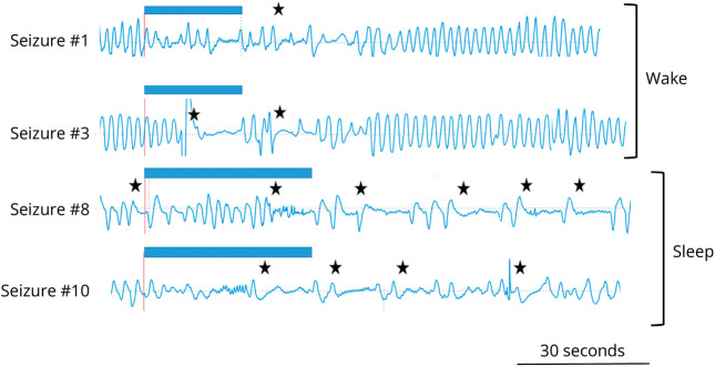
Results: A total of 69 patients (mean age 35.7 years; 42% female) with 406 focal-onset seizures were analyzed. ICA was recorded in 71 seizures (17%) in 27 patients. PICA was recorded in 24 seizures in 12 patients (10 with temporal lobe epilepsy) corresponding to 5.9% of all recorded seizures. Notably, PICA was observed only in seizures showing ictal apnea (in 33.8%). In 11 seizures with PICA, a single apneic event starting in the ictal and extending to the postictal period was observed. In 13 seizures, multiple apneic events were present in the postictal period (range 2-8). Seizures with PICA showed a longer peri-ictal apnea time (mean 75 seconds vs 40 seconds; p = 0.007) and a longer time to restore a regular rhythmic breathing after seizure termination (mean 173 seconds vs 42 seconds; p < 0.001) than seizures with self-limiting ictal apnea. Amygdala volumes ipsilateral to the epileptogenic zone were larger in patients with ICA/PICA compared with controls and patients without seizure-related apnea.

Discussion: PICA occurs in approximately 6% of focal seizures and is associated with extended apnea time and an enlarged amygdala ipsilaterally to the epileptogenic zone. Our data support the existence of a continuum from ictal to PICA and highlight the importance of cardiorespiratory recordings in LTVEM.


Conflict of interest statement

S. Meletti received research grant support from the Ministry of Health (MOH); and has received personal compensation as a scientific advisory board member for UCB, Jazz Pharmaceuticals, and EISAI. A.E. Vaudano has received speaker's or consultancy fees from Angelini. All other authors report no relevant disclosures. Go to Neurology.org/N for full disclosures.

Figures

Figure 1
Figure 1. Study Flowchart
(A) Flowchart of the study population according to inclusion/exclusion criteria (Methods in text). (B) Proportion of seizures with ICA of all captured seizures per individual patient in whom we recorded an ictal apnea event (n = 27). In most patients (from 1 to 19), ICA was observed in all seizures. (C) Proportion of seizures with PICA in each patient in whom we recorded at least 1 episode of ictal apnea (n = 27). Persistent postictal apnea was observed in 12 patients, showing intrapatient variability among seizures. ICA = ictal central apnea; PICA = postictal central apnea.
Figure 2
Figure 2. Features of Ictal and Postictal Apnea
(A) A compressed 2-minute polygraphic recording of ictal apnea extending into the postictal period. The patient (MO#12) had a left frontotemporal lobe seizure (indexed here by the Fp1-F7 EEG channel). The blue bar at the top marks scalp EEG seizure onset and offset. The black star marks the onset of apnea that precedes the EEG seizure onset by 20 seconds. Note that apnea persisted after the EEG seizure termination for approximately 15 seconds. Then, an irregular breathing pattern was evident with 2 more brief apnea episodes in the postictal period (black stars). Oxygen saturation shows a progressive decrease with a nadir below 80% during the postictal period. (B) Percentage of seizures with self-limiting ICA and wit persistent PICA among all seizures with breathing alterations. (C) Duration of breathing alterations in seizures with ICA and PICA (details in text). ICA = ictal central apnea; PICA = postictal central apnea; Resp = thoracoabdominal respiration; SpO2 = peripheral oxygen saturation.
Figure 3
Figure 3. Persistent and Prolonged Postictal Apnea Episodes
(A–C) Three consecutive compressed 3-minute peri-ictal polygraphic recording periods. The patient (MO#11) had a right frontotemporal lobe seizure (indexed here by the Fp2-F8 EEG channel). An ictal apnea followed by several postictal apnea episodes is evident (Resp channel) extending for several minutes after seizure termination. (A), EEG and the mylohyoid channels show muscle activity related to oroalimentary automatisms. The blue bar at the top marks scalp EEG seizure onset and offset. The black star marks the onset of ictal and postictal apnea episodes. Oxygen saturation showed a decrease between 85% and 80% that remained in this range until minute 8 of the postictal period. Milo = mylohyoid muscle; Resp = thoracoabdominal respiration; SpO2 = peripheral oxygen saturation.
Figure 4
Figure 4. Seizure-Related Apnea During Wake and Sleep
Respirogram during 4 different seizures in a young patient with focal left temporal lobe seizures due to herpes virus encephalitis (MO#01). Seizures 1 and 3 that occurred during wake were shorter (blue bars: scalp EEG seizure duration) and characterized by brief postictal apnea periods while seizures 8 and 10 that occurred during sleep showed prolonged and persistent apnea events. In all 4 seizures, the patient was not actively tested.
Figure 5
Figure 5. Amygdala Volumes in Patients With and Without Seizure-Related Apnea
(A) The amygdala ipsilateral to the epileptic focus is presented. The comparisons between patients' groups and controls are reported in t values; blue colors represent an amygdala atrophy compared with controls while red colors indicate an increase in volume compared with controls. (B) The box-and-whisker plots show the z-scores of ipsilateral whole amygdala volume and ipsilateral basolateral amygdala volume in PICA, ICA, and no-ICA groups compared with controls. The boxes' central line marks the mean; upper and lower edges of the box (the hinges) mark the 25th and 75th percentiles (the central 50% of the values fall within the box). The open circles represent each individual patient's volume. The dashed line on the 0 indicates the mean volume of control population. Finally, the asterisks on the top of the boxes show the significant differences with the control group while lines and asterisks below the boxes show the significant differences across patient groups. AAA = anterior amygdaloid area; AB = accessory basal nucleus; Ba = basal nucleus; CAT = cortico-amygdaloid transition area; Ce = central nucleus; Co = cortical nucleus; ICA = ictal central apnea; La = lateral nucleus; Me = medial nucleus; PICA = postictal central apnea; PL = paralaminar nucleus.

References

    1. Harden C, Tomson T, Gloss D, et al. Practice guideline summary: sudden unexpected death in epilepsy incidence rates and risk factors: report of the Guideline Development, Dissemination, and Implementation Subcommittee of the American Academy of Neurology and the American Epilepsy Society. Neurology. 2017;88(17):1674-1680. doi: 10.1212/WNL.0000000000003685 - DOI - PubMed
    1. Shorvon S, Tomson T. Sudden unexpected death in epilepsy. Lancet. 2011;378(9808):2028-2038. doi: 10.1016/S0140-6736(11)60176-1 - DOI - PubMed
    1. Dlouhy BJ, Gehlbach BK, Richerson GB. Sudden unexpected death in epilepsy: basic mechanisms and clinical implications for prevention. J Neurol Neurosurg Psychiatry. 2016;87(4):402-413. doi: 10.1136/jnnp-2013-307442 - DOI - PubMed
    1. Ryvlin P, Nashef L, Lhatoo SD, et al. Incidence and mechanisms of cardiorespiratory arrests in epilepsy monitoring units (MORTEMUS): a retrospective study. Lancet Neurol. 2013;12(10):966-977. doi: 10.1016/S1474-4422(13)70214-X - DOI - PubMed
    1. So EL, Sam MC, Lagerlund TL. Postictal central apnea as a cause of SUDEP: evidence from near-SUDEP incident. Epilepsia. 2000;41(11):1494-1497. doi: 10.1111/j.1528-1157.2000.tb00128.x - DOI - PubMed

MeSH terms