. 2025 Jul 14;9(8):vlaf028.
doi: 10.1093/immhor/vlaf028.

Exogenous arginine differentially regulates inflammatory cytokine and inducible nitric oxide synthase expression in macrophages

Affiliations

Exogenous arginine differentially regulates inflammatory cytokine and inducible nitric oxide synthase expression in macrophages

Kelsey Stayer et al. Immunohorizons. .

Abstract

Immune dysfunction and late mortality from multiorgan failure are hallmarks of severe sepsis. Arginine, a semi-essential amino acid important for protein synthesis, immune response, and circulatory regulation, is deficient in sepsis. However, arginine supplementation in sepsis remains controversial due to the potential to upregulate inducible nitric oxide synthase (iNOS)-mediated excessive nitric oxide (NO) generation in macrophages, leading to vasodilation and hemodynamic catastrophe. Citrulline supplementation has been considered an alternative to replenishing arginine via de novo synthesis, orchestrated by argininosuccinate synthase 1 (ASS1) and argininosuccinate lyase (ASL). However, the functional relevance of the ASS1-ASL pathway in macrophages after endotoxin stimulation is unclear but it is crucial to consider amino acid restoration as a tool for treating sepsis. We demonstrate that lipopolysaccharide (LPS)-mediated iNOS, ASS1, and ASL protein expression and nitric oxide generation were dependent on exogenous arginine in RAW 264.7 macrophages. Exogenous citrulline was not sufficient to restore nitric oxide generation in arginine-free conditions. Despite the induction of iNOS and ASS1 mRNA in arginine-free conditions, exogenous arginine was necessary and citrulline was not sufficient to overcome eIF2-α (elongation initiation factor 2-α)-mediated translational repression of iNOS and ASS1 protein expression. Moreover, exogenous arginine, but not citrulline, selectively modified the inflammatory cytokine and chemokine expression profile of the LPS-activated RAW 264.7 and bone marrow-derived macrophages. Our study highlights the complex, differential regulation of proinflammatory cytokine expression, and NO generation by exogenous arginine in macrophages.

Keywords: cell activation; cytokines; endotoxin shock; monocytes/macrophages; nitric oxide.


Conflict of interest statement

The authors declare no conflict of interest.

Figures

Figure 1.
Figure 1.
LPS treatment induces dose-dependent induction of iNOS, ASS1, and ASL protein expression in macrophages. (A) Total protein extracts of RAW 264.7 macrophages cultured in the standard media (400 μM ARG, 0 μM CIT) and treated for 24 h with increasing concentrations of LPS were analyzed by western blotting for expression of iNOS, ARG-1, ASS1, and ASL. (B) Densitometric analysis of band intensity was performed. Bar values represent the mean ± SD of n = 3/group with *P < 0.05, **P < 0.01, and ***P < 0.001 representing statistical significance as determined by 1-way ANOVA with Sidak’s multiple comparisons test. (C) Cell culture supernatant from RAW 264.7 macrophages cultured in standard media treated with LPS (1 μg/ml) for 24 h was analyzed by Griess assay to quantify nitrite production. Bar values represent the mean ± SD of n = 3/group ***P < 0.001 representing statistical significance as determined by unpaired Student t-tests.
Figure 2.
Figure 2.
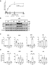
Exogenous arginine is necessary for iNOS, ASS1, and ASL protein induction and NO generation in macrophages. (A) The cell culture supernatant of RAW 264.7 macrophages cultured in arginine-free media or media with increasing concentrations of ARG or CIT (0 to 1000 μM) and treated for 24 h with LPS (1.0 μg/ml) was analyzed by Griess assay to quantify nitrite production. Graph values represent n = 3/group and EC50 and R2 values shown are representative of the 4-parameter logistic regression curve for the LPS ARG group. (B) Total protein extracts isolated from macrophages cultured in arginine-free (AF) media (0 μM ARG, 0 μM CIT) or supplemented media (125 μM ARG or 125 μM CIT) and treated with LPS (1.0 μg/ml) for 24 h were analyzed by western blotting for iNOS, ARG-1, ASS1, and ASL protein expression. (C) Densitometric analysis of band intensity was performed. Bar values rep7resent the mean ± SD of n = 3/group with *P < 0.05, **P < 0.01, and ***P < 0.001 representing statistical significance as determined by 1-way ANOVA with Sidak’s multiple comparisons test. (D) The relative mRNA expression of iNOS, ARG-1, ASS1, and ASL was analyzed by qRT-PCR in macrophages cultured in AF media or ARG- or CIT-supplemented media and treated with LPS (1.0 μg/ml) for 24 h. Bar values represent the mean ± SD of n = 3/group with *P < 0.05, **P < 0.01, and ***P < 0.001 representing statistical significance as determined by 1-way ANOVA with Sidak’s multiple comparisons test.
Figure 3.
Figure 3.
LPS-induced iNOS and ASS1 protein expressions are enhanced by exogenous arginine and not citrulline in bone marrow-derived macrophages. (A) Total protein extracts isolated from bone-marrow-derived macrophages cultured in arginine-free (AF) media (0 μM ARG, 0 μM CIT) or supplemented media (125 μM ARG or 125 μM CIT) and treated with LPS (1.0 μg/ml) for 24 h were analyzed by western blotting for iNOS, ARG-1, ASS1, and ASL protein expression. (B) Densitometric analysis of band intensity was performed. Bar values represent the mean ± SD of n = 3/group with *P < 0.05, **P < 0.01, and *** P < 0.01 representing statistical significance as determined by 1-way ANOVA with Sidak’s multiple comparisons test.
Figure 4.
Figure 4.
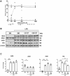
Exogenous arginine and not citrulline selectively regulates LPS-mediated phosphorylation and activation of STAT3. (A) RAW 264.7 macrophages were cultured in arginine-free (AF) media (0 μM ARG, 0 μM CIT) or supplemented media (125 μM ARG or 125 μM CIT) and treated with LPS (1.0 μg/ml) for 30 minutes. Total protein extracts were analyzed by western blotting for expression of enzymes involved in major proinflammatory signal transduction pathways. (B) Densitometric analysis of band intensity was performed. Bar values represent the mean ± SD of n = 3/group with *P < 0.05, **P < 0.01, and ***P < 0.001 representing statistical significance as determined by 1-way ANOVA with Sidak’s multiple comparisons test.
Figure 5.
Figure 5.
Exogenous arginine and not citrulline is necessary for the suppression of eIF2-α phosphorylation. (A) RAW macrophages were cultured in arginine-free media (0 μM ARG, 0 μM CIT) or supplemented media (125 μM ARG or 125 μM CIT) and treated with LPS (1.0 μg/ml) for 30 minutes or 24 h. Total protein extracts were analyzed by Western blotting for expression of enzymes involved in translational regulation and the ER stress response. (B) Densitometric analysis of band intensity was performed. Bar values represent the mean ± SD of n = 3/group with **P < 0.01 representing statistical significance as determined by 1-way ANOVA with Sidak’s multiple comparisons test.
Figure 6.
Figure 6.
Citrulline supplementation in the presence of arginine does not affect iNOS protein expression or NO generation in macrophages. (A) RAW 264.7 macrophages were cultured in arginine-free media (AFM; 0 μM ARG, 0 μM CIT) supplemented with increasing concentrations of arginine or citrulline (0 to 1000 μM) or the standard media (SM; 400 μM ARG, 0 μM CIT) supplemented with increasing concentrations of citrulline (0 to 1000 μM) and treated for 24 h with LPS (1.0 µg/ml). Cell culture supernatant was analyzed by Griess assay to quantify nitrite production. Graph values represent n = 3/group. (B) Macrophages were cultured in the standard media, or the standard media supplemented with citrulline (125 or 1000 μM), and treated with LPS (1.0 μg/ml) for 24 h. Total protein extracts were analyzed by Western blotting for expression of iNOS, ARG-1, ASS1, and ASL. (C) Densitometric analysis of band intensity was performed. Bar values represent the mean ± SD of n = 3/group with *P < 0.05, **P < 0.01, and ***P < 0.001 representing statistical significance as determined by 1-way ANOVA with Sidak’s multiple comparisons test.
Figure 7.
Figure 7.
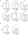
Exogenous arginine differentially regulates pro- and anti-inflammatory cytokines secreted by macrophages. (A) RAW 264.7 macrophages were cultured in arginine-free media (0 μM ARG, 0 μM CIT) or supplemented media (125 μM ARG or 125 μM CIT) and treated with LPS (1.0 μg/ml) for 6 h. The supernatant was analyzed by a bead-based discovery assay for quantification of secreted pro-inflammatory modulators and (B) anti-inflammatory modulators. Bar values represent the mean ± SD of n = 3/group with **P < 0.01, and ***P < 0.001 representing statistical significance as determined by 1-way ANOVA with Sidak’s multiple comparisons test.
Figure 8.
Figure 8.
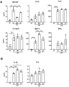
Exogenous arginine differentially regulates pro- and anti-inflammatory cytokines secreted by bone marrow-derived macrophages. (A) Bone marrow-derived macrophages were cultured in arginine-free media (0 μM ARG, 0 μM CIT) or supplemented media (125 μM ARG or 125 μM CIT) and treated with LPS (1.0 μg/ml) for 6 h. The supernatant was analyzed by a bead-based discovery assay for quantification of secreted pro-inflammatory modulators and (B) anti-inflammatory modulators. Bar values represent the mean ± SD of n = 3/group with *P < 0.05, **P < 0.01, and ***P < 0.001 representing statistical significance as determined by 1-way ANOVA with Sidak’s multiple comparisons test.

Similar articles

References

    1. Singer M et al. The Third International Consensus Definitions for Sepsis and Septic Shock (Sepsis-3). JAMA. 2016;315:801–810. - PMC - PubMed
    1. Gotts JE, Matthay MA. Sepsis: pathophysiology and clinical management. BMJ. 2016;353:i1585. - PubMed
    1. Cavaillon JM, Singer M, Skirecki T. Sepsis therapies: learning from 30 years of failure of translational research to propose new leads. EMBO Mol Med. 2020;12:e10128. - PMC - PubMed
    1. Rudd KE et al. Global, regional, and national sepsis incidence and mortality, 1990–2017: analysis for the Global Burden of Disease Study. Lancet. 2020;395:200–211. - PMC - PubMed
    1. Mira JC et al. Sepsis pathophysiology, chronic critical illness, and persistent inflammation-immunosuppression and catabolism syndrome. Crit Care Med. 2017;45:253–262. - PMC - PubMed

Publication types

MeSH terms

LinkOut - more resources