. 2025 Jul 19;53(14):gkaf710.
doi: 10.1093/nar/gkaf710.

FEN1 is critical for rapid single-strand break repair in G1 phase

Affiliations

FEN1 is critical for rapid single-strand break repair in G1 phase

Kamila Burdova et al. Nucleic Acids Res. .

Abstract

Flap endonuclease 1 (FEN1)-dependent long-patch repair has been considered a minor sub-pathway of DNA single-strand break repair (SSBR), activated only when short-patch repair is not feasible. However, the significance of long-patch repair in living cells remains unclear. Here, we employed human RPE-1 cells with FEN1 deletion to compare the requirements for short- and long-patch pathways for the rapid repair of various types of DNA single-strand breaks (SSBs). We found that SSBs arising from abortive topoisomerase 1 activity are repaired efficiently without FEN1. In contrast, the rapid repair of SSBs arising during base excision repair following treatment with methyl methanesulphonate (MMS) or following treatment with hydrogen peroxide (H2O2) exhibits an unexpectedly high dependence on FEN1. Indeed, in G1 phase, FEN1 deletion slows the rate of SSBR to a similar or even greater extent than deletion of the short-patch repair proteins XRCC1 or POLβ. As expected, the combined deletion of FEN1 with XRCC1 or POLβ has an additive or synergistic effect, severely attenuating SSBR rates after MMS or H2O2 exposure. These data highlight an unanticipated requirement for FEN1 in the rapid repair of SSBs in human cells, challenging the prevailing view that long-patch repair is a minor sub-pathway of SSBR.


Conflict of interest statement

None declared.

Figures

Graphical Abstract
Graphical Abstract
Figure 1.
Figure 1.
FEN1 facilitates efficient DNA SSBR in human cells. (A) DNA strand breaks in asynchronous wild-type (RPE-1), FEN1-deleted (koFEN1), and XRCC1-deleted (koFEN1) RPE-1 cells, measured by alkaline comet assays in untreated cells (NT) or after treatment with 0.1 mg/ml MMS for 15 min at 37°C. (B) DNA strand breaks in asynchronous wild-type (RPE-1), FEN1-deleted (koFEN1), and XRCC1-deleted (koXRCC1) RPE-1 cells, measured by alkaline comet assays in untreated cells (NT) or after treatment with 100 μM hydrogen peroxide (H2O2) on ice for 10 min, followed by release to drug-free media at 37°C for the indicated times. Tail moments are shown relative to values immediately after H2O2 treatment (absolute tail moments are provided in Supplementary Fig. S1D). (C) DNA strand breaks in asynchronous wild-type (RPE-1), FEN1-deleted (koFEN1), and XRCC1-deleted (koXRCC1) RPE-1 cells, measured by alkaline comet assays in untreated cells (NT) or after treatment with 10 or 30 μM CPT for 45 min at 37°C. (D) Levels of mono- and poly-ADP-ribosylated proteins (MAR/PAR) in wild-type (RPE-1), FEN1-deleted (koFEN1), and XRCC1-deleted (koXRCC1) RPE-1 cells in untreated conditions (NT) or after treatment with 0.1 mg/ml MMS at 37°C for the indicated times. Levels of FEN1, XRCC1, and histone H3 are shown as controls. (E) Levels of mono- and poly-ADP-ribosylated proteins (MAR/PAR) in wild-type (RPE-1), FEN1-deleted (koFEN1), and XRCC1-deleted (koXRCC1) RPE-1 cells in untreated conditions (NT) or after treatment with H2O2 as described in panel (B).
Figure 2.
Figure 2.
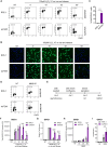
FEN1 is required for rapid rates of SSBR in the G1 phase. (A) Detection of chromatin-associated ADP-ribosylation (MAR/PAR) by indirect immunofluorescence in asynchronous wild-type (RPE-1) and FEN1-deleted (koFEN1) RPE-1 cells. Cells were left untreated (NT) or treated with 100 μM H2O2 on ice for 10 min, followed by incubation in drug-free media at 37°C for the indicated times. Contour plots show ADP-ribosylation levels (MAR/PAR) versus DNA content (DAPI). (B) Representative images of asynchronous cells shown in panel (A). Chromatin-bound nuclear mono- and poly-ADP-ribosylated proteins (MAR/PAR), visualized after detergent extraction, are show in green; nuclei are stained with DAPI (blue). (C) Quantification of MAR/PAR-positive 2n (G1 phase) cells in samples treated with 100 μM H2O2 on ice for 10 min, followed by incubation in drug-free media at 37°C for 60 min, as shown in panel (A). (D) Detection of chromatin-associated ADP-ribosylation (MAR/PAR) by indirect immunofluorescence in asynchronous cells, untreated (NT) or treated with 0.1 mg/ml MMS for 60 min. Contour plots of ADP-ribosylation versus DNA content (DAPI) are shown. (E) Schematic representation of the G0/G1 synchronization protocol. (F) Quantification of chromatin-associated ADP-ribosylation (MAR/PAR) by indirect immunofluorescence in G0/G1-synchronized cells, untreated (NT) or treated with 100 μM H2O2 on ice for 10 min, followed by incubation in drug-free media at 37°C for the indicated times. (G) DNA strand breaks in G1 phase wild-type (RPE-1), FEN1-deleted (koFEN1), and XRCC1-deleted (koXRCC1) RPE-1 cells, measured by alkaline comet assays after treatment or not (NT) with 100 μM H2O2 on ice for 10 min, followed by release into drug-free media at 37°C for the indicated times. Tail moments are presented relative to values immediately after H2O2 treatment (absolute tail moments are shown in Supplementary Fig. S2E). (H) Quantification of chromatin-associated ADP-ribosylation (MAR/PAR) by indirect immunofluorescence in G0/G1-synchronized cells, untreated (NT) or treated with 0.1 mg/ml MMS for the indicated times. (I) DNA strand breaks in G1 phase wild-type (RPE-1) and FEN1-deleted (koFEN1) RPE-1 cells, measured by alkaline comet assays in untreated cells (NT) or after treatment with 0.1 mg/ml MMS for 15 min at 37°C.
Figure 3.
Figure 3.
FEN1-dependent repair is the dominant pathway for rapid SSBR in G1 phase. (A) Levels of FEN1, XRCC1, and POLβ in whole-cell extracts from the indicated wild-type (RPE-1) and single/double-knockout RPE-1 cells, detected by western blotting. Levels of histone H2B and importin β were used as loading controls. (B) Quantification of chromatin-associated ADP-ribosylation (MAR/PAR) by indirect immunofluorescence in G0/G1-synchronized wild-type (RPE-1), FEN1-deleted (koFEN1), POLβ-deleted (koPOLβ), and FEN1/POLβ-deleted (koFEN1/POLβ) RPE-1 cells treated with 0.1 mg/ml MMS for the indicated times. (C) DNA strand breaks in G1-phase wild-type (RPE-1) and indicated single/double-knockout RPE-1 cell lines, measured by alkaline comet assays in untreated cells (NT) or after treatment with 0.1 mg/ml MMS for 15 min at 37°C, followed by incubation in drug-free media for the indicated times. Absolute tail moments are shown (relative tail moments are presented in Supplementary Fig. S3A). (D) DNA strand breaks in G1-phase wild-type (RPE-1) and the indicated single/double-knockout RPE-1 cell lines, measured by alkaline comet assays in untreated cells (NT) or after treatment with 50 μM H2O2 on ice for 10 min, followed by incubation in drug-free media at 37°C for the indicated times. Tail moments are presented relative to the values immediately after H2O2 treatment (absolute tail moments are shown in Supplementary Fig. S3C).
Figure 4.
Figure 4.
Recruitment of short-patch and long-patch repair proteins during SSB repair. (A) Western blot analysis of proteins in whole-cell lysate or in the detergent-extracted fraction of whole-cell extract (chromatin fraction; CH) from G0/G1-synchronized wild-type (RPE-1) and XRCC1-deleted (koXRCC1) RPE-1 cells after treatment with 0.1 mg/ml MMS for 1 h at 37°C. Where indicated, cells were pre-treated with 10 μM PARP inhibitor (PARPi; olaparib) for 15 min before MMS treatment. (B) Western blot analysis of protein extracts as above, after treatment of the indicated RPE-1 cells with 100 μM H2O2 in serum-free media on ice for 10 min, followed by release into fresh full media at 37°C for 20 min. (C) Western blot analysis of protein extracts after treatment of the indicated RPE-1 cells with 100 μM H2O2 as above. Where indicated, cells were pre-treated with 10 μM PARP inhibitor (PARPi; olaparib) for 15 min before H2O2 treatment. (D) Western blot analysis of protein extracts as above, following treatment of RPE-1 cells expressing HA-tagged POLD1 with 0.1 mg/ml MMS for 1 h at 37°C. Where indicated, cells were pre-treated with 10 μM PARP inhibitor (PARPi; olaparib) for 15 min before MMS treatment.

Similar articles

References

    1. Caldecott KW Causes and consequences of DNA single-strand breaks. Trends Biochem Sci. 2024; 49:68–78. 10.1016/j.tibs.2023.11.001. - DOI - PubMed
    1. Milano L, Gautam A, Caldecott KW DNA damage and transcription stress. Mol Cell. 2024; 84:711–3. 10.1016/j.molcel.2023.11.014. - DOI - PubMed
    1. Caldecott KW, Ward ME, Nussenzweig A The threat of programmed DNA damage to neuronal genome integrity and plasticity. Nat Genet. 2022; 54:115–20. 10.1038/s41588-021-01001-y. - DOI - PubMed
    1. Caldecott KW DNA single-strand break repair and human genetic disease. Trends Cell Biol. 2022; 32:733–45. 10.1016/j.tcb.2022.04.010. - DOI - PubMed
    1. Gohil D, Sarker AH, Roy R Base excision repair: mechanisms and impact in biology, disease, and medicine. Int J Mol Sci. 2023; 24:14186. 10.3390/ijms241814186. - DOI - PMC - PubMed

MeSH terms