Review
. 2025 Oct:60:102479.
doi: 10.1016/j.tranon.2025.102479. Epub 2025 Jul 22.

Understanding the heterogeneity of pancreatic ductal adenocarcinoma

Affiliations
Review

Understanding the heterogeneity of pancreatic ductal adenocarcinoma

Juan Iovanna et al. Transl Oncol. 2025 Oct.

Abstract

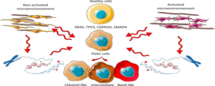
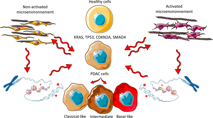
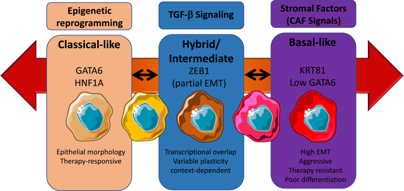
Pancreatic ductal adenocarcinoma (PDAC) is a highly aggressive, treatment-resistant cancer characterized by extensive inter- and intra-tumoral heterogeneity. Although over 95 % of cases harbor KRAS mutations and commonly altered tumor suppressors like TP53, SMAD4, and CDKN2A, these genetic changes alone do not fully explain PDAC variability. We propose a paradigm shift: PDAC heterogeneity is not solely genetic but also shaped by epigenetic regulation and the tumor microenvironment. Traditional transcriptomic classifications define PDAC into fixed subtypes, primarily classical and basal-like, but we argue these are not static categories. Instead, PDAC phenotypes exist along a dynamic continuum influenced by stromal interactions and epigenetic cues. This model challenges the binary classification view. We show that transitions from classical to basal-like states are gradual and reversible, driven by tumor-stroma crosstalk and chromatin remodeling. Such plasticity underpins tumor adaptation, resistance, and progression. Embracing this dynamic framework offers novel therapeutic opportunities.

Keywords: Epigenetics; Metastases; PDAC; Tumor heterogeneity; Tumor microenvironment; Tumor progression.


Conflict of interest statement

Declaration of competing interest The authors disclose no conflicts of interest.

Figures

Image, graphical abstract
Graphical abstract
Fig 1
Fig. 1
Cell phenotype is determined by the complex interaction between the tumor microenvironment and the Epigenetic Regulation of Epithelial Cells rather than by the Accumulation of Genetic Mutations. The schematic representation illustrates how mutations in driver genes can trigger the malignant transformation of pancreatic cells without directly influencing their phenotype. In contrast, the stroma plays a crucial role in phenotype determination through direct interactions with tumor cells or by modulating epigenetics, thereby influencing cellular plasticity and tumor progression.
Fig 2
Fig. 2
A. Single-cell analysis reveals basal, classical, and chimeric phenotypes within the same PDAC Tumor. Single-cell RNA sequencing (scRNA-seq) analysis of PDAC samples reveals the coexistence of basal, classical, and chimeric phenotypes within the same tumor. B. Genome-Wide Mutational Analysis Indicates no Significant Correlation Between Specific Genetic Mutations and PDAC Phenotypes. The mutational landscape of key driver genes KRAS, TP53, SMAD4, and CDKN2A was examined across 39 patient-derived xenografts (PDXs) previously classified as Basal or Classical. Lateral bars represent the -log10 p-value from Fisher’s exact test, assessing the association between mutations and the consensus multiomics classification. Statistically significant associations (p ≤ 0.05, indicated by a red threshold line) are highlighted, with bars colored orange for basal and blue for classical subtypes. C. Proportion of Each Mutant Analyzed on the TGAC cohort. No significant association was found between mutations with the phenotype of the PDAC. D. Distribution of PDAC Subtypes Across Disease Stages. The proportion of Basal and Classical PDAC subtypes was assessed through transcriptomic analysis of samples from different disease stages, including Operable (n=35), Locally Advanced (n=244), Metastatic (n=76), and Liver Metastases (n=88). This classification provides insights into the distribution of molecular subtypes across the clinical spectrum of PDAC.
Fig 3
Fig. 3
Phenotypic continuum model of PDAC. This schematic represents a dynamic, non-binary model of PDAC cellular states, emphasizing phenotypic plasticity across a spectrum from classical to basal-like subtypes. In contrast to traditional classifications that treat subtypes as mutually exclusive, this continuum highlights reversible and transitional cellular identities, influenced by epigenetic reprogramming, TGF-β signaling, and stromal-derived cues such as those from cancer-associated fibroblasts (CAFs). On the left, the classical phenotype is characterized by epithelial morphology, strong expression of GATA6 and HNF1A, and greater responsiveness to conventional therapies. In the center, an intermediate or hybrid state emerges, marked by partial EMT (epithelial–mesenchymal transition), expression of S100A2 and ZEB1, and a context-dependent transcriptional program. These cells exhibit high plasticity, enabling them to shift toward either end of the spectrum depending on environmental pressures. On the right, the basal-like phenotype exhibits mesenchymal morphology, expression of markers such as KRT81, TP63, and low GATA6, along with features of high EMT, invasiveness, and resistance to therapy. Bidirectional arrows between these states indicate that phenotypic transitions are reversible, reflecting the underlying plasticity of PDAC cells. This flexibility poses a major challenge for therapeutic targeting, as tumor cells can dynamically adapt to selective pressures, including treatment and microenvironmental changes.

Similar articles

References

    1. Ying H., Kimmelman A.C., Bardeesy N., Kalluri R., Maitra A., DePinho R.A. Genetics and biology of pancreatic ductal adenocarcinoma. Genes Dev. 2025;39:36–63. doi: 10.1101/gad.351863.124. - DOI - PMC - PubMed
    1. Nicolle R., Blum Y., Marisa L., Loncle C., Gayet O., Moutardier V., Turrini O., Giovannini M., Bian B., Bigonnet M., et al. Pancreatic adenocarcinoma therapeutic targets revealed by tumor-stroma cross-talk analyses in patient-derived xenografts. Cell Rep. 2017;21:2458–2470. doi: 10.1016/j.celrep.2017.11.003. - DOI - PMC - PubMed
    1. Bailey P., Chang D.K., Nones K., Johns A.L., Patch A.M., Gingras M.C., Miller D.K., Christ A.N., Bruxner T.J.C., Quinn M.C., et al. Genomic analyses identify molecular subtypes of pancreatic cancer. Nature. 2016;531:47–52. doi: 10.1038/nature16965. - DOI - PubMed
    1. Collisson E.A., Sadanandam A., Olson P., Gibb W.J., Truitt M., Gu S., Cooc J., Weinkle J., Kim G.E., Jakkula L., et al. Subtypes of pancreatic ductal adenocarcinoma and their differing responses to therapy. Nat. Med. 2011;17:500–503. doi: 10.1038/nm.2344. - DOI - PMC - PubMed
    1. Moffitt R.A., Marayati R., Flate E.L., Volmar K.E., Loeza S.G.H., Hoadley K.A., Rashid N.U., Williams L.A., Eaton S.C., Chung A.H., et al. Virtual microdissection identifies distinct tumor- and stroma-specific subtypes of pancreatic ductal adenocarcinoma. Nat. Genet. 2015;47:1168–1178. doi: 10.1038/ng.3398. - DOI - PMC - PubMed