. 2025 Sep 3;45(36):e0055252025.
doi: 10.1523/JNEUROSCI.0055-25.2025.

Asymmetries in Foveal Vision

Affiliations

Asymmetries in Foveal Vision

Samantha K Jenks et al. J Neurosci. .

Abstract

Visual perception is characterized by known asymmetries in the visual field; human's visual sensitivity is higher along the horizontal than the vertical meridian, and along the lower than the upper vertical meridian. These asymmetries decrease with decreasing eccentricity from the periphery to the center of gaze, suggesting that they may be absent in the [Formula: see text] foveola, the retinal region used to explore scenes at high-resolution. Using high-precision eyetracking and gaze contingent display, allowing for accurate control over the stimulated foveolar location despite the continuous eye motion at fixation, we investigated fine visual discrimination at different isoeccentric locations across the foveola and parafovea in 12 human observers (both sexes). Although the tested foveolar locations were only [Formula: see text] away from the center of gaze, we show that, similar to more eccentric locations, humans are more sensitive to stimuli presented along the horizontal than the vertical meridian. Whereas the magnitude of this asymmetry is reduced in the foveola, the magnitude of the vertical meridian asymmetry is comparable but, interestingly, the asymmetry is reversed: stimuli presented slightly above the center of gaze are more easily discerned than when presented at the same eccentricity below the center of gaze. Therefore, far from being uniform, as often assumed, foveolar vision is characterized by perceptual asymmetries. Further, these asymmetries differ not only in magnitude but also in direction compared to those present just ∼[Formula: see text] away from the center of gaze, resulting in overall different foveal and extrafoveal perceptual fields.


Conflict of interest statement

The authors declare no competing financial interests.

Figures

Figure 1:
Figure 1:. Experimental protocol.
A-Subjects fixated monocularly (left eye patched) on a central fixation marker while stimuli were presented. The target, a small bar tilted ± 45 deg, appeared briefly (50ms) at one of 4 possible locations and subjects were asked to report its orientation once the response cue was presented. A total of eight locations were tested. Cardinal and intercardinal locations were tested in separate blocks. B-Stimuli spatial arrangement and dimensions in the foveola (left; n = 12) and parafovea (right; n = 8) conditions. In the latter condition, stimuli were magnified according to the cortical magnification factor (Rovamo and Virsu 1979). C-In a preliminary session, the stimulus contrast was determined for the foveola and the parafovea conditions separately, so that overall performance across cardinal and intercardinal locations yielded ≈70% correct responses. Error bars are 95% confidence intervals. D-The target was 20 arcmin from the center of the display. The box and whisker plot shows that the average gaze distance from the target was approximately 20 arcminutes for different target locations in the foveola condition. Triangles represent the two subjects under conditions of retinal stabilization (see methods for detail). Error bars are 95% confidence intervals.
Figure 2:
Figure 2:. Horizontal-Vertical Meridian Asymmetry.
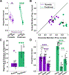
A and B-Average performance along the horizontal (pooled left and right locations) and vertical meridian (pooled top and bottom locations) across subjects for the foveola and the parafovea. In A, lines represent individual observers; the two observers who viewed stimuli under retinal stabilization conditions are marked by dashed lines. Asterisks denote a statistically significant difference (*P<0.05, **P ≤ 0.01, ***P ≤ 0.001, paired t-test p = 0.02 foveola, p = 0.0004 parafovea). C-Magnitude and direction of the horizontal-vertical meridian asymmetry. Asterisks indicate a statistically significant difference from zero (one sample t-test p = 0.02 foveola, p = 0.0004 parafovea). D-Average performance along the horizontal meridian compared to the upper and lower vertical meridian performance for the foveola and the parafovea. Asterisks denote statistically significant differences (one-way ANOVA, p = 0.0001 foveola, p < 0.0001 parafovea). All Error bars are 95% confidence intervals. Triangles represent the two observers tested under conditions of retinal stabilization (see methods for detail).
Figure 3:
Figure 3:. Vertical Meridian Asymmetry.
Conventions are the same as in Fig. 2. A and B-Average performance along the upper and lower vertical meridian across subjects. In A, asterisks denote a statistically significant difference (paired t-test, p = 0.001 foveola, p = 0.002 parafovea). C-Magnitude and direction of the vertical meridian asymmetry. Asterisks indicate a statistically significant difference from zero (one sample t-test, p = 0.001 foveola, p = 0.002 parafovea). All error bars are 95% confidence intervals.
Figure 4:
Figure 4:. Performance fields in the foveola and parafovea.
A-The visual performance field in the foveola (purple) and parafovea (green). Each angle represents one tested location. Numbers along the radial direction represent percent correct, and the shaded regions represent SEM. The lines connect the average performance across subjects for the different locations tested. B-Magnitude and direction of the asymmetry between the upper intercardinal locations (locations at 45 and 135 deg) and the lower intercardinal locations (locations at 225 and 315 deg). Error bars are 95% confidence intervals. Triangles represent the two subjects tested under conditions of retinal stabilization (see methods for detail).

Update of

References

    1. Abrams J, Nizam A, Carrasco M (2012) Isoeccentric locations are not equivalent: The extent of the vertical meridian asymmetry. Vision Research 52:70–78. - PMC - PubMed
    1. Anstis SM (1974) Chart demonstrating variations in acuity with retinal position. Vision Research 14:589–592. - PubMed
    1. Anton-Erxleben K, Carrasco M (2013) Attentional enhancement of spatial resolution: linking behavioural and neurophysiological evidence. Nature Reviews Neuroscience 14:188–200. - PMC - PubMed
    1. Atchison DA, Pritchard N, Schmid KL (2006) Peripheral refraction along the horizontal and vertical visual fields in myopia. Vision Research 46:1450–1458. - PubMed
    1. Azzopardi P, Cowey A (1993) Preferential representation of the fovea in the primary visual cortex. Nature 361:719–721. - PubMed

LinkOut - more resources