. 2025 Jul 22;11(1):81.
doi: 10.1038/s41540-025-00559-1.

Complex-mediated evasion: modeling defense against antimicrobial peptides with application to human-pathogenic fungus Candida albicans

Affiliations

Complex-mediated evasion: modeling defense against antimicrobial peptides with application to human-pathogenic fungus Candida albicans

Yann Bachelot et al. NPJ Syst Biol Appl. .

Abstract

Understanding the complex interplay between host and pathogen during infection is critical for developing diagnostics and improving therapeutic interventions. Among the diverse arsenal employed by the host, antimicrobial peptides (AMP) play a key role in the defense against pathogens. We propose an immune evasion mechanism termed "Complex-mediated evasion" (CME), that allows pathogens to protect themselves against AMP and investigate it through mathematical modeling and computer simulations. To achieve CME, we hypothesize that the pathogen secretes defense molecules that bind AMP. When bound within the complex, AMP are unable to harm the pathogen. Due to molecular gradients, complexes may diffuse away from the pathogen, enhancing the protective effect of the mechanism by decreasing the concentration of AMP in the vicinity of the pathogen. We establish a mathematical model to (i) explore the sensitivity of the mechanism to various parameters and (ii) simulate the immune evasion of the human-pathogenic fungus Candida albicans.


Conflict of interest statement

Competing interests: The authors declare no competing interests.

Figures

Fig. 1
Fig. 1. Complex-mediated evasion mechanism.
The pathogen cell (yellow) is surrounded by AMP (blue) and secretes defense molecules (red). These molecules bind to AMP forming complexes in the extracellular space, preventing AMP from harming the pathogen cell.
Fig. 2
Fig. 2. Visual representations of an example simulation for dynCME.
The parameters used for the dynCME example simulation are presented in Supplementary Table 2. a Visual representation of the system as a simulation snapshot at t=0.75s. The system is defined as a three-dimensional cube, with the pathogen cell modeled as a sphere centered in the environment. AMP (blue), defensive (red), and complexes (purple) molecules diffuse in the extracellular space and interact with each other as well as with the pathogen cell (yellow). b Spatiotemporal distributions of AMP, defense molecules, and complexes during t=3.0s represented as two-dimensional kymographs. The x-axis indicates the distance to the pathogen cell (spatial axis), while the y-axis corresponds to the time (temporal axis).
Fig. 3
Fig. 3. Reaction rate parameter screening of conCME model.
Screening over six reaction rate parameters as presented in Supplementary Table 1. The color bar shows the sAMPcon score on a log scale. For example, the orange color corresponds to a score of 10%, indicating that 10% of the initial concentration of AMP has been taken up by the pathogen.
Fig. 4
Fig. 4. Parameters effect of conCME.
Partial dependence plots. Each subplot corresponds to one reaction rate parameter, with the mean score sAMPcon (%) computed for each unique parameter value screened.
Fig. 5
Fig. 5. Comparison of two regimes: Diffusion of complexes promoting CME (RD<0, blue) or inhibiting CME (RD>0, orange).
a Distribution of the parameter values in both regimes. b Spatial distribution of the formation of complexes. The dotted lines represent the median value.
Fig. 6
Fig. 6. Parameter screening for dynCME.
Screening over the three parameters UA,SD, FA. The color bar shows the score sAMPdyn on a log scale. Low values correspond to the regime of immune evasion and are shown in dark blue, whereas high values are shown in yellow. The results presented here correspond to the parameter subspace kon=102μm3s1,koff=101s1,kdeg=0s1 and t*=103s.
Fig. 7
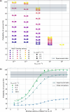
Fig. 7. Relation between the simulated concentration of LL-37 taken up by the pathogen and its experimentally measured probability of survival.
The x-axis represents the simulated concentration of LL-37 that was taken up during 1.5 h using conCME. The y-axis represents the probability of survival, which was measured experimentally in the survival assays performed in ref. , shown with standard deviation. The colors refer to the screened values of the uptake rate used in the simulations. The different experimental conditions, corresponding to the initial concentration of LL-37 in the system, are indicated on the right side of the plot. The dotted lines represent the exponential decay fitted (see Supplementary Table 5) to the experimental and simulated data that allow us to relate the concentration of LL-37 that was taken up by the pathogen cell to its probability of survival.
Fig. 8
Fig. 8. Simulation outcome for different numbers of LL-37 binding sites on Msb2*.
a. The simulated survival probability of C. albicans is plotted as a function of three parameters: the uptake rate ULL37 (represented on x-axis), the number of binding sites used in simulation (indicated by different colors), and the association rate kon (shown with markers’ shapes). The gray solid line and the shaded area, respectively, represent the mean and standard deviation of the experimental data. b. The survival probability of the pathogen is plotted against the number of LL-37 binding sites on Msb2* for kon=101μm3s1. The different colors refer to different values of the uptake rate ULL37.
Fig. 9
Fig. 9. ConCMEval simulation outcome for different parameters for the pathogen C. albicans.
The color bar shows the probability of survival of the pathogen for a particular parameter set. Higher values (dark blue) correspond to the immune-evasion regime, with high chances for the pathogen to survive, whereas lower values (orange) correspond to a probability of survival of around 30%. Simulations that agree with experimental data (probability of survival between 50 and 60%) are highlighted with a black box. Parameter values used for the simulations are presented in Supplementary Table 4.
Fig. 10
Fig. 10. DynCMEval simulation outcome for different parameters for the pathogen C. albicans and its comparison to conCMEval simulations.
a Survival probabilities of C. albicans for different parameter sets. The color shows the survival probability of the pathogen for a particular parameter set. Higher values (dark blue) correspond to the immune-evasion regime, with high chances for the pathogen to survive, whereas lower values (yellow) correspond to a probability of survival for the pathogen cell close to 0%. Parameter values used in the simulations are presented in Supplementary Table 4. b Boxplot of the probability of survival of the parameter screening for the conCMEval simulations (blue), and for the different values of FLL37 of the dynCMEval simulations. The black line on the boxes indicates the median value. The comparison between the two models was performed by a Kruskal-Wallis test. The significance shown by *** indicates Pval<0.001, and nsPval>0.05.

Similar articles

References

    1. Meylan, E., Tschopp, J. & Karin, M. Intracellular pattern recognition receptors in the host response. Nature442, 39–44 (2006). - PubMed
    1. Zasloff, M. Antimicrobial peptides of multicellular organisms. Nature415, 389–395 (2002). - PubMed
    1. Bahar, A. A. & Ren, D. Antimicrobial peptides. Pharmaceuticals6, 1543–1575 (2013). - PMC - PubMed
    1. Huan, Y., Kong, Q., Mou, H. & Yi, H. Antimicrobial peptides: classification, design, application and research progress in multiple fields. Front. Microbiol.11, 1–21 (2020). - PMC - PubMed
    1. Tsai, P. W., Yang, C. Y., Chang, H. T. & Lan, C. Y. Human antimicrobial peptide LL-37 inhibits adhesion of Candida albicans by interacting with yeast cell-wall carbohydrates. PLoS ONE6, 17755 (2011). - PMC - PubMed

MeSH terms

Substances

LinkOut - more resources