. 2025 Jul 23;16(1):6771.
doi: 10.1038/s41467-025-61757-3.

Genotype B3.13 influenza A(H5N1) viruses isolated from dairy cattle demonstrate high virulence in laboratory models, but retain avian virus-like properties

Affiliations

Genotype B3.13 influenza A(H5N1) viruses isolated from dairy cattle demonstrate high virulence in laboratory models, but retain avian virus-like properties

Thomas P Fabrizio et al. Nat Commun. .

Abstract

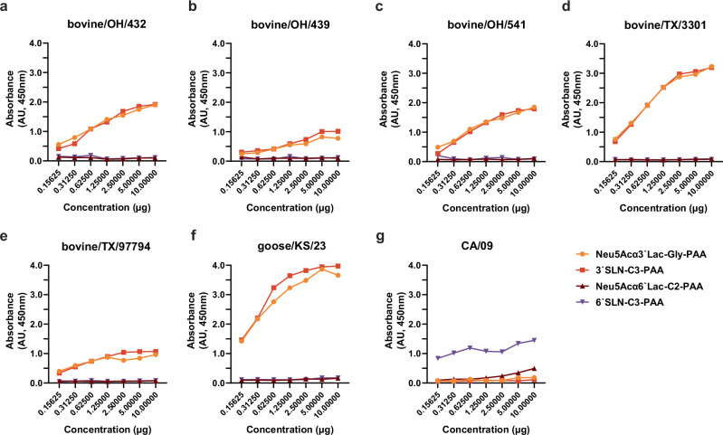
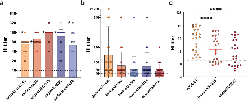
In March 2024, clade 2.3.4.4b highly pathogenic avian influenza A(H5N1) viruses were first detected in U.S. dairy cattle. Similar viruses have since caused 70 zoonotic human infections. To assess changes to zoonotic potential, we characterized A(H5N1) clade 2.3.4.4b viruses isolated from cows' milk and birds. Bovine-derived viruses are lethal in mice and ferrets and transmit to direct but not airborne contact ferrets. All viruses replicate in human bronchial epithelial cells despite preferentially binding avian virus-like receptors. The bovine-derived viruses remain susceptible to FDA-approved antivirals, and they are inhibited by sera from ferrets vaccinated with WHO-recommended candidate vaccine viruses (CVV) or human sera from clade 2.3.4.4c vaccinees. While 2.3.4.4b viruses induce severe disease in mammalian models, they retain many avian virus-like characteristics. Combined, we conclude that the risk of contemporary bovine-derived viruses to humans not in contact with affected animals is low. However, heightened vigilance remains essential to promptly detect and respond to any changes.


Conflict of interest statement

Competing interests: The authors declare no competing interests.

Figures

Fig. 1
Fig. 1. Replication growth kinetics of bovine HPAI A(H5N1) 2.3.4.4b viruses in vitro.
a MDCK. b NHBE cells were inoculated at MOI of 0.005 with three representative bovine A(H5N1) 2.3.4.4b, concurrently circulating avian A(H5N1), and human A(H1N1)pdm09 viruses and incubated at 37 °C. The supernatants from MDCK virus-infected cells and apical surface washes from NHBE inserts were collected at the indicated time points and virus titers were determined by TCID50 assay. The data are shown as the mean ± SD from quadruplicate wells. Statistical significance was determined by two-way ANOVA with Tukey’s post-test for multiple comparisons.
Fig. 2
Fig. 2. Receptor binding specificity of bovine HPAI A(H5N1) 2.3.4.4b viruses.
a–e The solid-phase binding assay with five representative bovine A(H5N1) 2.3.4.4b viruses. f concurrently circulating avian A(H5N1) virus. g and human A(H1N1)pdm09 viruses to biotinylated sialylglycopolymers (Neu5Acα3’Lac-Gly-PAA, and 3’SLN-C3-PAA) that are the avian influenza virus preferred receptors and biotinylated sialylglycopolymers (Neu5Acα6’Lac-C2-PAA, and 6’SLN-C3-PAA) that are the human influenza virus preferred receptors. The data are shown as the mean ± SD from duplicate wells and represent one of two independent experiments.
Fig. 3
Fig. 3. Pathogenicity of bovine HPAI A(H5N1) 2.3.4.4b viruses in mice.
a, b BALB/c mice (6-8 weeks old) were lightly anesthetized and inoculated IN with 10-fold-serial dilutions of bovine/OH/439 (H5N1) and bovine/TX/98638 (H5N1) viruses to determine MLD50 for each (n = 3/virus dose). c Virus titer in tissues from infected mice was determined by TCID50 assay in MDCK cells shown as the mean ± SD with individual mouse data represented by shapes. Immunohistochemistry of fixed tissues from A(H5N1) 2.3.4.4b virus-infected mice (n = 3/group) showed virus antigen in nasal epithelium (d, scale bar, 50 µm), lung (e, scale bar, 200 µm), brain (f, scale bar, 2 mm), spinal cord (g, scale bar, 1 mm), liver (h, scale bar, 100 µm), and brown adipose tissue (BAT) (i, scale bar, 100 µm).
Fig. 4
Fig. 4. Pathogenicity and transmission of bovine HPAI A(H5N1) 2.3.4.4b virus in ferrets.
Ferrets (n = 3) were lightly anesthetized and IN inoculated with 104 TCID50 units of bovine/OH/439 (H5N1) virus in 500 μl of PBS. a Body weights (% starting weight) and b survival were monitored daily. c Virus titers in nasal washes and d various tissues were determined by TCID50 assay in MDCK cells. At 24 hpi, donor ferrets were placed in direct contact (DC) or aerosol contact (AC) with naïve uninfected ferrets (n = 3). Shapes and colors differentiate between inoculated, DC and AC ferrets. a data are represented as lines for individual ferrets. The data in panels (c) and (d) are shown as the mean ± SD with individual ferret data represented by shapes. The dotted line indicates limit of detection for the assay (1.0 log10TCID50/mL). Weight loss data for AC animals was excluded from panel a to improve visualization of data points for infected animals in the Donor and DC groups.
Fig. 5
Fig. 5. Experimental infection of chickens with bovine HPAI A(H5N1) 2.3.4.4b viruses.
Chickens (6 weeks old) were inoculated intra-tracheally with 106 EID50/0.3 mL of goose/KS/930F (H5N1) or bovine/OH/439 (H5N1) viruses (n = 6/group). After 16 hpi, two donor chickens from each experimental group were placed in contact with three naive direct contact chickens. The chickens were monitored twice daily for disease symptoms and survival.
Fig. 6
Fig. 6. Antigenic and serologic characterization of bovine HPAI A(H5N1) 2.3.4.4b viruses.
a Antigenic properties of 16 bovine HPAI A(H5N1) 2.3.4.4b viruses were determined in HI assay with post-infection ferret antisera generated against the WHO-recommended CVVs of clade 2.3.4.4b [Astrakhan/3212 (H5N8), ck/Ghana/21 (H5N1), wigeon/SC/345 (H5N1),], and 2.3.4.4c [gyrfalcon/41088 (H5N8)] and representative avian eagle/FL/W22 (H5N1) virus. The data are shown as the mean ± SD with HI titers for each virus as individual circles. The dotted line indicates limit of detection for the assay (HI titer = 10). (★) indicates the endpoint titer of homologous ferret sera to the indicated CVV. b The presence of cross-reactive HA antibodies against bovine A(H5N1) 2.3.4.4b viruses was examined in a set of human sera obtained from a Phase 1 clade 2.3.4.4c H5 vaccine trial. Individuals (aged 18 to 50 years) were vaccinated with adjuvanted 15 µg of the HA derived from gyrfalcon/41088 (H5N8) 2.3.4.4c antigen (n = 20). The data are shown as the mean ± SD. c A set of human sera (n = 24) was obtained from BioIVT (individuals, aged 18 to 46 years, vaccination history unknown). NA activity inhibiting (NI) antibody levels in human serum samples against N1 NA protein derived from CA/09 (H1N1)pdm09, bovine/OH/439 (H5N1), and eagle/FL/W22 (H5N1) viruses (as measured by ELLA assay). The data are shown as geometric mean titer (line) of the individual IC50 NI titers (dots). Statistical significance was determined by two-way ANOVA with Tukey’s post-test for multiple comparisons.****p < 0.0001.

Similar articles

References

    1. WHO. Avian Influenza Weekly Update # 990: 21 March 2025. 4 (2025).
    1. Leonard, J. H. et al. Notes from the field: seroprevalence of highly pathogenic avian influenza a(H5) virus infections among bovine veterinary practitioners — United States, September 2024. MMWR and Morbidity and Mortality Weekly Report 74 10.15585/mmwr.mm7404a2 (2025). - PMC - PubMed
    1. Gomaa, M. et al. We are underestimating, again, the true burden of H5N1 in humans. Bmj Glob. Health8, e13146 (2023). - PMC - PubMed
    1. WHO. Global Influenza Program, (https://www.who.int/publications/m/item/cumulative-number-of-confirmed-h..., 2024).
    1. CDC. H5N1 Bird Flu Detections across the United States in Backyard and Commercial Poultry, https://www.cdc.gov/bird-flu/situation-summary/data-map-commercial.html (2024).

MeSH terms

Substances

LinkOut - more resources