. 2025 Jul 22;15(1):26589.
doi: 10.1038/s41598-025-12323-w.

Shear stress-triggered adenosine triphosphate regulates angiogenic properties of human periodontal ligament stem cells

Affiliations

Shear stress-triggered adenosine triphosphate regulates angiogenic properties of human periodontal ligament stem cells

Maythwe Kyawsoewin et al. Sci Rep. .

Abstract

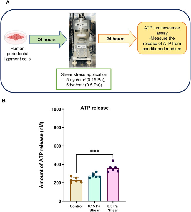
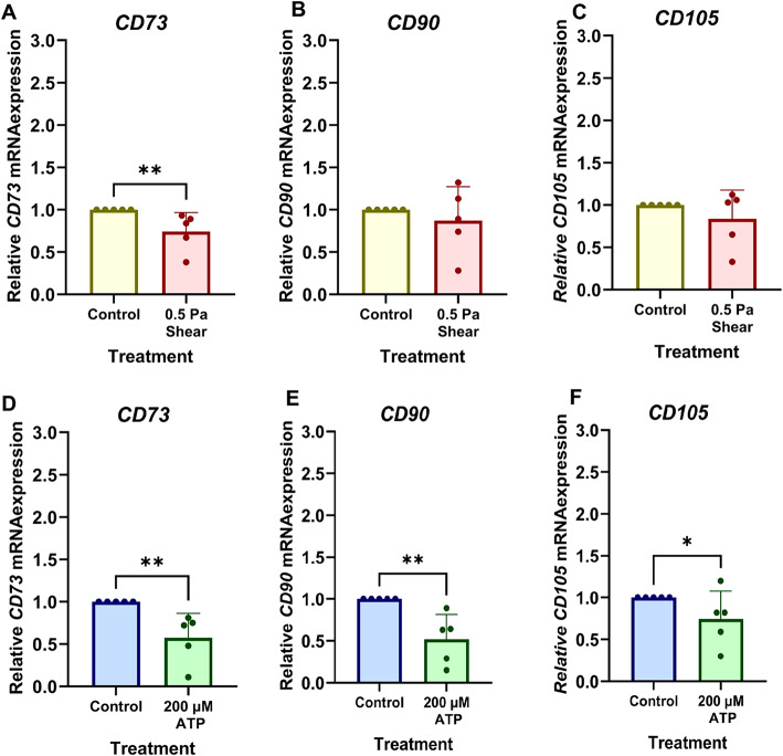
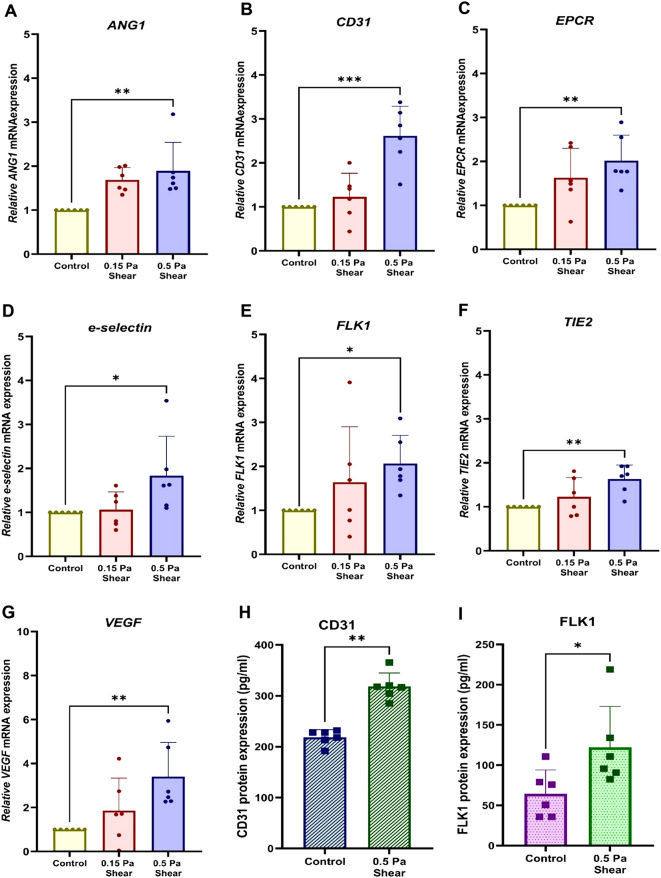
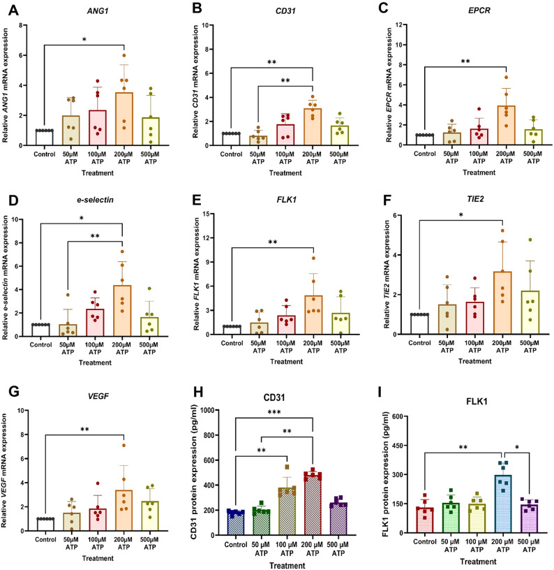
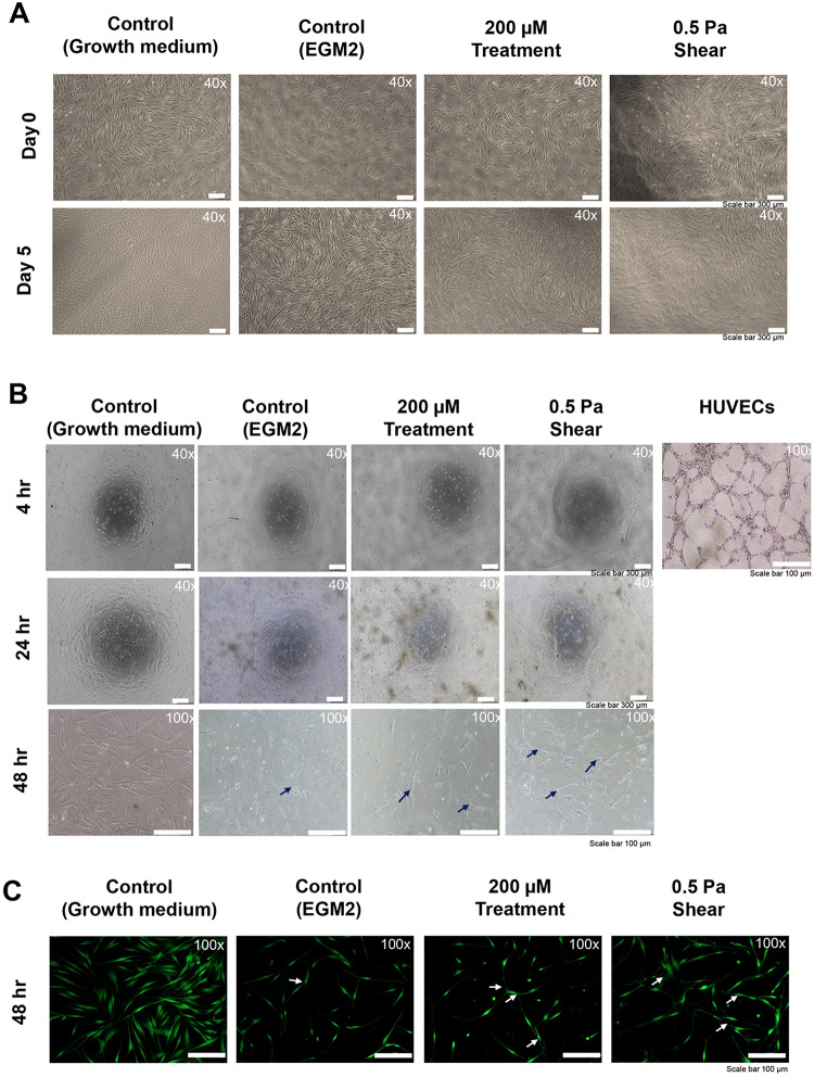
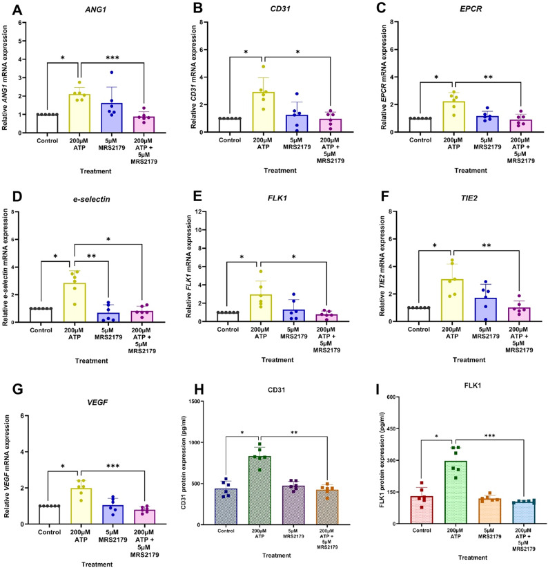
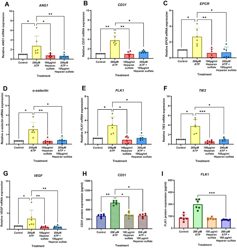
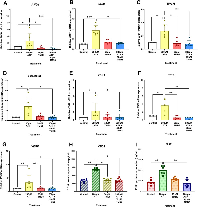
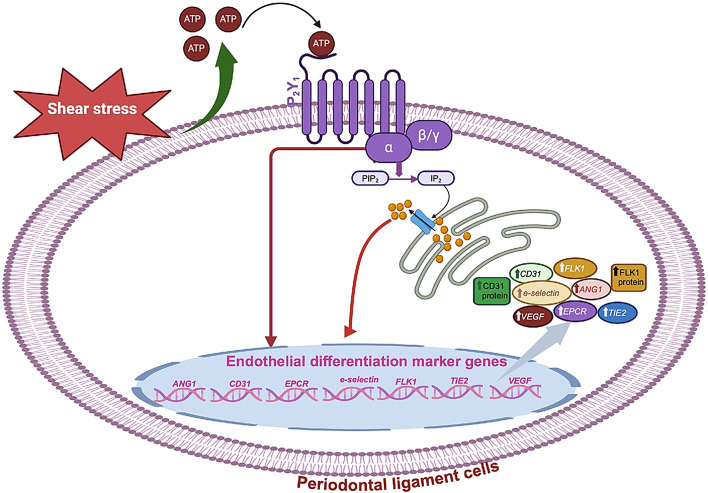
Mechanical forces stimulate human periodontal ligament stem cells (HPDLSCs) to release extracellular adenosine triphosphate (eATP). The eATP impacts various functions of HPDLSCs, i.e., immunosuppression and inflammation. eATP has been reported to promote the angiogenesis of pulmonary vascular endothelial cells. Shear force, one of the mechanical forces involved in orthodontic tooth movement, influences osteogenic differentiation and ECM remodeling of HPDLSCs. However, the relationship between shear force and the impact of eATP on endothelial differentiation and angiogenic characteristics of HPDLSCs remains unclear. This study aimed to determine the response of HPDLSCs on endothelial differentiation and angiogenic properties after shear stress loading and eATP treatment as well as explored the mechanism of eATP-involved in angiogenic responses of HPDLSCs. Shear stress application at 5 dyn/cm2 for 24 h stimulated the release of ATP by HPDLSCs. Both shear stress and 200 µM eATP promoted the expression of endothelial differentiation and angiogenic markers (ANG1, CD31, EPCR, e-selectin, FLK1, TIE2, VEGF). Blockade of specific P2Y1 receptor and intracellular calcium signaling attenuated eATP-induced expression of endothelial differentiation and angiogenic expression. Both shear stress and eATP have stimulatory effects on endothelial differentiation and angiogenesis in HPDLSCs through P2Y1-intracellular calcium signaling.

Keywords: Angiogenesis; Endothelial differentiation; Extracellular ATP; Human periodontal ligament stem cells; Shear stress.


Conflict of interest statement

Declarations. Competing interests: The authors declare no competing interests.

Figures

Fig. 1
Fig. 1
Human periodontal ligament stem cells (HPDLSCs) respond to shear force application by releasing ATP into the extracellular environment. (A) Shear force at the magnitude of 1.5 dyn/cm2 (0.15 Pa) and 5 dyn/cm2 (0.5 Pa) was applied to HPDLSCs for 24 h. (B) After 24-hour application, the amount of ATP release is measured by ATP bioluminescence assay. Data were statistically analyzed by Mann-Whitney U test. (n=6) ** represents P<0.001.
Fig. 2
Fig. 2
Shear force and the released eATP downregulated the expression of stemness markers by HPDLSCs. (A-C) Real-time PCR analysis of CD73, CD90,CD 105 mRNA expression after 24-hour application of shear force at 0.5 Pa. (D-F) mRNA expression of CD73>, CD90 and CD105 after 24-hour ATP treatment. Data were statistically analyzed by Mann-Whitney U test. (n=6) *, **, and *** represent P<0.05, P<0.001 and P<0.0001, respectively.
Fig. 3
Fig. 3
Shear force induced the expression of endothelial differentiation and angiogenic markers by HPDLSCs. (A-G) Real-time PCR analysis of ANG1, CD31, EPCR, e-selectin, FLK1, TIE2, and VEGF mRNA expression after 24-hour application of shear force at the magnitudes of 1.5 dyn/cm2 (0.15 Pa) and 5 dyn/cm2 (0.5 Pa). (H-I) The expression of CD31 and FLK1 proteins was detected by ELISA analysis. Data were statistically analyzed by Mann-Whitney U test. (n=6) *,**, and *** represent P<0.05, P<0.001 and P<0.0001, respectively.
Fig. 4
Fig. 4
Extracellular ATP (eATP) induced the expression of endothelial differentiation and angiogenic markers by HPDLSCs. HPDLSCs were treated with various concentrations of eATP for 24 h. Expression of endothelial differentiation and angiogenic markers by HPDLCs were examined after eATP treatment. (A-G) mRNA expression of ANG1, CD31,EPCR,e-selectin,FLK1, TIE2, and VEGF were analyzed by real-time PCR. (H-I) Protein expression of CD31 and FLK1 were measured by ELISA. Data were statistically analyzed using Kruskal Wallis test followed by Dunn’s multiple comparison tests (n=6) *,**, and *** represent P<0.05, P<0.001 and P<0.0001, respectively.
Fig. 5
Fig. 5
Endothelial differentiation and Matrigel tube formation validated the role of shear stress and eATP treatment on endothelial differentiation and angiogenesis of HPDLSCs. HPDLSCs were treated with either 0.5 Pa shear stress or 200 µM eATP for 24 h, after which they were incubated in EGM2 for 5 days. (A) Changes in morphology were observed using an inverted microscope. (B) A Matrigel tube formation assay was conducted and incubated for 48 h, with observations made under a microscope to assess tube formation. HUVECs used as a positive control (C) Calcein AM fluorescence staining was performed to validate tube formation. Arrows pointed out the tube-like structures.
Fig. 6
Fig. 6
P2Y1 receptor activation is associated with eATP-induced endothelial differentiation and angiogenesis of HPDLSCs. HPDLSCs were pre-treated with MRS 2179 30 min before the 24-hour eATP application. (A-G) mRNA expression of ANG1, CD31, EPCR, e-selectin,FLK1,TIE2, and VEGF was analyzed using real-time PCR. (H-I) the protein expression of CD31 and FLK1 were evaluated using ELISA analysis. Data were statistically analyzed using Kruskal Wallis test followed by Dunn’s multiple comparison tests (n=6) *,**, and *** represent P<0.05, P<0.001 and P<0.0001, respectively.
Fig. 7
Fig. 7
Inositol 1-4-5 triphosphate (IP3) regulates eATP-induced endothelial differentiation and angiogenic marker expression by HPDLSCs. Pre-treatment with IP3 inhibitor (100 µg/ml Heparan sulfate) was applied to human periodontal ligament cells 30 min before 200 µM eATP 24 h-treatment. (A-G) mRNA expression of ANG1, CD31,EPCR, e-selectin, FLK1,TIE2, and VEGF was analyzed using real-time PCR. (H-I) the protein expression of CD31 and FLK1 were evaluated using ELISA analysis. Data were statistically analyzed using Kruskal Wallis test by Dunn’s comparison test (n=6). The values are presented as the mean values±SD. *,**, and ***represent P<0.05, P<0.001 and P<0.0001, respectively.
Fig. 8
Fig. 8
The involvement of intracellular calcium signaling in eATP-induced endothelial differentiation and angiogenic markers expression by HPDLSCs. Pre-treatment with intracellular calcium mobilization inhibitor (50 µM TMB8) was applied to human periodontal ligament cells 30 min before 200 µM eATP 24 h-treatment. (A-G) mRNA expression of ANG1, CD31, EPCR, e-selectin, FLK1, TIE2, and VEGF was analyzed using real-time PCR. (H-I) the protein expression of CD31 and FLK1 were evaluated using ELISA analysis. Data were statistically analyzed using Kruskal Wallis test by Dunn’s comparison test (n=6). The values are presented as the mean values±SD.*,**, and *** represent P<0.05, P<0.001 and P<0.0001, respectively.
Fig. 9
Fig. 9
Schematic diagram demonstrating shear stress influencing endothelial differentiation of HPDLSCs through eATP-P2Y1 binding signaling pathway. HPDLSCs respond to shear stimulation by releasing ATP into the extracellular environment. The released ATP is involved in the induction of angiogenic properties of HPDLSCs through the P2Y1-intracellular calcium signaling pathway. The figure is created in BioRender. Osathanon, T. (2025) https://BioRender.com/ tt7y20y.

Similar articles

References

    1. Kanjanamekanant, K., Luckprom, P. & Pavasant, P. Mechanical stress-induced interleukin-1beta expression through adenosine triphosphate/P2X7 receptor activation in human periodontal ligament cells. J. Periodontal Res.48, 169–176. 10.1111/j.1600-0765.2012.01517.x (2013). - PubMed
    1. Luckprom, P., Kanjanamekanant, K. & Pavasant, P. Role of connexin43 hemichannels in mechanical stress-induced ATP release in human periodontal ligament cells. J. Periodontal Res.46, 607–615. 10.1111/j.1600-0765.2011.01379.x (2011). - PubMed
    1. Takahara, N. et al. Real-time imaging of ATP release induced by mechanical stretch in human airway smooth muscle cells. Am. J. Respir. Cell Mol. Biol.51, 772–782. 10.1165/rcmb.2014-0008OC (2014). - PubMed
    1. Shi, Q. et al. Fluid shear stress promotes periodontal ligament cells proliferation via p38-Amot-Yap. Cell. Mol. Life Sci.79, 551. 10.1007/s00018-022-04591-w (2022). - PMC - PubMed
    1. Suwittayarak, R. et al. Shear stress enhances the Paracrine-Mediated immunoregulatory function of human periodontal ligament stem cells via the ERK signalling pathway. Int. J. Mol. Sci.2310.3390/ijms23137119 (2022). - PMC - PubMed

Substances