

Development of lesion and organ negative cast modelling technique for quality assurance and optimization of nuclear medicine images
- PMID: 40695971
- PMCID: PMC12284253
- DOI: 10.1038/s43856-025-01009-z
Development of lesion and organ negative cast modelling technique for quality assurance and optimization of nuclear medicine images
Abstract
Background: Nuclear medicine imaging allows for a wide variety of data acquisition and image generation methods in the clinical setting. Imaging phantoms are routinely used to evaluate and optimize image quality and quantitative accuracy of features, but few phantoms realistically model the anatomy or heterogeneity of target regions within patient images, such as tumours that are commonly observed in oncology. We developed a negative cast modelling (NCM) technique which enables applications such as non-standard shape tumour phantoms, organ phantoms for radiation dosimetry, and quality control phantoms with small lesions.
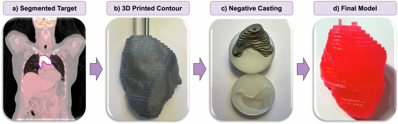
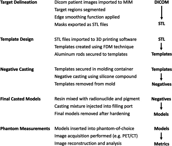
Methods: Tumour templates were derived from segmented PET images of primary mediastinal B-cell lymphoma (PMBCL) patients. Lesion segmentations were saved and 3D-printed. Negatives were developed using silicone-based molding materials, and final models cast using a composition of liquid plastic, pigment, and PET radiotracer. Images of lesions were acquired using the GE DMI PET/CT scanner, and image features were quantified.
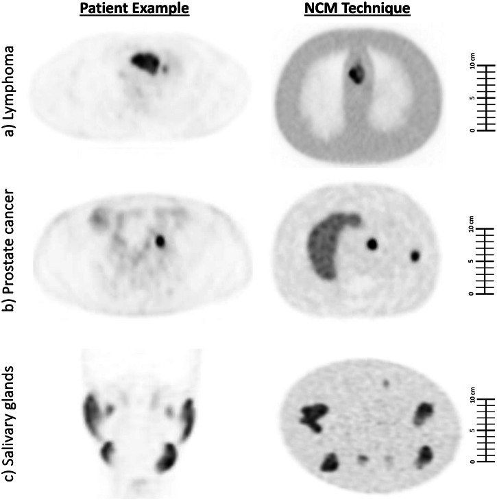
Results: Mean absolute error (MAE) for tumour volume between the original template and casted models is 13.8%, indicating that the method is reasonably accurate. The high viscosity of the liquid plastic used in the casting process establishes non-uniform tumour models, which is very useful in practice for evaluating image features related to heterogeneity. PET images using the NCM method is determined to be highly realistic by an experienced nuclear medicine physician, due to the non-standard shapes that can be established within the tumours.
Conclusions: The NCM method has potential to enable more realistic phantom studies within nuclear medicine imaging. The cost for the lymphoma tumour phantom study is less than $400 USD, making it feasible for large-scale studies.
Plain language summary
Radioactive substances are used to diagnose and treat disease. Special testing devices called phantoms are used to ensure that the scanners used to detect the radioactive substances work properly. These are usually simple shapes, such as spheres, which do not accurately represent the complexity of real human anatomy. In this study, we developed a technique that uses medical images from patients to create phantoms with more realistic shapes, such as irregular tumors, salivary glands, and very small lesions. These phantoms are made using a low-cost molding and casting process and can be reused. By improving how scanners are tested and optimized, this approach may lead to better diagnosis and treatment for conditions such as cancer.
© 2025. The Author(s).
Conflict of interest statement
Competing interests: The authors declare no competing interests. Ethics: This work depicts compounds pertaining to patent WO 2017/117687 A1, which entitles F.B. to royalties upon licensing.
Figures


Similar articles
-
Cost-effectiveness of using prognostic information to select women with breast cancer for adjuvant systemic therapy.Health Technol Assess. 2006 Sep;10(34):iii-iv, ix-xi, 1-204. doi: 10.3310/hta10340. Health Technol Assess. 2006. PMID: 16959170
-
A rapid and systematic review of the clinical effectiveness and cost-effectiveness of paclitaxel, docetaxel, gemcitabine and vinorelbine in non-small-cell lung cancer.Health Technol Assess. 2001;5(32):1-195. doi: 10.3310/hta5320. Health Technol Assess. 2001. PMID: 12065068
-
Short-Term Memory Impairment.2024 Jun 8. In: StatPearls [Internet]. Treasure Island (FL): StatPearls Publishing; 2025 Jan–. 2024 Jun 8. In: StatPearls [Internet]. Treasure Island (FL): StatPearls Publishing; 2025 Jan–. PMID: 31424720 Free Books & Documents.
-
Sexual Harassment and Prevention Training.2024 Mar 29. In: StatPearls [Internet]. Treasure Island (FL): StatPearls Publishing; 2025 Jan–. 2024 Mar 29. In: StatPearls [Internet]. Treasure Island (FL): StatPearls Publishing; 2025 Jan–. PMID: 36508513 Free Books & Documents.
-
Can a Liquid Biopsy Detect Circulating Tumor DNA With Low-passage Whole-genome Sequencing in Patients With a Sarcoma? A Pilot Evaluation.Clin Orthop Relat Res. 2025 Jan 1;483(1):39-48. doi: 10.1097/CORR.0000000000003161. Epub 2024 Jun 21. Clin Orthop Relat Res. 2025. PMID: 38905450
References
-
- World Nuclear Association. Radioisotopes in Medicine. January 10, Accessed May 29, 2025. https://world-nuclear.org/information-library/non-power-nuclear-applicat... (2025).
-
- Weber, W. A. et al. The future of nuclear medicine, molecular imaging, and theranostics. J. Nucl. Med.61, 263S–272S (2020). - PubMed
-
- Walker M. Quantitative Dynamic 3D PET Scanning of the Body and Brain Using LSO Tomographs. University of Manchester; 2009.
LinkOut - more resources
Full Text Sources
Miscellaneous
