. 2025 Jul 22;15(1):26625.
doi: 10.1038/s41598-025-12005-7.

CXCL12 chemokine dimer signaling modulates acute myelogenous leukemia cell migration through altered receptor internalization

Affiliations

CXCL12 chemokine dimer signaling modulates acute myelogenous leukemia cell migration through altered receptor internalization

Donovan Drouillard et al. Sci Rep. .

Abstract

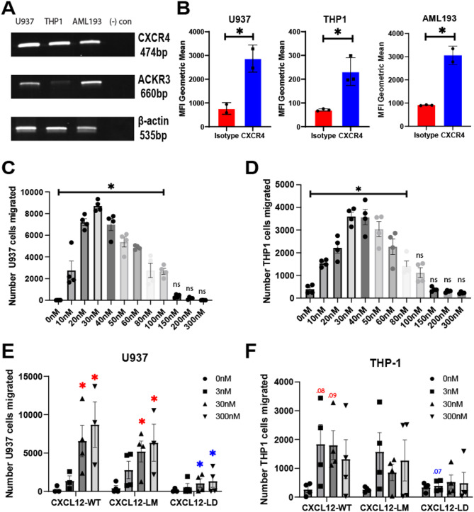
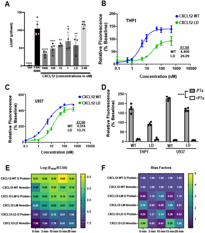
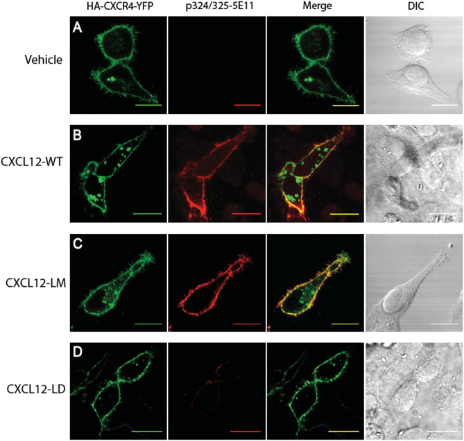
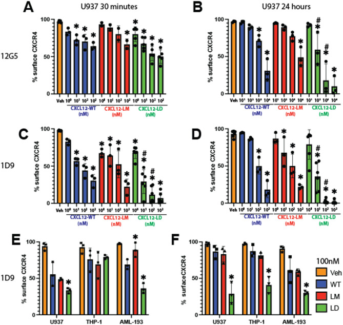
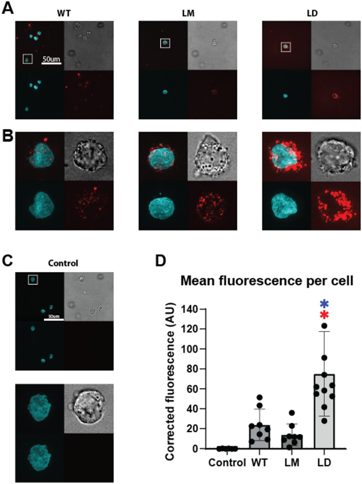
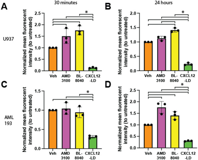
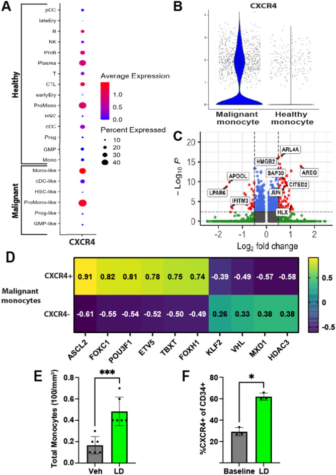
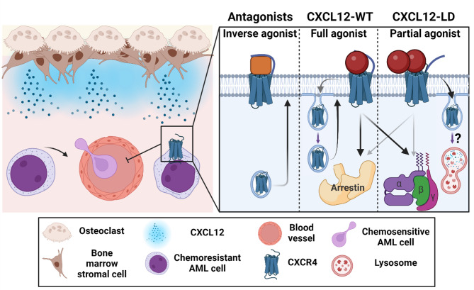
Acute myeloid leukemia (AML) is a malignancy of immature myeloid blast cells with stem-like and chemoresistant cells being retained in the bone marrow through CXCL12-CXCR4 signaling. Current CXCR4 inhibitors that mobilize AML cells into the bloodstream have failed to improve patient survival, likely reflecting persistent chemokine receptor localization on target cells. Here we characterize the signaling properties of CXCL12-locked dimer (CXCL12-LD), a bioengineered variant of the naturally occurring oligomer of CXCL12. CXCL12-LD, in contrast to wild-type or locked monomer variants, was unable to induce chemotaxis in AML cells. CXCL12-LD binding to CXCR4 decreased G protein, β-arrestin, and intracellular calcium mobilization signaling pathways, and indicated the locked dimer is a partial agonist of CXCR4. Despite these partial agonist properties, CXCL12-LD increased CXCR4 internalization compared to wild-type and monomeric CXCL12. Analysis of a previously published AML transcriptomic data showed CXCR4 positive AML cells co-express genes involved in survival, proliferation, and maintenance of a blast-like state. The CXCL12-LD partial agonist effectively mobilized stem cells into the bloodstream in mice suggesting a potential role for their use in targeting CXCR4. Together, our results suggest that enhanced internalization by CXCL12-LD partial agonist can avoid pharmacodynamic tolerance and may identify new avenues to better target GPCRs.

Keywords: Acute myeloid leukemia; CXCR4; Cell signaling; Chemokines; GPCR; Receptor internalization.


Conflict of interest statement

Declarations. Competing interests: MBD and FP have financial interests in Protein Foundry, LLC and Xlock Biosciences, Inc. The remaining authors have no conflicts to disclose.

Figures

Fig. 1
Fig. 1
CXCR4 expression in AML cell lines. (A) RT-PCR of CXCR4, ACKR3, and β actin in three human AML cell lines U937, THP-1, and AML-193. Data representative of 3 separate analyses. (B) Flow cytometry results of the three AML cell lines stained with either anti-CXCR4 clone 12G5 or control isotype antibody. * = P ≤ 0.05 by Student’s t-test. n = 2–3 independent analyses. Values are mean ± SEM. (C) U937 or (D) THP-1 cell chemotaxis from the top chamber of a transwell filter towards the lower chamber containing the indicated concentration of CXCL12-WT chemokine. All values under significance bar represent a significant increase by one-way ANOVA with Dunnett’s multiple comparison test to 0 nM vehicle treated. (E) Total number of U937 or (F) THP-1 migrated from the top chamber of a transwell chamber towards the lower chamber containing the indicated concentration of CXCL12-WT, CXCL12-LM, or CXCL12-LD. Migrating cells in panels C-F were enumerated after a 2 h incubation using flow cytometry. Values represent mean ± SEM, n = 3–4 independent biological replicates. Red asterisks or numbers indicate adjusted p value by two-way ANOVA with Tukey’s multiple comparisons test to that respective variant’s 0 nM (e.g. CXCL12-WT 30 nM to CXCL12-WT 0 nM) while blue indicates significance compared to the same dose of a different variant (e.g. CXCL12-LD 30 nM to CXCL12-WT 30 nM).
Fig. 2
Fig. 2
CXCL12-LD is a balanced partial agonist. (A) cAMP levels in THP-1 cells stimulated with 10 mM IBMX and 1 µM forskolin followed by treatment with various compounds measured by a competitive enzyme immunoassay. All significance values are in relation to FSK/IBMX by two-way ANOVA with Dunnett’s multiple comparison test. (B, C) Calcium flux in THP-1 and U937 cells treated with either CXCL12-WT or CXCL12-LD. n = 3. (D) AML cells were pre-treated with pertussis toxin prior to treatment with CXCL12 variants. All pertussis toxin pre-treated values are significantly different by Student’s t-test when compared to the same form of CXCL12 without pertussis toxin. (E) The log(Emax/EC50) of the CXCL12 variants from net BRET in HEK293-CXCR4-Luc cells transfected with β-arrestin or Gαi Venus transducer. n = 6, EC50 values calculated from non-linear curve fit, all r2 values > 0.82. (F) Bias factors for CXCL12-WT, -LM, and -LD at all timepoints tested from HEK293 BRET studies calculated using the calculator found on the Biased Signaling Atlas. A bias factor > 5 is indicative of biased agonism.
Fig. 3
Fig. 3
Differential CXCR4 C-terminal phosphorylation in response to CXCL12 variants. (A) HeLa cells transiently transfected with HA-CXCR4-YFP were stimulated with vehicle, (B) CXCL12-WT, (C) CXCL12-LM, or (D) CXCL12-LD. The far left panels show visualization of YFP-CXCR4 and the middle left panel is visualization of the phosphorylated Ser324/325 residues on CXCR4. Co-localization of the YFP-CXCR4 and phosphorylated CXCR4 appear yellow in the merge panel on the middle right. Differential image contrast (DIC) images are shown on the far right panels. Shown are the representative micrographs from three independent experiments. Bars = 20 μm.
Fig. 4
Fig. 4
CXCL12-LD promotes internalization of CXCR4. (A) Percent of CXCR4 positive U937 cells detected by flow cytometry with the 12G5 CXCR4 antibody after 30 min or (B) 24 h of incubation with various concentrations of CXCL12-WT (blue), -LM (orange), or -LD (green). *Represent significance when comparing to vehicle and # represents significance when comparing to respective dose of CXCL12-WT (e.g. CXCL12-LD 10 nM to CXCL12-WT), adjusted P ≤ 0.05 by two-way ANOVA with Tukey’s multiple comparison test. (C) The percent of CXCR4 positive U937 cells detected by flow cytometry with the 1D9 CXCR4 antibody after 30 min or (D) 24 h of incubation with various concentrations of CXCL12 variants. (E) Three different leukimic cell lines treated with CXCL12-WT, -LM, or -LD and stained with the 1D9 antibody after 30 min of treatment or (F) 24 h. Significance represents comparison of CXCL12-variant to CXCL12-WT in the same cell line by one-way ANOVA with Dunnett’s multiple comparison test, P ≤ 0.05.
Fig. 5
Fig. 5
Labeled CXCL12 ligands show internalization with CXCL12-LD. (A) U937 cells were treated with 200 nM of AzDye647-conjugated CXCL12 variants (red, bottom right of one image) of their respective variant for 30 min before fixation and staining with Hoechst 3342 (cyan, bottom left). Brightfield is shown in the top right and the merged image is the top left. Images shown are at 100X. Scale bar is 50 μm. (B) Enhancement on the cutout of the images in the white square from (A). (C) Untreated U937 cells shown under the same conditions as (A) and (B). (D) Quantification of the corrected (background substracted) average red AzDye647 fluorescence intensity inside of cells. Red asterisks indicate significance compared to control and blue asterisks indicate signficance compared to CXCL12-WT by one-way ANOVA with Tukey’s multiple comparisons test, P ≤ 0.05.
Fig. 6
Fig. 6
CXCR4 surface upregulation seen with AMD3100 and BL-8040 but not CXCL12-LD. (A) U937 cells were treated with either 1 µM AMD3100, 100 nM BL-8040, or 100 nM CXCL12-LD and immunostained for surface CXCR4 with the 1D9 clone. Mean fluorescent intensity (MFI) values were normalized against vehicle-treated (PBS) U937 cells stained for surface CXCR4. Treatment was for 30 min or (B) 24 h. (C) CXCR4 inhibitor treatment of AML-293 cells for 30 min or (D) 24 h under the same conditions seen in 6 A and 6B. * P ≤ 0.05 by one-way ANOVA with Tukey’s multiple comparisons test.
Fig. 7
Fig. 7
CXCL12-LD increases the frequency of hematopoietic progenitor cells in peripheral blood. (A) Dot plot of CXCR4 expression in the bone marrow aspirates of 16 AML patient’s pre-treatment. Each of the “-like” cells had a confirmed mutation in a targeted DNA sequence to confirm their malignancy. (B) The “Mono” and “Mono-like” cell types were further analyzed for CXCR4 expression. CXCR4 was elevated in the Mono-like (Malignant monocyte) compared to the Mono (Healthy monocyte) by MAST test (P ≤ 0.05). (C) The malignant monocyte population from Fig. 6B was grouped by CXCR4 expression. Differentially expressed genes by MAST test were found and plotted on the volcano plot. Grey dots represent non-significant genes by both log2FC or adjusted p-value, green dots represent genes with significant adjusted p-value but not log2FC, blue dots represent genes with significant log2FC but not adjusted p-value, and red dots represent genes with both significant log2FC and adjusted p-value. Log2FC threshold was 0.5 and adjusted p-value threshold was 0.005. (D) The malignant monocytes sorted by CXCR4 expression were analyzed using the CollecTRI R package to estimate the transcription factor activity based on weighted mean gene expression with the obtained t-values of the slope plotted. Positive values represent inferred transcription factor activity while negative values represent inferred inactive transcription factor activity. (E) Wildtype C57BL/6 mice were given 200 µL 5 µM CXCL12-LD or vehicle PBS. The frequency of total monocytes (E) and CD34 + CXCR4 + progenitor cells (F) in peripheral blood was measured two hours after treatment. Values are mean ± SD from 3–6 separate mice.
Fig. 8
Fig. 8
CXCL12-LD functions differently than currently available CXCR4 inhibitors. Left, CXCL12 produced by bone marrow stromal cells maintains chemoresistant CXCR4-expressing AML blasts in the marrow. If CXCR4 expression is lost or the receptor is inhibited, the blasts are mobilized into the bloodstream. Right, 2nd generation CXCR4 inhibitors are shown to be inverse agonists of CXCR4 that inhibit internalization and result in surface receptor upregulation. In the middle, CXCL12-WT as a balanced agonist activating both β-arrestin and the G-protein signaling pathways. Signaling with CXCL12-WT results in receptor internalization and recycling. On the right, CXCL12-LD is a balanced partial agonist whose signaling results in greater receptor internalization compared to CXCL12-WT, possibly through increased receptor lysosomal degradation rather than endosomal recycling. Created in BioRender. Drouillard, D. (2025) https://BioRender.com/t28xc88.

Update of

Similar articles

References

    1. Veldkamp, C. T., Peterson, F. C., Pelzek, A. J. & Volkman, B. F. The monomer-dimer equilibrium of stromal cell-derived factor-1 (CXCL 12) is altered by pH, phosphate, sulfate, and heparin. Protein Sci.14, 1071–1081. 10.1110/ps.041219505 (2005). - PMC - PubMed
    1. Veldkamp, C. T. et al. Structural basis of CXCR4 Sulfotyrosine recognition by the chemokine SDF-1/CXCL12. Sci. Signal.1, ra4. 10.1126/scisignal.1160755 (2008). - PMC - PubMed
    1. Ziarek, J. J. et al. Sulfopeptide probes of the CXCR4/CXCL12 interface reveal oligomer-specific contacts and chemokine allostery. ACS Chem. Biol.8, 1955–1963. 10.1021/cb400274z (2013). - PMC - PubMed
    1. Roy, I. et al. Pancreatic Cancer cell migration and metastasis is regulated by Chemokine-Biased agonism and bioenergetic signaling. Cancer Res.75, 3529–3542. 10.1158/0008-5472.CAN-14-2645 (2015). - PMC - PubMed
    1. Roy, I., Getschman, A. E., Volkman, B. F. & Dwinell, M. B. Exploiting agonist biased signaling of chemokines to target cancer. Mol. Carcinog.56, 804–813. 10.1002/mc.22571 (2017). - PMC - PubMed