

Ephaptic conduction molding memory engrams
- PMID: 40696380
- PMCID: PMC12285051
- DOI: 10.1186/s12915-025-02323-7
Ephaptic conduction molding memory engrams
Abstract
Background: Memories are programmed in the brain as connected neuronal ensembles called engrams. However, the method by which the brain forms engrams during memory encoding is not understood.
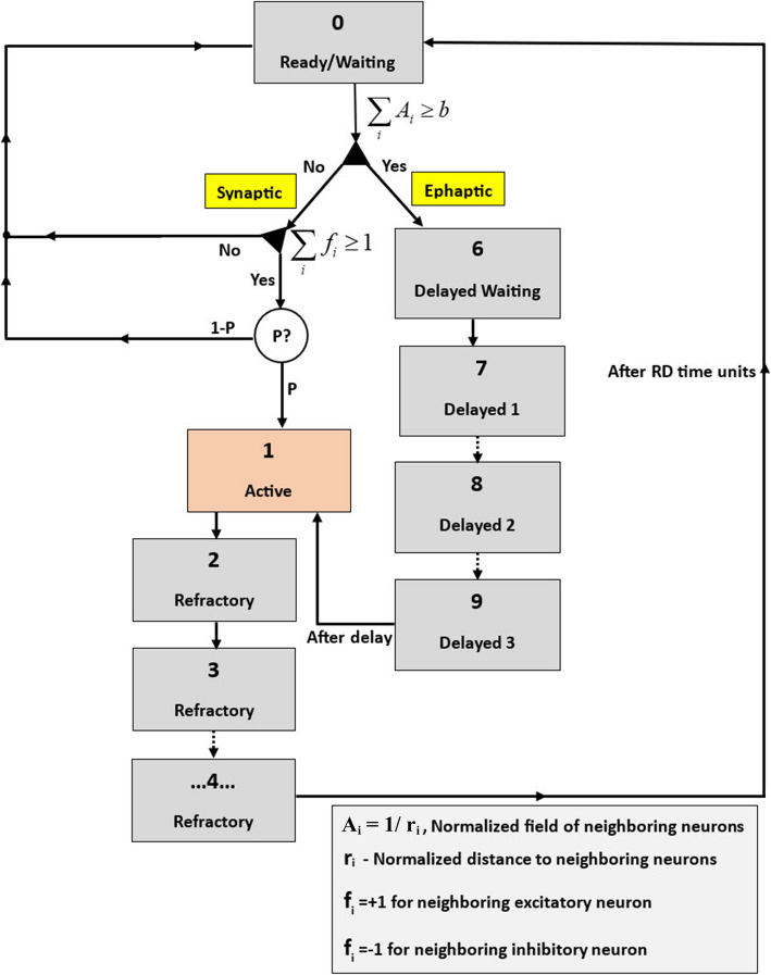
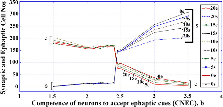
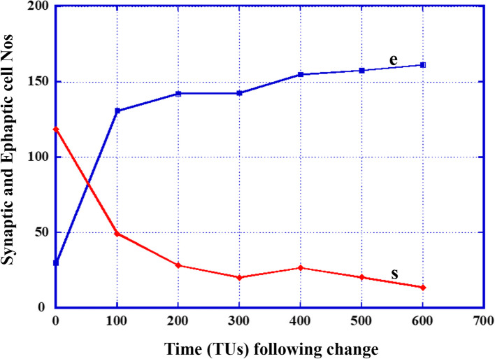
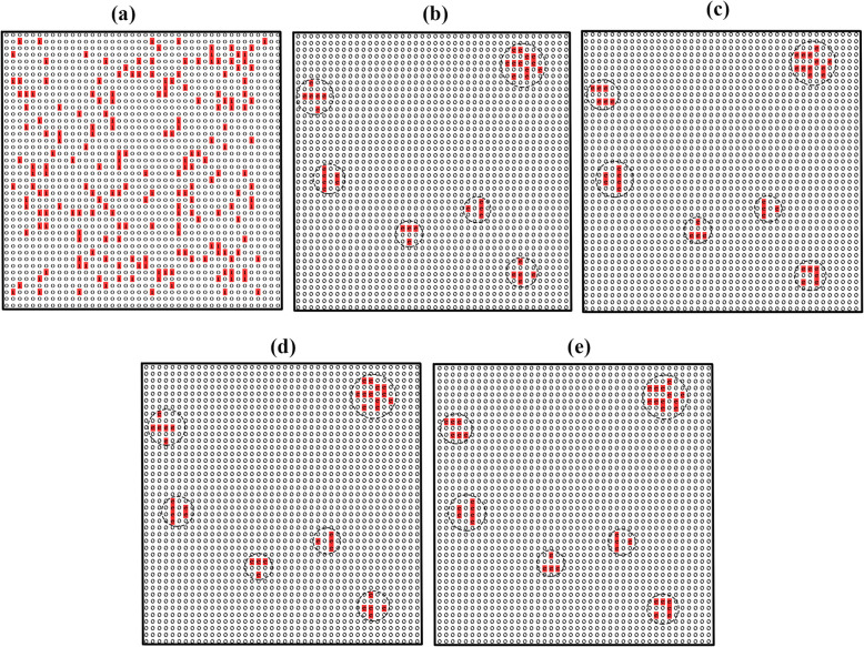
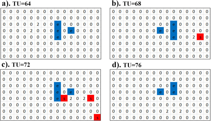
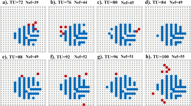
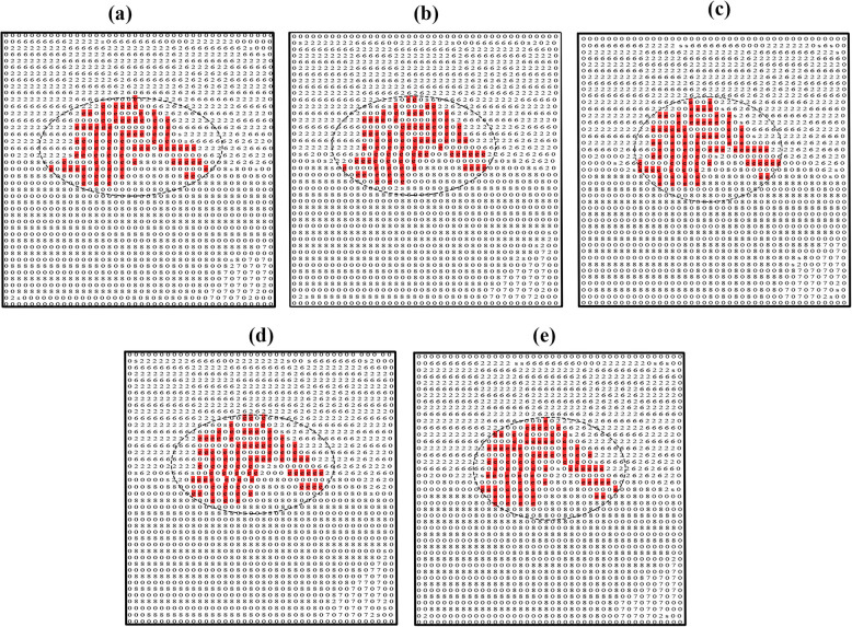
Results: We have created a mechanistic mathematical model showing a possible method of the encoding process. Our model is based on the cellular automata approach, which can specifically distinguish between neurons operated on by the synaptic and those operated on by the ephaptic modes. This feature allows us to confirm that the ephaptic mode induces the formation of repeating collections of operating neurons (sub-engrams) that can become memory-preserving entities, and the synaptic influence is manifested by molding these sub-engrams by pruning small ones and size-increasing and rounding larger ones to form the engrams' final structures.
Conclusions: Ephaptic and synaptic dual-participation in the memory encoding process was exhibited. The sequence of activities was unveiled. We also speculate on possible procedures the brain can employ to enable the ephaptic mode to overtake the normal, synaptic-dominating one.
Keywords: Engram formation; Ephaptic domination; Memory encoding; Sub-engrams; Synaptic molding.
© 2025. The Author(s).
Conflict of interest statement
Declarations. Ethics approval and consent to participate: Not applicable. Consent for publication: Not applicable. Competing interests: The authors declare no competing interests.
Figures
Similar articles
-
Short-Term Memory Impairment.2024 Jun 8. In: StatPearls [Internet]. Treasure Island (FL): StatPearls Publishing; 2025 Jan–. 2024 Jun 8. In: StatPearls [Internet]. Treasure Island (FL): StatPearls Publishing; 2025 Jan–. PMID: 31424720 Free Books & Documents.
-
Selective inhibition in CA3: A mechanism for stable pattern completion through heterosynaptic plasticity.PLoS Comput Biol. 2025 Jul 7;21(7):e1013267. doi: 10.1371/journal.pcbi.1013267. eCollection 2025 Jul. PLoS Comput Biol. 2025. PMID: 40623085 Free PMC article.
-
Ensembles and engrams in mouse cortical and sub-thalamic brain regions supporting context and memory recall.bioRxiv [Preprint]. 2025 Jul 4:2025.06.29.662192. doi: 10.1101/2025.06.29.662192. bioRxiv. 2025. PMID: 40631237 Free PMC article. Preprint.
-
Technological aids for the rehabilitation of memory and executive functioning in children and adolescents with acquired brain injury.Cochrane Database Syst Rev. 2016 Jul 1;7(7):CD011020. doi: 10.1002/14651858.CD011020.pub2. Cochrane Database Syst Rev. 2016. PMID: 27364851 Free PMC article.
-
Comparison of self-administered survey questionnaire responses collected using mobile apps versus other methods.Cochrane Database Syst Rev. 2015 Jul 27;2015(7):MR000042. doi: 10.1002/14651858.MR000042.pub2. Cochrane Database Syst Rev. 2015. PMID: 26212714 Free PMC article.
References
-
- Anastassiou CA, Perin R, Markram H, Koch C. Ephaptic coupling of cortical neurons. Nat Neurosci. 2011;14(2):217–23. 10.1038/nn.2727. - PubMed
MeSH terms
LinkOut - more resources
Full Text Sources
Medical
