. 2025 Jul 22;20(1):89.
doi: 10.1186/s13062-025-00666-3.

CLIC2 regulates immunosuppression and macrophage differentiation in genomically stable gastric cancer

Affiliations

CLIC2 regulates immunosuppression and macrophage differentiation in genomically stable gastric cancer

Viviana Longo et al. Biol Direct. .

Abstract

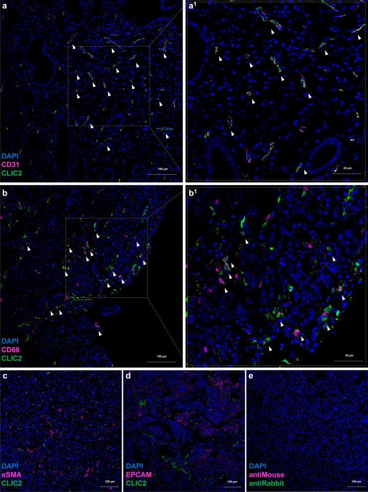
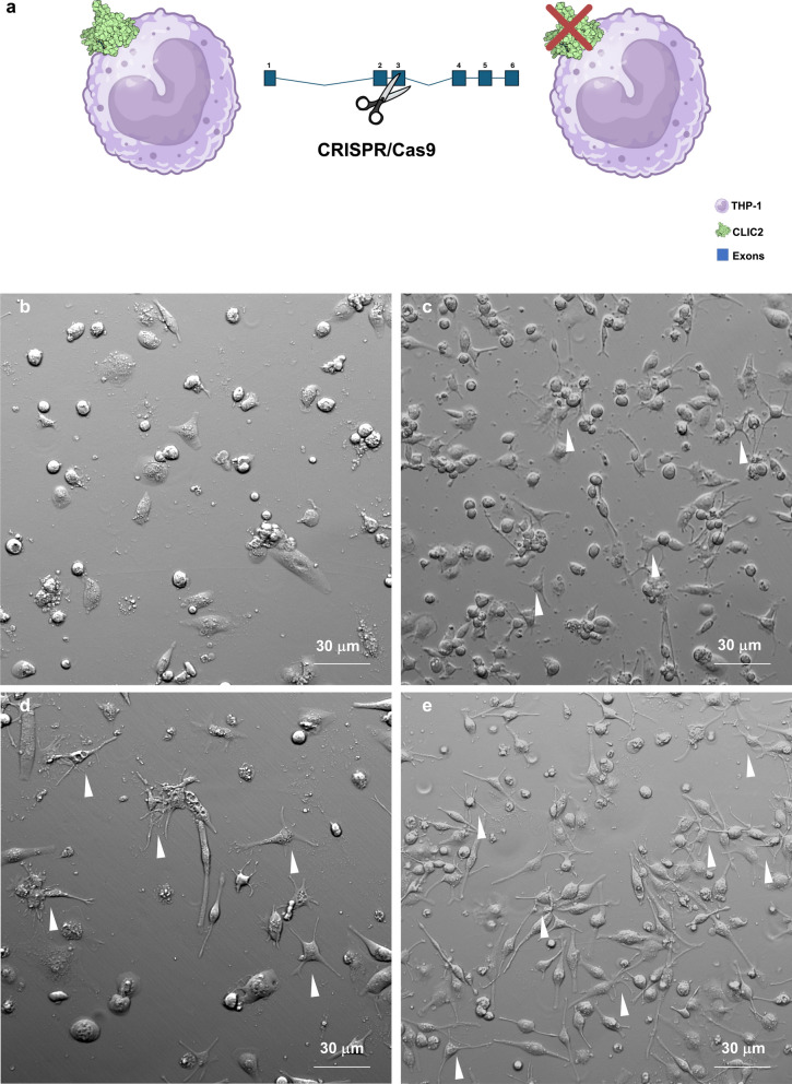
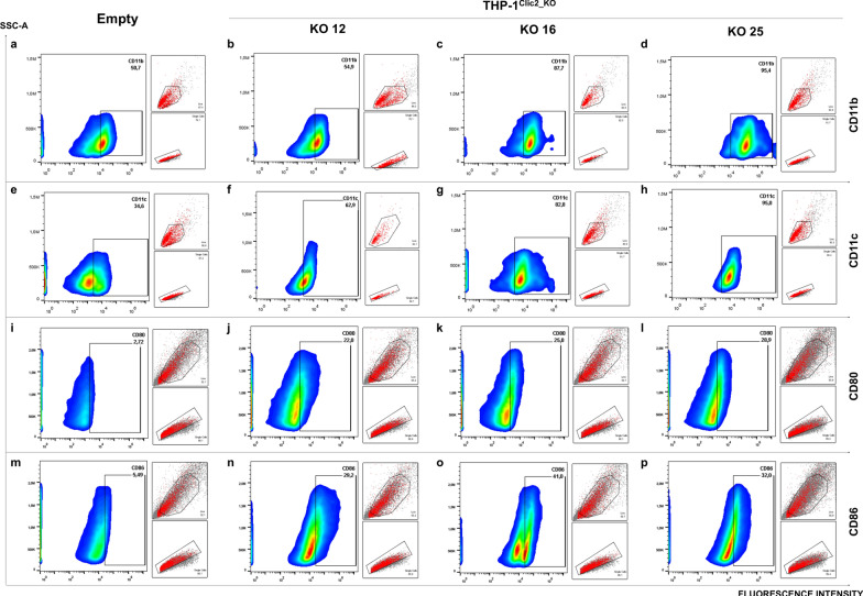
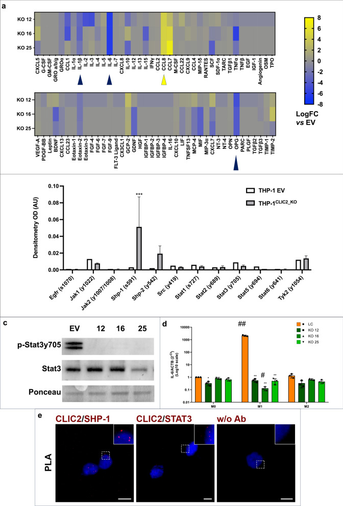
Chloride intracellular channels (CLICs) are a family of six evolutionarily conserved proteins with diverse functions. Previously, we identified CLIC2, as the fifth-ranked master regulator associated with diffuse-type gastric cancer (dGC) showing increased expression in tumors. Here we used bulk, as well as single cell sequencing datasets of dGC, to demonstrate for the first time a direct association of CLIC2 with the microsatellite stable GC and, furthermore, the expression of CLIC2 in macrophages (MCs), and endothelial cells (ECs) populating gastric tissue. We generated CLIC2 knock-out THP-1 monocytic cells (THP-1CLIC2_KO) determining that while CLIC2 deletion had no observable effect on monocytes, THP-1CLIC2_KO macrophages exhibited significant morphological changes, including increased membrane protrusions, and upregulated expression of CD11b, CD11c, CD80, and CD86 markers. Furthermore, cytokine secretion profiling of THP-1CLIC2_KO differentiated cells revealed elevated secretion of CCL8, alongside reduced secretion of IL-1β, IL-6, and osteoprotegerin (OPG). Additionally, we observed increased phosphorylation of Shp1 phosphatase with the concomitant absence of Stat3 phosphorylation, which impaired downstream signaling, in line with the evidence that Clic2 interacts with both Shp1 and Stat3. Based on these findings, we suggest that CLIC2 plays a pivotal role in regulating monocyte-to-macrophage differentiation by modulating the Stat3 signaling pathway, thus enhancing gastric cancer progression by establishing a tumor-permissive microenvironment.

Keywords: Clic2; Differentiation; Gastric cancer; Gastric microenvironment; Macrophages; Signaling; Tumor microenvironment.


Conflict of interest statement

Declarations. Ethics approval and consent to participate: Human biopsies were collected from patients affected by diffuse-type gastric cancer, in accordance with the approved guidelines of the World Medical Association’s Declaration of Helsinki and informed consent and ethical committee authorization with protocol number 20180042426 of the COMITATO ETICO UNICO REGIONALE PER LA BASILICATA. Competing interests: The authors declare no competing interests.

Figures

Fig. 1
Fig. 1
CLIC2 regulon and gene correlates with dGC and with genomically stable disease. a Heatmap visualization of the CLIC2 regulon gene core, showing expression levels of genes in relation to Lauren classification and master regulator activation. b Relative expression of the CLIC2 regulon in relation to the microsatellite stable gastric cancer (MSS) or microsatellite instable, low or high, gastric cancer (MSI-L and MSI-H). Orange boxes indicate patients expressing the active core of the CLIC2 regulon, green boxes indicate patients expressing genes not composing the CLIC2 regulon core, violet boxes indicate patients expressing the inactive core of the CLIC2 regulon. c Relative expression of the CLIC2 gene in relation to the microsatellite stable gastric cancer (MSS) or microsatellite instable, low or high, gastric cancer (MSI-L and MSI-H). Orange boxes indicate patients expressing the active core of the CLIC2 regulon, green boxes indicate patients expressing genes not composing the CLIC2 regulon core, violet boxes indicate patients expressing the inactive core of the CLIC2 regulon. d Gene network visualization of genes composing CLIC2 regulon. White circles indicate genes of the regulon, gray rhombus indicate categories from MSigDB, grey circles are secondary genes intercepted during analysis of connections, green lines are genetic interactions and grey lines are gene connections on reactome, MSigDB, InterPro and Wikipathways
Fig. 2
Fig. 2
CLIC2 expression is gastric cancer tissues is restricted to endothelial cells and macrophages. Analysis of TCGA STAD dataset to infer tumor purity, applying ESTIMATE algorithm and correlating CLIC2 gene expression with a immune score, b stromal score, c ESTIMATE score. Gene expression based single-cell clustering of d normal gastric mucosa and e diffuse gastric cancer (see legend for more detail about color coding of cell populations; EC—Endothelial Cells, GMC—Gland Mucous Cells, PC—Proliferative Cells, PMC—Pit Mucous Cells, SM—Smooth Muscle Cells). CLIC2 presence (violet dots) or absence (grey dots) in f normal gastric mucosa and g diffuse gastric cancer single-cell clusters identified in d and e. h Odd ratio of cell populations presence in gastric cancer compared to normal samples
Fig. 3
Fig. 3
In dGC samples CLIC2 is expressed by tissue endothelial cells and macrophages. Confocal microscopy images (10X magnification) of immunofluorescence staining of representative tissue slides from 7 dGC patient samples. Images show co-staining of Clic2 with a CD31 as endothelial cells marker, b CD68 as pan-macrophages marker, c αSMA as fibroblast marker and d Epcam as epithelial cells marker. Panels “1” show a magnification of respective areas. White arrows indicate cells co-expressing both assayed proteins. e Negative controls
Fig. 4
Fig. 4
CLIC2 follows the canonical secretory pathway. Clic2 intracellular staining in THP-1 cells differentiated to unpolarized macrophages visualized by confocal microscospy (63X magnification). Dapi staining of nuclei is showed in blue (a1, b1, c1), Clic2 signal is showed in green (a2, b2, c2), cell compartments are showed in red (a3 for endoplasmic reticulum using anti-KDEL; b3 for Golgi apparatus using anti-GM130; c3 for plasma membrane using anti-Integrin β1). Merged signals are showed in a4, b4, c4. For the staining showed in panels a and b, differentiated cells were fixed and permeabilized, for the staining of panel c, differentiated cells were fixed without permeabilization
Fig. 5
Fig. 5
CLIC2 influences macrophages differentiation. a CLIC2 expression was abrogated in THP-1 cells using CRISPR/Cas9, then phenotype and molecular profiling was carried out. Control cells (b) and Clic2 knock out clones (c—KO 12; d—KO 16; e—KO 25), were differentiated to unpolarized macrophages and then observed to the microscope. Gradient contrast images (20X) shows altered morphology of THP-1CLIC2_KO cells after induction of differentiation to M0 macrophages. White arrows evidences dendritic-like macrophages
Fig. 6
Fig. 6
THP-1CLIC2_KO macrophages display enhanced differentiation and increased antigen presentation markers. THP-1 control cells (a, e, i, m) and three THP-1.CLIC2_KO clones (bd; fh jl; np), were differentiated to unpolarized macrophages and analyzed by flow cytometry for common membrane markers such as CD11b (ad), CD11c (eh), CD80 (il), CD86 (mp). Graphs show fluoresce intensity in relation to SSC-A physical parameter, smaller boxes display gating strategies (first gating on live cells FSC-A/SSC-A, second gating on single cells FSC-A/FSC-H)
Fig. 7
Fig. 7
THP-1CLIC2_KO macrophages can positively influence the tumor microenvironment. a THP-1 control cells and three THP-1CLIC2_KO clones were differentiated to unpolarized macrophages and 80 cytokines were simultaneously measured in their culture media using antibody arrays. Dot blot signals of all samples were normalized and compared to those of controls, then LogFC of THP-1CLIC2_KO clones vs controls was calculated. Blue arrows indicate cytokines secreted at least −2 LogFC from all three clones respect to controls. Yellow arrows indicate cytokines secreted at least + 2 LogFC from all three clones respect to controls. The details of data normalization are reported in materials and methods. Statistical significance was calculated using GraphPad Prism 10, unpaired t-test of densitometry of CLIC2-KO clones vs LC (CCL7 p = 0.5188; CCL8 p = 0.0126; IL-1β p = 0.0066; IL-6 p < 0.0001; OPG p = 0.0003). b THP-1 control cells and three THP-1CLIC2_KO clones were differentiated to unpolarized macrophages and Jak/Stat pathway phosphorylation was explored using antibody arrays. Densitometric analysis was carried out and mean of KO clones signals were visualized as histograms. Statistical analysis was performed with GraphPad Prism 10, 2-way Anova, Shp-1 p = 0.0007. c Western blot analysis of Stat3 phosphorylation in THP-1 controls and THP-1CLIC2_KO clones differentiated to M0 macrophages. d Real-time PCR analysis of IL6 transcription in THP-1 controls and THP-1CLIC2_KO clones differentiated to M0 macrophages and further polarized to M1 or M2 phenotype. e Proximity Ligation Assay (PLA) to assess Clic2/Shp-1 and Clic2/Stat3 proximity in fixed and permeabilized THP-1 cells differentiated to unpolarized macrophages. (e1, e2) Representative images of PLA signals (red puncta) in THP-1 cells differentiated to unpolarized macrophages. The primary antibodies used are indicated in red. Nuclei were counterstained with DAPI (blue). In (e3) “w/o Ab” indicates the negative control performed in the absence of primary antibodies. Scale bars, 10 µm

References

    1. Laurén P. The two histological main types of gastric carcinoma: diffuse and so-called intestinal-type carcinoma. Acta Pathol Microbiol Scand. 1965;64:31–49. - PubMed
    1. Iyer P, Moslim M, Farma JM, Denlinger CS. Diffuse gastric cancer: histologic, molecular, and genetic basis of disease. Transl Gastroenterol Hepatol. 2020;5:52–52. - PMC - PubMed
    1. Bass AJ, et al. Comprehensive molecular characterization of gastric adenocarcinoma. Nature. 2014;513:202–9. - PMC - PubMed
    1. Yamaguchi H, Hsu J-M, Sun L, Wang S-C, Hung M-C. Advances and prospects of biomarkers for immune checkpoint inhibitors. Cell Rep Med. 2024;5: 101621. - PMC - PubMed
    1. Russi S, et al. Gene regulatory network characterization of gastric cancer’s histological subtypes: distinctive biological and clinically relevant master regulators. Cancers. 2022;14:4961. - PMC - PubMed

MeSH terms

Substances