. 2025 Jul 22;23(1):810.
doi: 10.1186/s12967-025-06880-x.

Hepatic stellate cell-specific miR-214 expression alleviates liver fibrosis without boosting steatosis and inflammation

Affiliations

Hepatic stellate cell-specific miR-214 expression alleviates liver fibrosis without boosting steatosis and inflammation

Fangqing Zhao et al. J Transl Med. .

Abstract

Background: Liver fibrosis is a progressive pathological process primarily driven by the transdifferentiation of hepatic stellate cells (HSCs) into myofibroblast-like cells which secrete excessive extracellular matrix (ECM). Although microRNAs (miRNAs) have emerged as key regulators of fibrogenesis, the therapeutic potential and mechanistic specificity of miR-214-3p (miR-214) in liver fibrosis remain insufficiently defined.

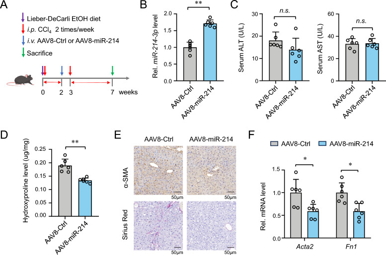
Methods: An adeno-associated virus (AAV)-based system was used to achieve either whole-liver or HSC-specific overexpression of miR-214 (via GFAP promoter) in a mouse model of alcohol-associated liver fibrosis induced by Lieber-DeCarli ethanol diet combined with low-dose CCl₄ injection. Liver fibrosis, steatosis, and inflammation were evaluated by biochemical assays, histology, immunostaining, and gene expression analyses. In vitro, stable miR-214 overexpression and knockdown in LX-2 cells were performed to assess effects on HSC proliferation, transdifferentiation, and ECM gene expression. MECP2 was identified as a direct functional target of miR-214 by bioinformatics and luciferase reporter assays.

Results: miR-214 expression was significantly downregulated during HSC activation in vitro and in fibrotic livers. Whole-liver overexpression of miR-214 alleviated liver fibrosis but caused undesirable steatosis and inflammation. Notably, HSC-specific miR-214 overexpression ameliorated liver fibrosis without inducing these adverse effects. Functionally, miR-214 inhibited HSC proliferation and ECM gene expression, while its inhibition promoted this process. Mechanistically, miR-214 exerts its anti-fibrosis function at least in part by directing targeting MECP2, a critical regulator for HSC activation.

Conclusions: These findings not only identify miR-214 as a promising antifibrotic agent, but also highlight the translational advantage of cell-specific miRNA delivery. HSC-targeted miR-214 gene therapy may offer a promising and safer approach for treating liver fibrosis.

Keywords: AAV; Gene therapy; Hepatic stellate cell; Liver fibrosis; MiR-214.


Conflict of interest statement

Declarations. Ethics approval and consent to participate: All animal experiments were approved by the Institutional Animal Care and Use Committee of Qingdao University (No. 20230901C577220240121151). All experimental procedures followed the approved guidelines. Consent for publication: All authors consent this manuscript for publication. Competing interests: The authors have no conflicts of interest to report.

Figures

Fig. 1
Fig. 1
miR-214 is downregulated in activated HSCs and fibrotic liver. A Morphological images of primary HSCs during spontaneous activation from day 0 to day 2. B RT-qPCR analysis of miR-214 expression in primary HSCs during day 0 to day 2. C RT-qPCR analysis of miR-214 expression in LX2 cells treated with 3 ng/ml TGFβ for 6 h and 12 h. D RT-qPCR analysis of miR-214 expression in LX2 cells treated with 0.3 and 3 ng/ml TGFβ for 12 h. E Experimental design scheme of the alcohol-related fibrosis (ALF) model, which was established by combining Lieber-DeCarli liquid ethanol diet and i.p. injection of low dose of CCl4 (0.6 ml/kg·b.w.). F RT-qPCR analysis of miR-214 expression in the liver of Ctrl and ALF group. G, H Correlation analysis between miR-214 expression and the expression of Acta2 (G) and Fn1 (H). Ctrl, pair-treated group; ALF, alcohol-related fibrosis. **, p < 0.01
Fig. 2
Fig. 2
Whole-liver miR-214 overexpression ameliorates liver fibrosis. A Schemic illustration of the experiment design. The fibrosis model was established by combining Lieber-DeCarli liquid ethanol diet and i.p. injection of CCl4. After 2 weeks, mice were administered with AAV8-miR-214 and AAV8-Ctrl via tail vain injection, and CCl4 i.p. was hold for 1 week. B RT-qPCR analysis of the expression of miR-214 in the liver. C Serum ALT and AST levels. D Liver hydroxyproline levels. E Sirius Red staining and αSMA immunohistochemical staining of the liver tissues. F RT-qPCR analysis of the expression of Acta2 and Fn1 genes. n.s., non-significant; *, p < 0.05; **, p < 0.01
Fig. 3
Fig. 3
Liver-wide miR-214 overexpression boosts liver steatosis and inflammation. A Liver weight. B Liver TG and TC levels. C H&E and Oil-Red O staining of the liver tissues. D RT-qPCR analysis of the gene expression related to fatty acid uptake and lipid metabolism. E F4/80 staining showing liver macrophage infiltration in the liver. F RT-qPCR analysis of the expression of inflammation-related genes. n.s., non-significant; *, p < 0.05; **, p < 0.01
Fig. 4
Fig. 4
HSC-specific miR-214 overexpression alleviates liver fibrosis. A Experimental design scheme. The fibrosis model was established by combining Lieber-DeCarli liquid ethanol diet and i.p. injection of CCl4. After 2 weeks, mice were administered with AAV8-GFAP-miR-214 and AAV8-GFAP-Ctrl via tail vain injection. B The expression level of miR-214 in the liver. C Serum ALT and AST levels. D The expression level of the fibrosis marker genes. E Sirius Red staining of liver tissues. F, G Western blotting showing the expression of proteins in the Smad signal pathway. n.s., non-significant; *, p < 0.05; **, p < 0.01
Fig. 5
Fig. 5
HSC-specific miR-214 overexpression does not promote steatosis and inflammation. A Liver TC and TG levels. B Oil-Red O staining of liver tissues. C RT-qPCR analysis of the expression of genes related to lipid metabolism. D F4/80 staining showing liver macrophages. E RT-qPCR analysis of the expression of inflammation-related genes. n.s., non-significant; *, p < 0.05; **, p < 0.01
Fig. 6
Fig. 6
miR-214 inhibits HSC proliferation. A Schematic illustrating the construction of miR-214 overexpression and inhibition plasmid. B RT-qPCR analysis of the expression of miR-214 in LX2 cells stably expressing (miR-214 OE) or inhibiting miR-214 (miR-214 TuD). miR-214 OE and miR-214 TuD lentivirus were packaged and LX2 cells were infected. Positive cells were selected by puromycin screening. C, D Cell proliferation ability of LX2 cells with miR-214 overexpression (C) or inhibition (D). E, F Colony images (E) and number/area analysis (F) of LX2 cells with miR-214-OE and miR-214 TuD. G, H Cell cycle analysis showing the cell cycle distribution of LX2 cells with miR-214 inhibition (G) or overexpression (H) group. **, p < 0.01
Fig. 7
Fig. 7
miR-214 inhibits ECM gene expression. A KEGG pathway enrichment analysis of miR-214 using the mirPath v.3 tool. B RT-qPCR analysis of the expression of ECM-related genes. C Immunoblotting analysis of the expression of ECM-related proteins. *, p < 0.05; **, p < 0.01
Fig. 8
Fig. 8
MECP2 is a potential target of miR-214. A Nuclear-cytoplasmic fractionation assay showing the cellular localization of miR-214 in LX2 cell. B Prediction of potential target genes of miR-214 using the TarBase, miRDB, TargetScan, and ENCORI online tools. C The expression levels of MECP2 in miR-214 overexpressing LX2 cells. D RT-qPCR analysis of the expression of Mecp2 in liver-wide and HSC-specific miR-214 overexpression group. E Cell ability analysis of MeCP2 overexpression in miR-214 OE LX2 cells. F Targeted binding site of miR-214 to MECP2 mRNA in human and mouse. G Luciferase reporter gene assay showing the direct binding of miR-214 to MECP2 mRNA. *, p < 0.05; **, p < 0.01

Similar articles

References

    1. Bataller R, Brenner DA. Liver fibrosis. J Clin Invest. 2005;115:209–18. - PMC - PubMed
    1. Unalp-Arida A, Ruhl CE. Liver fibrosis scores predict liver disease mortality in the United States population. Hepatology. 2017;66:84–95. - PMC - PubMed
    1. Friedman SL, Pinzani M. Hepatic fibrosis 2022: unmet needs and a blueprint for the future. Hepatology. 2022;75:473–88. - PMC - PubMed
    1. Senoo H. Structure and function of hepatic stellate cells. Med Electron Microsc. 2004;37:3–15. - PubMed
    1. Mederacke I, Hsu CC, Troeger JS, Huebener P, Mu X, Dapito DH, Pradere JP, Schwabe RF. Fate tracing reveals hepatic stellate cells as dominant contributors to liver fibrosis independent of its aetiology. Nat Commun. 2013;4:2823. - PMC - PubMed

MeSH terms

LinkOut - more resources