. 2025 Dec;47(1):2536730.
doi: 10.1080/0886022X.2025.2536730. Epub 2025 Jul 23.

Exploring the comorbidity mechanisms of ITGB2 in rheumatoid arthritis and membranous nephropathy through integrated bioinformatics analysis

Affiliations

Exploring the comorbidity mechanisms of ITGB2 in rheumatoid arthritis and membranous nephropathy through integrated bioinformatics analysis

Wenlong Cao et al. Ren Fail. 2025 Dec.

Abstract

Background: Patients with rheumatoid arthritis (RA) are more likely to comorbid renal diseases, with membranous nephropathy (MN) being the most common. This study aimed to explore the common pathogenesis between RA and MN using integrated bioinformatics analysis.

Methods: Bulk and single-cell RNA sequencing datasets were obtained from the Gene Expression Omnibus and ImmPort databases. Differential expressed genes (DEGs) were identified and enrichment analysis was performed. Topology analysis and the random forest algorithm were applied to identify hub genes. The single-sample Gene Set Enrichment Analysis method was used to assess immune infiltration. Single-cell RNA sequencing analysis was employed to compare the transcript levels of key gene across different cell types. Pseudotime analysis was conducted using Monocle3, and cellular communication was analyzed with CellChat. The L1000FWD database was used to identify potential drugs, and molecular docking was performed.

Results: 66 common upregulated DEGs were identified, primarily associated with leukocyte migration and the chemokine signaling pathway. ITGB2 was finally identified as the shared pathogenic gene of both RA and MN. ITGB2 was predominantly expressed in macrophages, and its expression increased as M0 macrophages differentiated into M1 macrophages. BAFF signaling between macrophages with high ITGB2 expression and B cells/plasma cells was enhanced. Small molecules targeting ITGB2, including LY-294002 and CP466722, may serve as potential drugs for both RA and MN.

Conclusion: As the pathogenic gene shared by both RA and MN, ITGB2 may play a role in M1 macrophage polarization and contribute to the maturation and differentiation of B cells through BAFF signaling.

Keywords: B-cell activating factor; Rheumatoid arthritis; bioinformatics; machine learning; macrophage polarization; membranous nephropathy.


Conflict of interest statement

The authors declare that they have no competing interests

Figures
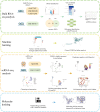
Figure 1.
Figure 1.
Flowchart of this study.
Figure 2.
Figure 2.
Identification of differentially expressed genes (DEGs) and functional enrichment analysis. (A and B) Volcano plots of DEGs in rheumatoid- arthritis (RA) and membranous nephropathy (MN) datasets. (C) Venn diagram to screen common DEGs in RA and MN. (D and E) Heatmaps of 68 common upregulated DEGs in RA and MN. (F) protein-protein interaction (PPI) network of 68 common upregulated DEGs. (G–I) Results of GO enrichment analysis results. (J) Results of KEGG pathway enrichment analysis.
Figure 3.
Figure 3.
Screening of the key gene and immune infiltration analysis. (A) Venn diagram to intersect the results from four topology algorithms (MCC, EPC, degree, MNC). (B) PPI network of 7 common hub genes in RA and MN. (C and D) Correlation between the total number of trees and the error rate in RA and MN datasets based on random Forest algorithm. (E and F) Relative importance scores of 7 hub genes in RA and MN. (G and H) Comparison of immune cell infiltration between diseased and normal samples in RA and MN. (I and J) Correlation analysis between the key gene (ITGB2) expression and immune cell infiltration. ns: no significance, *p < 0.05, **p < 0.01, ***p < 0.001.
Figure 4.
Figure 4.
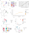
Overview of the scRNA-seq data of RA. (A) The UMAP plot classified by cell type. (B)Proportional representation of each cell type in RA samples. (C) Expression of gene markers in different cells. (D) ITGB2 expression in different cells. (E) Expression of markers associated with macrophage polarization. (F) The UMAP plot of macrophages classified by cell type.
Figure 5.
Figure 5.
Overview of the scRNA-seq data of MN. (A) The UMAP plot classified by cell type. (B) Proportional representation of each cell type in MN samples. (C) Expression of gene markers in different cells. (D) Expression of ITGB2 in different cells. (E) Expression of gene markers in different myeloid cells. (F) The UMAP plot of myeloid cells classified by cell type. (G) ITGB2 expression in different myeloid cells. (H) Expression of markers associated with macrophage polarization. (I) The UMAP plot of macrophages classified by cell type.
Figure 6.
Figure 6.
Pseudotime analysis of macrophage differentiation in RA and MN. (A–D, G–J) Pseudotime trajectory of macrophages. (E and K) Change of ITGB2 expression with pseudotime trajectory in different types of macrophages. (F and L) Expression of ITGB2 in different types of macrophages.
Figure 7.
Figure 7.
Cellular communication analysis in RA and MN. (A and F) Comparison of overall intensity of cellular communication between ITGTB2(+) group and ITGTB2(−) group. ITGB2(−) macrophages were not included in ITGB2(+) group while ITGB2(+) macrophages were not included in ITGB2(−) group. (B and G) Intensity of signals in each cell type. (C, D, H, I) Differences in cellular communication between the two groups when macrophages act as signal receivers or senders. (E and J) Expression of TNFSF13B in different types of macrophage.
Figure 8.
Figure 8.
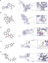
Molecular docking of candidate small molecule drugs with ITGB2. 3D structures and molecular docking of (A) CP466722, (B) emetine, (C) LY-294002, (D) mirin and (E) A-443644.

Similar articles

References

    1. Ronco P, Debiec H.. Pathophysiological advances in membranous nephropathy: time for a shift in patient’s care. Lancet. 2015;385(9981):1983–1992. doi: 10.1016/S0140-6736(15)60731-0. - DOI - PubMed
    1. McGrogan A, Franssen CF, de Vries CS.. The incidence of primary glomerulonephritis worldwide: a systematic review of the literature. Nephrol Dial Transplant. 2011;26(2):414–430. doi: 10.1093/ndt/gfq665. - DOI - PubMed
    1. Couser WG. Primary membranous nephropathy. Clin J Am Soc Nephrol. 2017;12(6):983–997. doi: 10.2215/CJN.11761116. - DOI - PMC - PubMed
    1. Smolen JS, Aletaha D, McInnes IB.. Rheumatoid arthritis. Lancet. 2016;388(10055):2023–2038. doi: 10.1016/S0140-6736(16)30173-8. - DOI - PubMed
    1. Smolen JS, Aletaha D, Barton A, et al. Rheumatoid arthritis. Nat Rev Dis Primers. 2018;4(1):18001. doi: 10.1038/nrdp.2018.1. - DOI - PubMed

MeSH terms