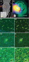
Radiation-induced changes of reactive astrocyte distribution in mice as a late response to partial-brain proton irradiation
- PMID: 40697178
- PMCID: PMC12305689
- DOI: 10.2340/1651-226X.2025.44056
Radiation-induced changes of reactive astrocyte distribution in mice as a late response to partial-brain proton irradiation
Abstract
Background and purpose: After proton therapy of brain tumors, several studies have reported late image changes in follow-up magnetic resonance imaging, which result from blood-brain barrier (BBB) disruption. Astrocytes play a central role in the formation and maintenance of the BBB. To study the late response to partial-brain proton irradiation, preclinical mouse data were utilized to investigate the spatial distribution and dose dependence of reactive astrocytes.
Material and methods: Previously, C57BL/6JRj mice were irradiated with protons targeting the right hippocampal region with single prescription doses of 45-85 Gy. After six months, mice were sacrificed and the excised brains axially cut into 3 µm thick slices and stained for glial fibrillary acidic protein (GFAP) to target astrocytes. Here, a workflow to segment the GFAP-positive area on slice images was established. The fraction of GFAP-positive area (GFAP+ fraction) was evaluated in the high-dose region in the right hemisphere and in the mirrored region in the left hemisphere. Dose distributions were simulated on pre-irradiation cone-beam computed tomography and co-registered to the histological slices.
Results: For all irradiated mice, the GFAP+ fraction in the right hemisphere was significantly increased compared to the left hemisphere and to a sham-irradiated mouse with a highly symmetric GFAP distribution. The GFAP+ fraction in the right hemisphere increased approximately linearly with prescription dose. For comparable doses, the cerebral cortex showed lower GFAP+ fractions than the midbrain.
Interpretation: GFAP upregulation correlated with dose level and distribution. In combination with other markers and timepoints, these findings contribute to a comprehensive understanding of cellular response.
Conflict of interest statement
The authors report there are no competing interests to declare.
Figures


Similar articles
-
Effects of Proton and Combined Proton and (56)Fe Radiation on the Hippocampus.Radiat Res. 2016 Jan;185(1):20-30. doi: 10.1667/RR14222.1. Epub 2015 Dec 31. Radiat Res. 2016. PMID: 26720797
-
Glial fibrillary acidic protein vs. S100B to identify astrocytes impacted by sex and high fat diet.Physiol Behav. 2025 Oct 1;299:115004. doi: 10.1016/j.physbeh.2025.115004. Epub 2025 Jun 18. Physiol Behav. 2025. PMID: 40541637
-
Transcription Factor EB Overexpression through Glial Fibrillary Acidic Protein Promoter Disrupts Neuronal Lamination by Dysregulating Neurogenesis during Embryonic Development.Dev Neurosci. 2025;47(1):40-54. doi: 10.1159/000538656. Epub 2024 Apr 18. Dev Neurosci. 2025. PMID: 38583418 Free PMC article.
-
Interventions for preventing and ameliorating cognitive deficits in adults treated with cranial irradiation.Cochrane Database Syst Rev. 2022 Nov 25;11(11):CD011335. doi: 10.1002/14651858.CD011335.pub3. Cochrane Database Syst Rev. 2022. PMID: 36427235 Free PMC article.
-
Autoimmune glial fibrillary acidic protein astrocytopathy misdiagnosed as intracranial infectious diseases: case reports and literature review.Front Immunol. 2025 Jan 22;16:1519700. doi: 10.3389/fimmu.2025.1519700. eCollection 2025. Front Immunol. 2025. PMID: 39911384 Free PMC article. Review.
References
-
- Bahn E, Bauer J, Harrabi S, Herfarth K, Debus J, Alber M. Late contrast enhancing brain lesions in proton-treated patients with low-grade glioma: clinical evidence for increased periventricular sensitivity and variable RBE. Int J Radiat Oncol. 2020;107(3):571–8. 10.1016/j.ijrobp.2020.03.013 - DOI - PubMed
MeSH terms
Substances
LinkOut - more resources
Full Text Sources
Medical
Miscellaneous

