

Pseudoachalasia as first manifestation of a diffusely infiltrative esophageal squamous cell carcinoma: A case report
- PMID: 40697215
- PMCID: PMC12278238
- DOI: 10.4251/wjgo.v17.i7.108162
Pseudoachalasia as first manifestation of a diffusely infiltrative esophageal squamous cell carcinoma: A case report
Abstract
Background: Pseudoachalasia closely mimics the clinical symptoms of idiopathic achalasia in both clinical symptoms and diagnostic findings, including those from high-resolution manometry and barium esophagography. The similarities often lead to misdiagnosis and the delay of appropriate treatment management. Although most malignancy-associated pseudoachalasia cases are attributed to adenocarcinoma at the gastroesophageal junction, pseudoachalasia due to esophageal squamous cell carcinoma (ESCC) should also be considered. However, the diffuse infiltrative growth patterns that can occur with ESCC can make diagnosis challenging.
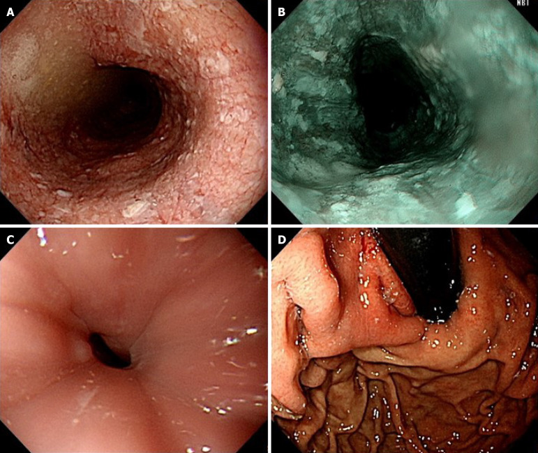
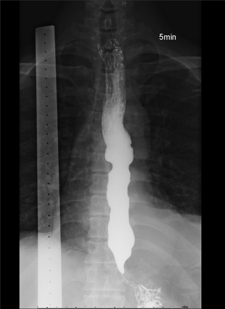
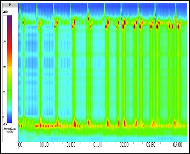
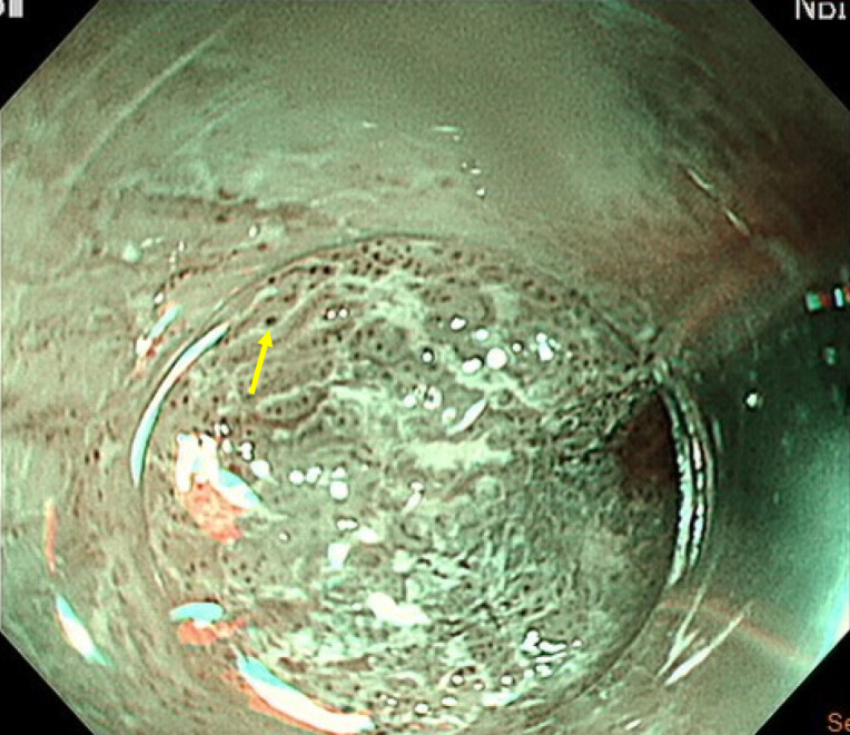
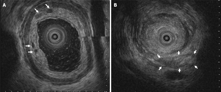
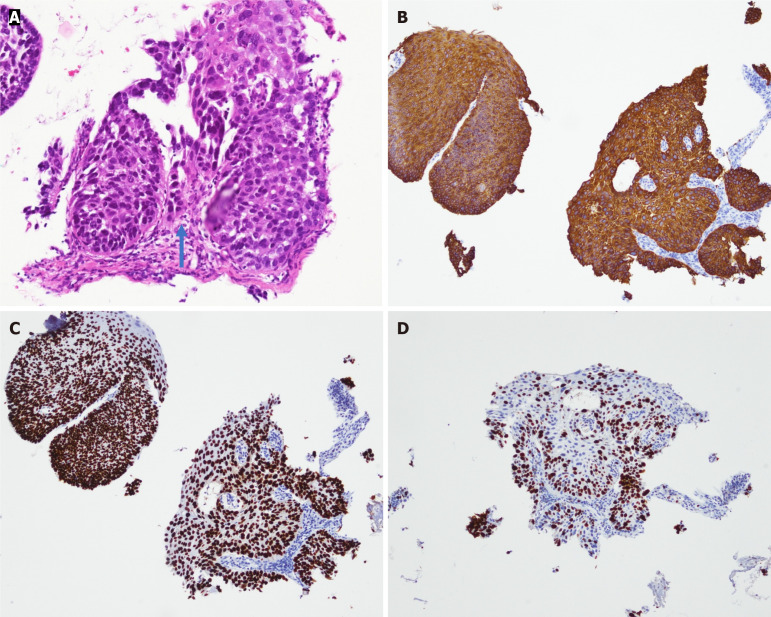
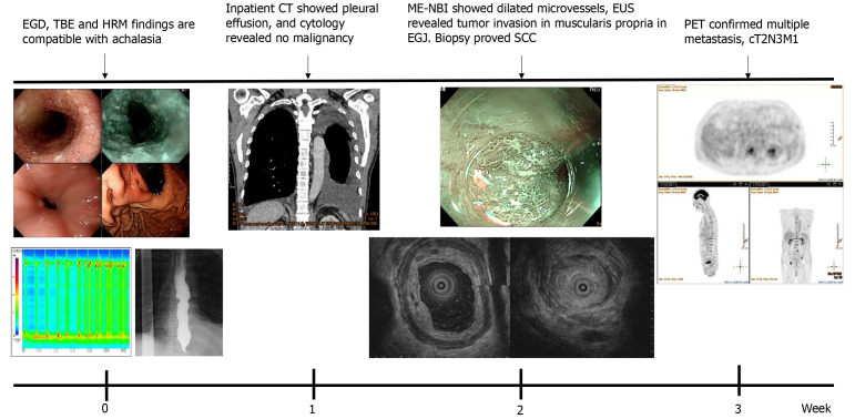
Case summary: We report the case of a 60-year-old man who presented with progressive dysphagia, weight loss, and nocturnal cough. Esophagogastroduodenoscopy, timed barium esophagogram, and high-resolution manometry were conducted. The results of these investigations supported a diagnosis of type II idiopathic achalasia. However, preoperative computed tomography revealed atypical findings, which prompted further evaluation. Repeat endoscopy with magnifying narrow-band imaging identified abnormal mucosal and vascular patterns, and endoscopic ultrasound demonstrated hypoechoic submucosal lesions with involvement of the muscularis propria. Targeted biopsies confirmed moderately differentiated ESCC. Positron emission tomography revealed extensive metastatic disease; therefore, the patient was diagnosed with stage IVB ESCC. Peroral endoscopic myotomy was aborted, and the patient was referred for palliative chemoradiotherapy.
Conclusion: Atypical malignant features should be critically examined. Multimodal tools such as magnifying narrow-band imaging and endoscopic ultrasound are essential for diagnosing pseudoachalasia.
Keywords: Achalasia; Case report; Endoscopic ultrasound; Esophageal squamous cell carcinoma; Magnifying endoscopy; Pseudoachalasia.
©The Author(s) 2025. Published by Baishideng Publishing Group Inc. All rights reserved.
Conflict of interest statement
Conflict-of-interest statement: All the authors report no relevant conflicts of interest for this article.
Figures
References
-
- Schizas D, Theochari NA, Katsaros I, Mylonas KS, Triantafyllou T, Michalinos A, Kamberoglou D, Tsekrekos A, Rouvelas I. Pseudoachalasia: a systematic review of the literature. Esophagus. 2020;17:216–222. - PubMed
-
- Haj Ali SN, Nguyen NQ, Abu Sneineh AT. Pseudoachalasia: a diagnostic challenge. When to consider and how to manage? Scand J Gastroenterol. 2021;56:747–752. - PubMed
-
- Ponds FA, van Raath MI, Mohamed SMM, Smout AJPM, Bredenoord AJ. Diagnostic features of malignancy-associated pseudoachalasia. Aliment Pharmacol Ther. 2017;45:1449–1458. - PubMed
-
- Uedo N, Fujishiro M, Goda K, Hirasawa D, Kawahara Y, Lee JH, Miyahara R, Morita Y, Singh R, Takeuchi M, Wang S, Yao T. Role of narrow band imaging for diagnosis of early-stage esophagogastric cancer: current consensus of experienced endoscopists in Asia-Pacific region. Dig Endosc. 2011;23 Suppl 1:58–71. - PubMed
Publication types
LinkOut - more resources
Full Text Sources
